

 PDF
PDF ePub
ePub Citation
 Print
Print


Abstract
Purpose
To report the results of ganglion cell analysis in a patient with optic tract syndrome who was previously diagnosed with glaucoma.
Case summary
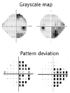
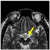
A 32-year-old male, who had been diagnosed with glaucoma 12 years ago, but had not visited an ophthalmology clinic since then, came to our clinic for evaluation of his glaucoma. Both eyes showed an increased cup-to-disc ratio and temporal pallor of the disc. Retinal nerve fiber layer (RNFL) optical coherence tomography showed thinning of the superior, inferior, and temporal peripapillary RNFL in both eyes. On ganglion cell analysis (GCA), ganglion cell layer thinning in the nasal region of the right eye and in the temporal region of the left eye was observed. The visual field test showed right incongruous homonymous hemianopsia. After the atrophic change of the left optic tract was confirmed by orbit magnetic resonance imaging, he was diagnosed with left optic tract syndrome.
Figures and Tables
Figure 1
Results of fundus photography. Both optic discs showed increased cup-disc ratio and mild temporal pallor.
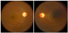
Figure 2
Results of RNFL optical coherence tomography (OCT). RNFL OCT showed thinning of superior, inferior and temporal RNFL of both eyes. RNFL = retinal nerve fiber layer; N = nasal; T = temporal.
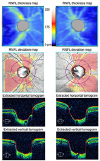
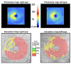
Figure 3
Results of ganglion cell analysis. On ganglion cell analysis, thinning of the ganglion cell layer was prominent in the nasal region of the right eye and the temporal region of the left eye respecting the vertical meridian.
References
1. el Gammal T, Brooks B, Harbour R, et al. MR of uncommon congenital and vascular lesions of the intracranial visual pathways. Neuroradiology. 1990; 32:488–491.


2. Margo CE, Hamed LM, McCarty J. Congenital optic tract syndrome. Arch Ophthalmol. 1991; 109:1120–1122.
3. Murphy MA, Grosof DH, Hart WM Jr. Congenital optic tract syndrome: magnetic resonance imaging and scanning laser ophthalmoscopy findings. J Neuroophthalmol. 1997; 17:226–230.
4. Lee EJ, Kim JH, Hwang JM. Congenital optic tract syndrome misdiagnosed with normal tension glaucoma. Graefes Arch Clin Exp Ophthalmol. 2016; 254:2481–2483.
5. Kim JY, Lee HJ, Kwag JY, Lee YH. The optical coherence tomography findings of optic tract syndrome. J Korean Ophthalmol Soc. 2013; 54:1144–1148.
6. Shin HY, Park HY, Choi JA, Park CK. Macular ganglion cell-inner plexiform layer thinning in patients with visual field defect that respects the vertical meridian. Graefes Arch Clin Exp Ophthalmol. 2014; 252:1501–1507.
7. Savino PJ, Paris M, Schatz NJ, et al. Optic tract syndrome. A review of 21 patients. Arch Ophthalmol. 1978; 96:656–663.
8. Unsöld R, Hoyt WF. Band atrophy of the optic nerve. The histology of temporal hemianopsia. Arch Ophthalmol. 1980; 98:1637–1638.
9. Newman SA, Miller NR. Optic tract syndrome. Neuro-ophthalmologic considerations. Arch Ophthalmol. 1983; 101:1241–1250.
10. Kupfer C, Chumbley L, Downer JC. Quantitative histology of optic nerve, optic tract and lateral geniculate nucleus of man. J Anat. 1967; 101(Pt 3):393–401.

11. Hsu CY, Lai YH, Hsu SY, et al. Optical coherence tomography (OCT) findings in patients with optic tract syndrome. Taiwan J Ophthalmol. 2011; 1:16–20.
 XML Download