

 PDF
PDF ePub
ePub Citation
 Print
Print


Abstract
Purpose
We report a case of acute dacryocystitis diagnosed with abscess and rupture of lacrimal sac and fistula to posterior orbit during the operation.
Case summary
A 71-year-old woman visited our clinic with edema and pain in the eyelid from three days ago. For past four months, there was viscous of the left eye and tears. The patient had severe conjunctival chemosis and hyperemia, compared with the left eyelid edema and redness. Orbital CT scan showed orbital cellulitis, which was followed by systemic antibiotics and steroid therapy. On the 4th day of therapy, orbital abscess formation was observed in orbit MRI and surgical drainage was planned. During surgery, we found rupture of the posterior part of lacrimal sac and fistula to posterior orbit. Pseudomonas aeruginosa was identified in the bacterial cultures, and after the administration of appropriate antibiotics, the disease showed improved progress, and then additional dacryocystorhinostomy was performed.
Conclusions
In our case, acute dacryocystitis rarely spread in orbit, which may lead to delayed diagnosis, orbital cellulitis and abscess, resulting in serious complications of vision threat. So, we think that it is necessary to consider surgical treatment more actively in the stage of chronic dacryocysitis.
References
1. Connell B, Kamal Z, McNabb AA. Fulminant orbital cellulitis with complete loss of vision. Clin Exp Ophthalmol. 2001; 29:260–1.

2. Mauriello JA Jr, Wasserman BA. Acute dacryocystitis: an unusual cause of life threatening orbital intraconal abscess with frozen globe. Ophthalmic Plast Reconstr Surg. 1996; 12:294–5.
3. Kikkawa DO, Heinz GW, Martin RT, et al. Orbital cellulitis and abdominal secondary to dacryocystitis. Arch Ophthalmol. 2002; 120:1096–9.
4. Ataullah S, Sloan B. Acute dacryocystitis presenting as an orbital abscess. Clin Experiment Ophthalmol. 2002; 30:44–6.
5. Choi YJ, Choi WC, Yang SW, Choi Y. A case of orbital abscess secondary to dacryocystitis. J Korean Ophthalmol Soc. 2005; 46:156–63.
6. Lee SM, Yen MT. Management of preseptal and orbital cellulitis. Saudi J Opthalmol. 2011; 25:21–9.
7. Weakley DR. Orbital cellulitis complicating strabismus surgery: a case report and review of the literature. Ann Ophthalmol. 1991; 23:454–7.
8. Bassetti M, Vena A, Croxatto A, et al. How to manage pseudomonas aeruginosa infections. Drugs Context. 2018; 7:212527.
9. Chen L, Silverman N, Wu A, et al. Intravenous steroids with abdominals for children with orbital cellulitis. Ophthalmic Plast Reconstr Surg. 2018; 34:205–8.
10. Goytia VK, Giannoni CM, Edwards MS. Intraorbital and abdominal extension of sinusitis: comparative morbidity. J Pediatr. 2011; 158:486–91.
11. Patt BS, Manning SC. Blindness resulting from orbital complications of sinusitis. Otolaryngol Head Neck Surg. 1991; 104:789–95.
12. Maheshwari R, Maheshwari S, Shah T. Acute dacryocystitis abdominal orbital cellulitis and abscess. Orbit. 2009; 28:196–9.
13. Martins MC, Ricardo JR, Akaishi P, et al. Orbital abscess abdominal to acute dacryocystitis: case report. Arq Bras Oftalmol. 2008; 71:576–8.
14. Coden DJ, Hornblass A, Haas BD. Clinical bacteriology of abdominal in adults. Ophthalmic Plast Reconstr Surg. 1993; 2:125–31.
Figure 1.
Initial photograph and gaze of extraocular movement. Clinical photograph shows lid swelling, proptosis, conjunctival chemosis, and hyperemia (A). Initial examination shows limitation of movement in all directions in the left eye (B).
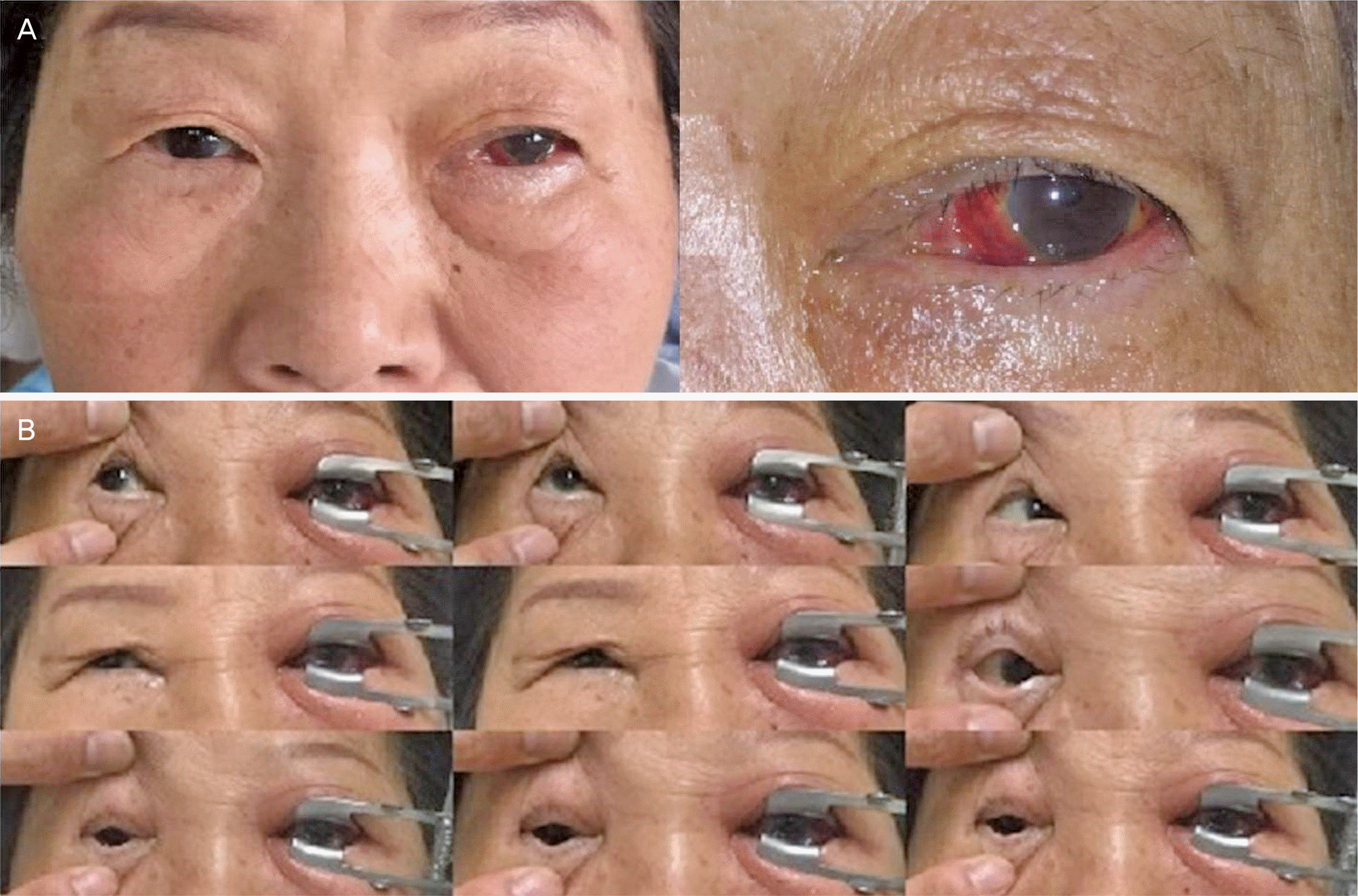
Figure 2.
Computed tomography (CT) image and magnetic resonance imaging (MRI) of the patient. Initial orbital CT scan shows low density lesion (arrow) at inferomedial aspect and the left eyeball was protruded by the lesion (A). There was no obvious evidence of inflammation near the fossa of lacrimal sac (B). Orbital enhanced fat-saturated T1-weighted MRI, four days after systemic antibiotics and steroid therapy, shows orbital abscess formation (arrow) in intraconal space (C).
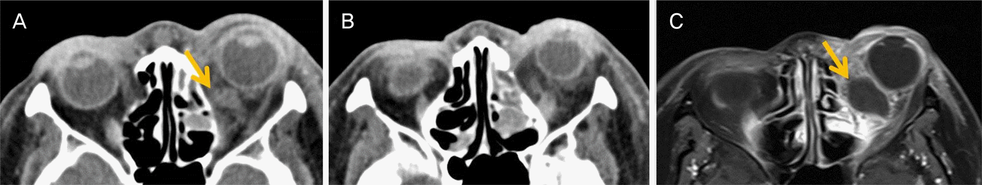
Figure 3.
Surgical procedures of the abscess drainage. Through an incision between the caruncle and plica semilunaris, soft tissue dissection is performed. Abscess drainage was done (A). Ruptured lacrimal sac (circle) and abscess (arrow) was found (B). The fistula from lacrimal sac to orbit was found using lacrimal probe (circle) (C).
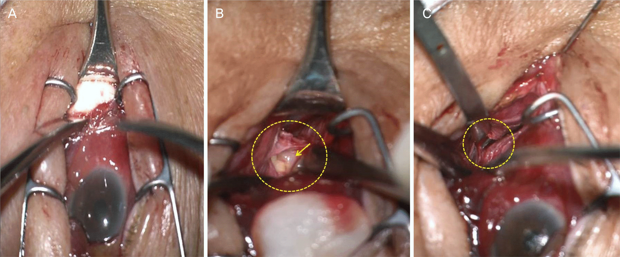
Figure 4.
Follow-up photograph and orbital computed tomography (CT). Clinical photograph, three months after abscess drainage, shows remarkable improvement of the lid swelling, proptosis and conjunctival chemosis (A). Orbital CT scan, three months after abscess drainage, shows marked decrease of the previous low density lesion (B).

 XML Download