

 PDF
PDF ePub
ePub Citation
 Print
Print


INTRODUCTION
Foreign body injections into breasts may produce local swelling of involved lymph nodes, which may be misdiagnosed as metastasis or malignancy (1). Contralateral internal mammary paraffin lymphadenopathy, mimicking breast cancer metastasis has not been described. Here, we report the case of a 58-year-old woman with history of interstitial mammoplasty via paraffin injection in both breasts, that presented with contralateral internal mammary lymphadenopathy, suspicious of breast cancer metastasis by magnetic resonance imaging (MRI) and positron emission tomography-computed tomography (PET-CT).
CASE REPORT
The 58-year-old woman concerned had history of liquid paraffin injection in both breasts 10 years previously, and had been diagnosed 2 years previously with left breast cancer and undergone left skin-sparing mastectomy, and left axillary lymph node dissection. Following complete tumor removal, bilateral breast reconstruction with submuscular silicone implants was performed. During surgery, most paraffinoma in both breasts was removed. Histopathological report of the left breast was invasive ductal carcinoma, not otherwise specified, of Bloom-Richardson grade 3, and 17 of 25 lymph nodes showed macrometastasis with extracapsular extension. TNM staging was pT2N3MX, and after surgery, she received adjuvant chemotherapy.
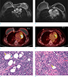
She was followed routinely by her oncologist for 2 years, with no evidence of breast cancer local recurrence or metastasis. However, she underwent breast MRI during routine follow-up a month prior to this presentation. Axial contrast-enhanced T1-weighted breast MR images showed two abnormally enlarged lymph nodes (0.8-cm and 1.0-cm) at the right first and third intercostal spaces (Fig. 1a, b). PET-CT was performed for further evaluation, and axial images showed increased 18F-fluorodeoxyglucose (FDG) uptake, confined to the same right internal mammary lymph nodes not observed in pre-operative PET-CT scan (Fig. 1c, d). SUV max values of two enlarged nodes were 1.6 and 3.2, respectively, and both were suspicious for breast cancer metastasis based on MRI and PET-CT findings. Nodes were not accessible by imaging guided percutaneous techniques, and thus, biopsy was performed by single-port video-assisted thoracoscopy. Biopsy specimen showed drained lipoid droplets into the lymph node. Multiple empty spaces containing fragments of refractile unstained material, showed a relatively thick and fibrotic wall. Macrophages with microvesicles were also dispersed in the lymph node (Fig. 1e, f). The picture was consistent with pathology of paraffin lymphadenopathy.
DISCUSSION
Interstitial mammoplasty by liquid paraffin injection, was used in the early 1900s until silicone was introduced (2). However, this method had serious complications, including local granulomatous reactions, material-induced mastitis, foreign body reactions, fibrosis, material migration, and autoimmune reactions (3). In addition, liquid paraffin was reported to interfere with physical examinations, mammography, and ultrasonography (45). Dynamic contrast enhanced MR imaging is a reliable imaging modality, in patients with foreign body injection (6).
Contralateral internal mammary silicone lymphadenopathy, as a complication of breast prosthesis implantation, has been described twice in literature (78).
This case demonstrates that in a patient with disrupted lymph drainage due to prior mastectomy and axillary node dissection for breast cancer, paraffin particles in breast parenchyma can migrate to internal mammary lymph nodes that imitate cancer metastasis. Although FDG positive lesions have high likelihood of harboring malignancy, false-positive uptake is not uncommon, and differential diagnosis of such lesions includes infection, inflammation, and granulomatous, including foreign body, related changes (9). Lymphadenopathy in patients with history of breast cancer raises concern of recurrence, and warrants prompt evaluation to prevent delays in diagnosis and treatment. Enlarged internal mammary nodes present a diagnostic challenge, due to interference by foreign body granuloma induced by paraffin injection on ultrasound, inaccessibility, and sample errors associated with imaging guided percutaneous needle biopsy (1). Video-assisted thoracoscopy provides reliable means of achieving accurate diagnosis.
In conclusion, we report a rare case of contralateral internal mammary paraffin lymphadenopathy suspicious of breast cancer metastasis, in a patient with history of breast cancer and previous interstitial mammoplasty by paraffin injection in both breasts.
 XML Download