

 PDF
PDF ePub
ePub Citation
 Print
Print


INTRODUCTION
Primary bone lymphoma is a rare disease, accounting for less than 5% of primary bone tumors and 3% of malignant bone tumors (12). Most of the primary bone lymphomas represent non-Hodgkin lymphoma, especially diffuse large B-cell lymphoma (DLBCL) (3). Clinically, bone lymphomas present as palpable masses with intermittent bone pain (4). According to a previous study (5), the metadiaphysis of the femur is the most common site involved. Other sites include pelvis, tibia, head and neck, and vertebra. Lymphoma localized to peripheral sites such as the feet and the hands, or the toes and the fingers, is rarely reported. Here, we report a very rare case of solitary primary bone lymphoma located in the middle phalanx of the third finger.
CASE REPORT
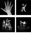
A 71-year-old male presented to our institution complaining of swelling in the right third finger, with a six-month history of symptoms. He had no specific medical history. Laboratory testing revealed elevated erythrocyte sedimentation rate and C-reactive protein (51 mm/h and 5.8 mg/dL, respectively). Plain radiograph revealed evidence of bony mass causing cortical disruption at the radial aspect of the third middle phalanx (Fig. 1a). The patient underwent magnetic resonance imaging (MRI) for further evaluation. T2-weighted MRI showed a well-defined mass of high signal intensity, involving the adjacent 3rd middle phalangeal bone with large soft tissue component. The mass was thought to have originated in the bone rather than soft tissue, because the center of the mass was near the cortex of the phalangeal bone and normal cortical bone was noted within the boundary of the mass. The mass abutted with the flexor tendon of the third finger (Fig. 1b). T1-weighted imaging revealed a mass of intermediate signal intensity (Fig. 1c). The contrast-enhanced MRI showed strong and homogeneous enhancement (Fig. 1d). Based on the imaging findings, the differential diagnosis included mimics of primary bone tumor such as glomus tumor or intraosseous epidermal inclusion cyst and metastatic bone tumor, despite its rarity. Total excision was performed for symptom relief.
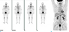
A yellowish-white soft tissue mass causing lysis of the middle phalangeal bone was visible in the operative field. Staining with Hematoxylin and Eosin (H&E) revealed diffuse infiltration of atypical cells (Fig. 2a). Immunohistochemical analysis showed positive test results for leukocyte common antigen (LCA) (Fig. 2b) and CD20, and negative findings for CD45R0, pan cytokeratin, synaptophysin, CD56, and CD99. Therefore, the final pathologic diagnosis was DLBCL. Chest and abdominal computed tomography (CT) scan, bone scan (Fig. 3a), and positron emission tomography (PET)-CT (Fig. 3b) were performed to determine the involvement of other organs. However, no evidence of lymphoma involvement was detected at any other site, nor was any evidence of disease spread to other organs detected during the 6 months of follow-up.
DISCUSSION
Lymphoma is the most common hematologic disorder of the lymphoreticular tissue. However, lymphomas can also involve organs outside of the lymphatic system, referred to as extranodal lymphoma. Extranodal lymphoma may occur in the gonads, gastrointestinal tract, lung, central nervous system and bone. In primary bone lymphoma, axial rather than appendicular skeleton is more commonly affected (6). Moreover, the small bones of the peripheral extremities are very rarely involved in bone lymphoma. To our knowledge, only few cases (7), which were confirmed as primary bone lymphoma, involved the hand.
The diagnostic criteria include the presence of lymphoma involving an osseous site, without evidence of any other organ involvement for at least 6 months after the initial diagnosis (8). The regional lymph node involvement cannot exclude a diagnosis of primary bone lymphoma. In our case, the palpable mass on the third finger was the initial manifestation of the disease, without any masses involving other organs on systemic evaluation. The patient visited our hospital in May, following initial plain radiograph at another hospital in January of the same year, showing the finger mass. Therefore, no evidence of metastatic spread was found about 6 months after the initial presentation. Thus, it is reasonable to define our case as primary bone lymphoma.
According to a previous report (6), the majority of primary bone lymphoma occurs in older adults, and more than 90% of patients are over the age of 30 years. In a large series, patients diagnosed with primary bone DLBCL were younger (median age, 56 years) than patients with nonosseous DLBCL and secondary bone lymphoma (9). In our case, the patient was 71 years old. Additionally, men are more frequently affected than women, with a ratio ranging from 1.2 to 1.8 (6). Femur is the most common site of involvement in nearly 50% of the cases. The pelvis is affected in about 20%, and other sites including the vertebra, ribs and tibia account for the remainder (5).
DLBCL is the most common histological subtype of primary bone lymphoma, as in our case (6). Rarely, follicular lymphoma, small lymphocytic lymphoma and marginal zone lymphoma may manifest as primary bone lymphoma.
Imaging findings of primary bone lymphoma vary, and may be non-specific in some patients (4). Although, the imaging findings of bone lymphoma exhibit a wide spectrum, a few features are characteristic of the disease. Typically, lymphomatous bone involvement is defined by a solitary lytic lesion near the end of a long bone with permeative or moth-eaten appearance, and aggressive periosteal reaction (10). In addition, extensive surrounding soft-tissue masses without significant cortical destruction has been reported exclusively in round cell tumors including bone lymphoma, multiple myeloma and Ewing sarcoma (3).
However, in our case, none of these typical imaging findings of bone lymphoma was detected. Therefore, the initial radiologic diagnosis was not focused on lymphoma, but rather on the more commonly encountered masses associated with the finger.
Here, we reported a very rare case of solitary primary bone lymphoma involving the third finger. Although rare with unusual location and imaging findings, the possibility of this disease should be considered to avoid misdiagnosis.

 XML Download