

 PDF
PDF ePub
ePub Citation
 Print
Print


Dear Editor:
Demand for new biomaterials that produce good results in clinical fields requiring tissue restoration and replacement is expected to continue to increase1. Poly-L-lactic acid (PLLA) has been widely used since it was approved by the Food and Drug Administration (U.S. FDA) as an injectable facial volumizer in the treatment of lipoatrophy due to its significant therapeutic effectiveness2. However, except for those clinical and histological reports, few data on the molecular biologic effects of PLLA have been reported3. In each of these studies, PLLA-induced mild inflammation and the paracrine effects of foreign body giant cells are presumed to be the central mechanistic feature, resulting in increases in the production of new collagen over the months. However, the exact mechanisms of how PLLA can increase collagen production are still unknown. Based on our clinical experiences, the facial volume-augmenting effect of PLLA was noticeable even at one month after injection, which is much faster than the rate reported in papers; thus, we hypothesized that PLLA could directly affect dermal fibroblasts34. The goal of this paper was to assess the effect of PLLA on collagen synthesis and related signal pathways in cultured dermal fibroblast.
Hs68 (human dermal fibroblast) and cell line were cultured and stimulated with PLLA. Vials of dry powder of PLLA (Sculptra®; Sanofi Aventis, Paris, France) were used in the present study. Five milliliters of sterile water was added to the dry powder of Sculptra® and finally adjusted to 0.1% concentration in the culture medium5. Type I collagen gene expression induced by PLLA was evaluated using real time RT-PCR, and we performed ELISA and Western-blot analyses to confirm the effect of PLLA on type I collagen synthesis.
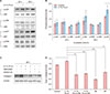
The dramatically increasing effects of PLLA on type I collagen gene expression were detected using real time RT-PCR at 48-hours incubation, suggesting that PLLA increased transcription of type I collagen gene within a relatively shorter period of time than expected (Fig. 1A). To examine whether the increase in collagen mRNA was followed by an increase in collagen protein synthesis, we measured the procollagen concentration in the medium according to the manufacturer's protocol (Takara Bio, Otsu, Japan). Consistent with the data on collagen gene expression, the amounts of procollagen were significantly increased by PLLA (Fig. 1B). In the Western blot analysis, treatment of Hs68 with 0.1% PLLA resulted in a significant increase in type I collagen in 48 hours (Fig. 1C, D). To dissect the signal transduction pathways involved in the up-regulation of type I collagen production, various signal molecules were tested (Supplementary Fig. 1). Only the anti-phospho p38 mitogen-activated protein kinase (p38 MAPK), JNK, and Akt antibodies were detected specifically as the phosphorylated form of p38, Akt, and JNK, respectively, after the PLLA treatment (Fig. 2A, B). Thus, we focused on the functional role of those molecules in the up-regulatory effect of PLLA. Pre-incubation of Hs68 cell with SB203580 (p38/Akt inhibitor) and SP600125 (JNK inhibitor) resulted in significant inhibition of PLLA-induced collagen synthesis (52% and 42%, respectively) (Fig. 2C, D). Together, these data demonstrated that PLLA has a direct stimulatory effect on collagen production, and these results were mediated through the activation of the signaling proteins of p38, Akt, and JNK.
It has been reported that one of the most important characteristics of biomaterials is their ability to trigger adequate levels of host inflammatory responses. Therefore, designing biomaterial-based therapies must be accompanied by biomaterial-immune system interactions6. PLLA has become one of the most promising biomaterials because of its advantages, such as biocompatibility, biodegradability, and minimal inflammatory inductive property4. PLLA is superior to conventional classic fillers in terms of stability and effect persistence; thus, it is thought that not only the face but also any part of the body will be used more as a supplement to fill tissue defects3. At the sites where the PLLA was administered, a weak inflammatory reaction, infiltration of foreign body giant cells, and elongated fibroblasts were observed, which are presumed to be the central mechanistic features4. Type I collagen statistically and significantly increased from baseline to 6 months after PLLA injection in human skin78. Moreover, the clinically volume-augmenting effect of PLLA could be confirmed even within one month after injection, which is much faster than the rate reported in an other study8. Therefore, it is assumed that there is another mechanism of collagen synthesis that enables early volumizing results. In this study, we have found that PLLA directly acts on dermal fibroblasts leading to a significant increase in type I collagen gene expression and protein synthesis. In addition, those reactions already began at 48-hours incubation and signal pathways through p38, Akt, and JNK were involved. These results supported by previously published reports suggest that MAPK and Akt pathways are involved in polymer-induced collagen synthesis. Ren et al.9 reported that both MAPK and Akt signaling pathways were activated by hydroxyapatite and PLLA, but its precise mechanisms of action remain to be elucidated. The Akt signaling pathway is involved in fibroblast migration and promotes collagen synthesis, resulting in cutaneous wound contraction10.
Our results provide the first evidence that PLLA directly stimulates dermal fibroblasts to increase collagen synthesis through activation of the p38, Akt, and JNK signaling pathways might be influenced by either an adjacent macrophage, which may serve as a late trigger of collagen synthesis or a limitation of the in vitro condition. However, it must be noted that the present study is limited in providing insights into the collagen synthesis effects of PLLA in an artificial in vitro system. As collagen synthesis is a complex in vivo event, many questions remain to be addressed. These in vitro data nevertheless provide a concept that may allow insights into the biologic effect of PLLA on fibroblast. Furthermore, P38, Akt, and JNK are signal transduction molecules that regulate cell growth, differentiation, and apoptosis. Hence, further research about the effects of PLLA on other extracellular matrix components is needed in the future.

 XML Download