

 PDF
PDF ePub
ePub Citation
 Print
Print


Dear Editor:
Acne vulgaris is a very common cutaneous disorder caused by hormone, sebum production, follicular hyperkeratinization, Propionibacterium acnes and inflammation. Over the last decade, it has become apparent that oxidative stress is linked to the pathogenesis of acne1. Sahib et al.2 reported that the oxidative stress of patients with acne was higher than in healthy individuals. Conversely, antioxidant activities have been found to be lower in patients with acne. Therefore, local and systemic antioxidants have been administered to control acne vulgaris3. There are numerous antioxidants; including lipoic acid, L-ascorbic acid, α-tocopherol, β-carotene and astaxanthin4. Lipoic acid is usually used as an antioxidant agent for the treatment of skin aging; however, reports on the treatment of acne with lipoic acid are limited. Lipoic acid, also known as alpha-lipoic acid; is an organosulfur compound derived from octanoic acid.
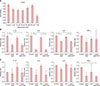
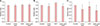
Primary human sebocytes were cultured using sebaceous glands obtained from occipital hair follicles. The sebocytes were cultured in Dulbecco's modified Eagle medium (DMEM; Hyclone Laboratories, Logan, UT, USA) in a humidified atmosphere with 5% CO2 at 37℃. The culture media were changed from DMEM to Epilife (MEPI500CA; Gibco BRL, Grand Island, NY, USA), and the sebocytes obtained after the second subculture were used. After the determination of alpha-lipoic acid (Sigma, St. Louis, MO, USA) concentration using the Cell Counting Kit-8 (CCK8; Dojindo, Kumamoto, Japan) assay, the human sebocyte culture media were supplemented with 50 µM of alpha-lipoic acid (Fig. 1A). Lipopolysaccharide (5 µg) (Sigma) or dihydrotestosterone (10−7 M) (Sigma) was added into the human sebocyte culture media. An enzyme-linked immunosorbent assay (ELISA) was used for the measurement of biomarker protein expression in cultured human sebocytes after treatment with alpha-lipoic acid (50 µM) and lipopolysaccharide (5 µg) or dihydrotestosterone (10−7 M). The ELISA was used for the analysis of biomarkers, including interleukin (IL): IL-1β, IL-6, IL-8 and tumor necrosis factor (TNF)-α (R&D Systems, Shanghai, China); according to the manufacturer's instructions. The TG-S reaction kit (Asan Pharm. Co., Seoul, Korea) was used for the detection of neutral lipids after the treatment of sebocytes with alpha-lipoic acid (50 µM), dihydrotestosterone (10−7 M), or a combination of lipoic acid (50 µM) and dihydrotestosterone (10−7 M). The BioVision lipid peroxidation assay kit (Biovision, Milpitas, CA, USA) was used for the sensitive detection of malondialdehyde assays. The ELISA data were evaluated using analysis of variance statistics (ver. 18.0; IBM Co., Armonk, NY, USA). The data were considered statistically significant when p<0.05.
The treatment of cultured human sebocytes with alpha- lipoic acid (50 µM) showed a statistically significant decrease in protein expression of IL-1β, IL-6 and IL-8 compared to control (p<0.05) (Fig. 1B). The treatment of cultured human sebocytes with alpha-lipoic acid (50 µM) and dihydrotestosterone (10−7 M) also showed a statistically significant decrease in protein expression of IL-1β, IL-6 and IL-8 compared to control (p<0.05) (Fig. 1B). In addition, the treatment of cultured human sebocytes with alpha- lipoic acid (50 µM) and dihydrotestosterone (10−7 M) showed a statistically significant decrease in protein expression of IL-6 and IL-8 compared to the treatment with dihydrotestosterone (10−7 M) (p<0.05) (Fig. 1B). The treatment of cultured human sebocytes with lipopolysaccharide (5 µg) insignificantly increased protein expression of IL-1β, IL-6 and IL-8 compared to control (Fig. 1C). The treatment of cultured human sebocytes with alpha-lipoic acid (50 µM) and lipopolysaccharide (5 µg) insignificantly decreased protein expression of IL-1β, IL-6 and IL-8 compared to control (Fig. 1C). In addition, the treatment of cultured human sebocytes with alpha-lipoic acid (50 µM) and lipopolysaccharide (5 µg) insignificantly decreased protein expression of IL-1β, IL-6, IL-8 and TNF-α compared to the treatment with lipopolysaccharide (5 µg) (Fig. 1C). There was no significant influence of alpha-lipoic acid (50 µM) and/or dihydrotestosterone (10−7 M) on the sebum production of cultured human sebocytes (Fig. 2A). Alpha-lipoic acid (50 µM) insignificantly decreased lipid peroxidation in cultured human sebocytes (Fig. 2B). Dihydrotestosterone (10−7 M) insignificantly increased lipid peroxidation in cultured human sebocytes (Fig. 2B). Lipid peroxidation in cultured human sebocytes after treatment with both alpha-lipoic acid (50 µM) and dihydrotestosterone (10−7 M) was insignificantly decreased compared to control or the treatment with dihydrotestosterone (10−7 M) (Fig. 2B). Lipid peroxidation in cultured human sebocytes after treatment with alpha-lipoic acid (50 µM) and lipopolysaccharide (5 µg) was insignificantly decreased (Fig. 2C).
Oxidative stress has been related to the pathogenesis of various skin diseases, including acne vulgaris. Potent reactive oxygen species can be generated in acne through damage to the follicular epithelium by inflammatory cells5. Oxidative stress in skin can also be induced by ultraviolet B irradiation6. In addition, lipopolysaccharide and Propionibacterium acnes can cause oxidative stress in the skin7. Antioxidants including lipoic acid can be used to treat patients with acne, however, it is unclear whether or not androgens are antioxidants. It was reported that testosterone can act as an anti-oxidizing agent in collagen-induced arthritis in rats8. On the contrary, it has also been reported that physiological levels of androgens can increase oxidative stress in androgen-responsive prostate carcinoma cells9. In this study, lipid peroxidation was decreased after treatment of cultured human sebocytes with alpha-lipoic acid. In addition, dihydrotestosterone slightly increased lipid peroxidation. Furthermore, alpha-lipoic acid decreased lipid peroxidation after treatment of cultured human sebocytes with dihydrotestosterone. In addition, treatment with both alpha-lipoic acid and lipopolysaccharide showed a decreased tendency towards lipid peroxidation compared to treatment with lipopolysaccharide alone. Inflammatory biomarkers, including IL-1β, IL-6, IL-8 and TNF-α, have been linked to the pathogenesis of inflammatory acne. Lipoic acid inhibited the expression of these inflammatory biomarkers. Despite the addition of dihydrotestoterone or lipopolysaccharide to the cultured human sebocytes, lipoic acid still inhibited the expression of the inflammatory biomarkers. Both dihydrotestosterone and lipopolysaccharide can upregulate the expression of inflammatory biomarkers in cultured human sebocytes10. The mechanism by which lipoic acid inhibits the expression of inflammatory biomarkers should be elucidated. Dihydrotestosterone is well known to influence sebum production and secretion. Human sebaceous glands, which are the sources of sebum production; express a variety of factors related to androgen biosynthesis. However, since primary cultured human sebocytes differ to sebocytes in vivo, cultured human sebocytes may show limited sebocyte differentiation even when incubated with androgens. Transcription factors such as CCAAT/enhancer binding proteins and peroxisome proliferator-activated receptors, in addition to androgens are very important for sebocyte production of sebum. In this study, dihydrotestosterone did not result in a significant increase in sebum production. Lipoic acid also did not influence sebum production in cultured human sebocytes. Therefore, we propose that lipoic acid is an antioxidant, and it acts as an anti-inflammatory agent.
 XML Download