

 PDF
PDF ePub
ePub Citation
 Print
Print


INTRODUCTION
Acne vulgaris is a very common skin disease of the pilosebaceous follicles characterized by comedones, papules, pustules, nodules, and cysts1. One of the main causative factors of acne is colonization of the follicular infundibulum and sebaceous duct by Propionibacterium acnes. P. acnes plays an important role in inducing an inflammatory event2. In addition, P. acnes can lead to the development of abnormal differentiation and keratinization in epidermal keratinocytes1. Therefore, P. acnes may act as a major causative factor in early inflammatory papules of acne.
The establishment of animal models for various diseases is helpful for expanding research fields and developing the production of new therapeutic modalities. Although there are various acne animal models, such as the Mexican hairless dog, the Rhino mouse, and the rabbit ear assay, elucidative models are still needed3. Previously, we reported the establishment of an acne mouse model; HR-1 mice develop acne-like inflammatory nodules on their backs following injection of P. acnes, in contrast to BALB/c, VDR k/o, and SCID mice4.
Bacteriophages are bacterial viruses consisting of proteins that encapsulate a DNA or RNA genome5; they are widely distributed in sites populated by bacteria. Bacteriophages replicate within bacteria after injecting their genome into the bacterial cytoplasm. They tend to kill only specific pathogens without damaging the normal flora; thus, they can be used as living drugs for bacterial infections, including acne6. Bacteriophages targeting P. acnes have been isolated from acne patients7.
In this study, we investigated the efficacy of bacteriophages in a P. acnes-induced acne mouse model by clinical examination, histopathology, and immunohistochemical studies.
MATERIALS AND METHODS
P. acnes culturing
P. acnes (ATCC 11828) were isolated from pustular lesions of Korean patients with moderate inflammatory acne. P. acnes from post log phase cultures were grown on brain heart infusion agar under anaerobic conditions at 37℃. P. acnes cells were harvested by centrifugation at 5,000×g for 10 minutes, washed three times with phosphate buffered saline (PBS), and resuspended to a concentration of 109 colony forming units/ml in PBS for the experiments.
Acne mouse model using HR-1 mice and injection of P. acnes and/or bacteriophages into HR-1 mice
Six-week-old female Hos:HR-1 mice (HR-1; SLC Inc., Hamamatsu, Japan) were maintained under conventional laboratory conditions following 1 week of acclimation. A live P. acnes suspension was injected intradermally (20 µl aliquots), using a 30-gauge needle, on both sides of the backs of three mice per group (group A, group B, and group C). Group A was used as the control; Group B was re-injected with P. acnes on the back 4 weeks following the first P. acnes injection; and Group C was injected with P. acnes and bacteriophages on the back 4 weeks following the first P. acnes injection. Bacteriophages were injected intradermally in 20 µl aliquots at the P. acnes injection site. This procedure was performed in accordance with the institutional guidelines for the care and use of laboratory animals (KNU-2016-0112).
Bacteriophage preparation
Bacteriophages (KNU, Daegu, Korea) against P. acnes were cultured and diluted using tryptone agar plates, tryptone soft agar tubes, and tryptone broth tubes. The bacteriophages were prepared at a concentration of 2.5×10⁶ plaque forming units (PFU)/ml. The concentration was determined using the following formula: PFU/ml=number of plaques/d×v (d: dilution, v: volume of diluted virus added to the plate).
Evaluation of clinical changes
The degree of clinical inflammatory changes was evaluated using digital photography at baseline and at week 1, 2, 3 and 4 post the first injection in group A and after the second injection in groups B and C.
Histological examination
Mouse tissue samples were collected at week 2 and 4 after the first injection in group A and after the second injection in groups B and C. Eight tissue samples were collected from group A, six from group B, and six from group C. Paraffin-embedded tissue sections (3-µm thick) were processed for hematoxylin and eosin staining. The histological changes were compared with an emphasis on changes in inflammation, epidermal/follicular wall thickness, and formation of cystic structures containing keratinized plugs (microcomedone-like cystic structures) in the dermis of groups A, B, and C.
Immunohistochemistry
Immunohistochemical staining was performed at week 2 after the first injection in group A and after the second injection in groups B and C using standard techniques. The primary antibodies included: integrin α6 (used at 1:150 dilution; Santa Cruz Biotechnology, Inc., Santa Cruz, CA, USA), CD4+ T cells (1:300 dilution; Abcam, Cambridge, UK), CD8+ T cells (1:100 dilution; Abcam), neutrophil (1:80 dilution; Abcam), myeloperoxidase (MPO, 1:200 dilution; Abcam), interleukin (IL)-1β (1:150 dilution; Abcam), matrix metalloproteinase (MMP)-2 (1:300 dilution; Abcam), MMP-3 (1:100 dilution; Abcam), MMP-9 (1:250 dilution; Abcam,), toll-like receptor (TLR)-2 (1:500 dilution; Abcam), and LL-37 (1:300 dilution; Abcam). The expression of inflammatory biomarkers per unit area in groups A, B, and C was measured using Image J (NIH Image, Bethesda, MD, USA).
Western blot analysis
Western blot analysis was performed at week 2 after the first injection in group A and after the second injection in groups B and C to detect the anti-immunoglobulin G (IgG) antibody against P. acnes. P. acnes was lysed in a lysis buffer (25 mM Tris-HCl [pH 7.2], 150 mM KCl, 0.1% sodium dodecyl sulfate, 1% Triton X-100, 5 mM EDTA [pH 8.0], and 2 mM phenylmethylsulfonyl fluoride) supplemented with a protease inhibitor cocktail. Equal protein amounts were resolved by sodium dodecyl sulfate-polyacrylamide gel electrophoresis, transferred to a polyvinylidene difluoride membrane, and blotted with the mouse serum obtained from each group, followed by enhanced chemiluminescence and autoradiography.
RESULTS
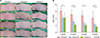
Changes in inflammatory nodules in acne mouse model groups A, B, and C
The size of the inflammatory nodules in all three groups decreased with time (Fig. 1A, B). There was a statistically significant difference in the size of the inflammatory nodules in groups B and C compared to group A at week 1, 2, 3, and 4. Group C showed a statistically significant decrease in inflammatory nodule size compared to group B at week 2 and 4.
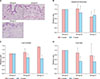
Characteristics of histopathological findings in groups A, B, and C
Epidermal thickness and the number and size of the microcomedone-like cysts were decreased in groups B and C compared to group A at week 4 (Fig. 2A~D). However, there were no statistically significant differences.
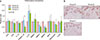
Expression of inflammatory biomarkers in groups A, B, and C
The immunohistochemistry results showed that infiltration of CD8 T cells and neutrophils and the expression of IL-1β, MMP-3 and MMP-9 in group B and group C were decreased when compared with group A (Fig. 3A). On the contrary, infiltration of CD4 T cells and the expression of LL-37 in group B and group C were increased when compared with group A (Fig. 3A). However, there were no statistically significant differences in all three groups (Fig. 3A). Integrin α6 expression was increased in group B and decreased in group C when compared with group A (Fig. 3A, B). Integrin α6 expression showed a statistically significant decrease in group B when compared with group B (p<0.05) (Fig. 3A).
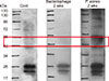
Western blot analysis using an anti-IgG antibody
No immunoreactivity for an anti-IgG antibody against P. acnes was observed at week 2 after the first injection in the group A or at week 2 after the second injection in the group C. However, western blot analysis could detect an IgG antibody component against P. acnes (approximately 47 kDa) at week 2 after the second injection in the group B (Fig. 4).
DISCUSSION
Topical and systemic medications have been used for the treatment of acne8. However, bacterial resistance to antibiotics and adverse retinoid events have led to the development of new therapeutic modalities for acne910; vaccination has been introduced as one of the promising therapeutics for acne11.
Killed-pathogen-based vaccines have been used as a vaccine for the treatment of acne12 and an intranasal killed whole P. acnes preparation has been tested12. P. acnes has been established as a potent immunomodulator13, which can produce antibodies against secretary enzymes or cell wall molecules14. However, antibodies against P. acnes do not completely protect against the recurrence of acne, most likely because of insufficient production of protective antibodies against P. acnes. This study also showed the production of antibody in mice after treatment with P. acnes. Bacteriophages inhibited the production of the antibody in mice after treatment with P. acnes.
Vaccination against cell wall-anchored sialidase has also been introduced15. Sialidase, a P. acnes virulence factor, is involved in degrading host tissue and inducing inflammation16. Nakatsuji et al.15 have reported that a sialidase-based acne vaccine showed good effects on P. acnes-induced ear inflammation. However, this vaccine may not be able to neutralize virulence factors secreted by P. acnes17. In addition, this vaccine may lack therapeutic effects for acne.
Monoclonal antibodies targeting Christie-Atkins-Munch-Peterson (CAMP) factor have demonstrated improvement of acne17. P. acnes possesses a CAMP that acts as a potential secretory virulence factor to enhance hemolysis and cytolysis1618. Nakatsuji et al.17 also investigated the treatment of acne using vaccines. They showed that passive immunization targeting secretory CAMP factors proteins can neutralize P. acnes virulence without disturbing other bacteria17. Active immunization using a CAMP factor-targeted vaccine and passive neutralization of the CAMP factor showed roughly equal potency in terms of improving P. acnes-induced inflammation.
TLR vaccines and antimicrobial peptides (AMPs) constitute other optional vaccines for the treatment of acne. Recent studies have shown that TLRs and AMPs play an important role in the pathogenesis of inflammatory acne. Therefore, regulation of TLRs and AMPs could provide different therapeutic options for acne19.
Bacteriophages targeting P. acnes are also promising acne vaccine candidates20. Bacteriophages have several advantages over antibiotics against P. acnes. They are environmentally friendly, rapidly processed, less expensive, and well tolerated5. In this study, we confirmed the potential of bacteriophages for the treatment of acne. The inflammatory nodules of HR-I mice decreased in size and microcomedone-like cysts was reduced in number and in size following bacteriophage treatment. In addition, epidermal thickness was decreased in bacteriophage treatment group. Epidermal hyperplasia can be induced by skin barrier disruption, calcium channel activation, metabotrophic receptor activation or unsaturated fatty acids21. Epidermal hyperplasia may be related to the induction of abnormal keratinization, which can be one of the causative factors of comedone formation. Furthermore, the decreased expression of integrin α6 after bacteriophage application shows its beneficial effect for the treatment of acne. Integrin α6 expression can be a response to inflammation and related to epidermal hyperplasia, leading to the abnormal differentiation2. Inflammatory biomarkers and cells, such as CD8 T cells, neutrophils, IL-1β, MMP-3 and MMP-9, were also decreased after bacteriophage treatment. Antimicrobial peptides can play a protective role against P. acnes or induce inflammatory signal in acne vulgaris22. In this study, the increased expression of LL-37 by P. acnes was not inhibited by bacteriophages.
Several parameters for bacteriophages need to be resolved for a comprehensive acne vaccine: delivery route, time point of delivery, dose, phage stability, infection rate, availability of highly efficient antibiotics, poor understanding of general aspects of phage life cycles, bacterial resistance to phage infection, and the possibility of temperate phages that integrate into the human genome23. Understanding the integration of temperate phages into the human genome is critical for the safety of phage therapy.
Recently, bacteriophages have been applied in a semi-solid preparation for the treatment of acne24. In addition, bacteriophages have also been formulated in a water-oil nanoemulsion25. Bacteriophages against Acinetobacter baumannii have been applied in humans in the form of an antiseptic gel or a paraffin-oil-based product26. In light of these reports, a variety of formulations for the treatment of acne using bacteriophages that target P. acnes should be examined.
In conclusion, further investigation of bacteriophages targeting P. acnes should be conducted to gain a better understanding of bacteriophage characteristics and to develop more beneficial vaccines for the treatment of acne.
 XML Download