

 PDF
PDF ePub
ePub Citation
 Print
Print


Dear Editor:
Fibroma of tendon sheath (FTS) is an uncommon soft tissue neoplasm belonging to the benign fibroblastic/myofibroblastic tumor group. It is manifested as an asymptomatic, firm, and well-demarcated nodule that grows slowly. FTS has a predilection to adhere to the tendon or tendon sheath of digits and palms, especially on the flexor surface1.
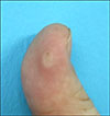
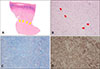
A 21-year-old man visited our department with a 2-month history of a solid mass on the volar aspect of his left thumb (Fig. 1). Physical examination revealed a hard hyperkeratotic papule, smaller than 1 cm. He reported mild tenderness without limitation in the range of motion. There was no history of trauma, but the lesion was trimmed at the local clinic due to suspicion of corn. To confirm a diagnosis, an incisional biopsy was performed. The specimen showed a well-circumscribed tumor in the deep dermis comprised of hyalinized collagenous fibers with haphazardly arrayed spindle-shaped cells and slit-like vascular spaces. The cells were immunoreactive for smooth muscle actin (SMA) and the fibrotic regions appeared blue in Masson's trichrome staining (Fig. 2). Based on the clinical and histopathologic findings, a diagnosis of FTS was established. The patient was requested to come in for further evaluation, but lost to follow-up.
FTS, also known as tenosynovial fibroma, accounts for approximately 3% of the incidence of all hand tumors2. It occurs 2.5 times more frequently on the right hand as well as on the volar surface and 10% of which is related to trauma, supporting a reactive fibrosing process1. It also has been described as a fibrotic neoplasm due to its chromosomal 2;11 translocation abnormality3. However, the pathogenesis regarding the origin has not been precisely established.
Considering the clinical and histological features, the differential diagnosis should include nodular fasciitis, giant cell tumor of tendon sheath (GCTTS), and desmoplastic fibroblastoma (DFB). Histopathologically, FTS is a well-lobulated encapsulated tumor, which consists of fibroblast-like spindle cells and numerous cleft-like vascular channels embedded in a dense hyalinized fibrous stroma. Composed of a predominantly paucicellular component, it is admixed with some hypercellular areas arranged in a storiform pattern. The matrix is stained blue with Masson's trichrome staining suggesting collagen alignment, and the tumor cells express myofibroblastic markers such as SMA and vimentin. GCTTS shows osteoclast-like multinucleated giant cells and stains positive for histiocytic determinants like CD684. Nodular fasciitis presents with a tissue-culture like growth pattern of randomly configured myofibroblasts. DFB and FTS show an identical cytogenetic aberration and similar histopathologic findings, so they are considered as parts of a morphological spectrum of a single entity. However, DFB commonly involves skeletal muscle or subcutaneous tissue of the extremities and trunk, and the vascular structure is not striking5.
Because a tumor firmly adhering to the tendon or tendon sheath shows a high recurrence rate, careful surgical excision is necessary for complete removal1. Herein, we report a rare case of FTS mimicking a corn or wart on the finger; therefore it is important to differentiate clinically and pathologically from other tumors.
 XML Download