

 PDF
PDF ePub
ePub Citation
 Print
Print


Gastric cancer is one of the most common types of cancer and is the second leading cause of cancer deaths worldwide [123]. It accounts for approximately 10% of newly diagnosed cancers [1], however, its diagnosis usually occurs at an advanced stage, which is one of the main reasons for its high mortality rate [4]. Therefore, the detection and diagnosis of gastric cancer at an early stage are important for its successful treatment. Studying models of early gastric cancer will help achieve this goal.
A significant decrease in Trefoil factor 1 (TFF1) expression occurs in approximately 50% of all human gastric cancers [56]. TFF1 encodes a small precursor protein of 84 amino acids, containing peptide sequence characteristics of secreted proteins [78]. The mature form of TFF1 is secreted by mucus-secreting pit cells in the corpus and pyloric antral region of the stomach, as a 60-amino-acid peptide consisting of a trefoil domain and a C-terminal dimerization domain [7]. It is implicated in many gastrointestinal cell biological processes, including repair, protection, cell proliferation, differentiation, and migration [910]. Depletion of TFF1 in mice results in the development of antropyloric adenoma, with severe hyperplasia and dysplasia of epithelial cells in the antropyloric region [48]. This indicates that Tff1 functions as a gastric tumor suppressor gene. Therefore, a Tff1-knockout (KO) mouse would a useful model to dissect the early events of gastric carcinogenesis and to investigate aspects of gastric cancer that are difficult to study in humans.
Clustered regularly interspaced short palindromic repeats (CRISPR)/CRISPR-associated nuclease 9 system (Cas9) technology is a useful tool to generate KO mouse models by simple genome editing. The Cas9 nuclease protein, derived from Streptococcus pyogenes, recognizes the protospacer adjacent motif (PAM) sequence, 5-NGG-3, in the target DNA. The sgRNA, consisting of a fusion of tracrRNA and crRNA, containing a sequence complementary to the 20 nucleotides upstream of the PAM, is required for recognizing the target site [11]. Cas9/sgRNA complexes generate double-strand breaks (DSB) by cleaving the target DNA, resulting in indel mutations and subsequently, loss of gene function. Furthermore, the use of multiple sgRNAs can efficiently induce large deletions in the target gene. In this study, to avoid any conflicts with patents or material transfer agreements, Tff1-KO mice were generated without any external genes (e.g. neomycin resistance) using the multiple sgRNAs and Cas9. This model will allow us to more accurately study the effects of genetic alteration during gastric tumorigenesis.
Materials and Methods
Animals
All mice were purchased from Taconic Biosciences (Dae Han Biolink, Co., Ltd., Republic of Korea) and were maintained in the specific pathogen-free (SPF) facility of the Yonsei Animal Research Center. Animal suffering was minimized and all experiments were performed in accordance with the Korean Food and Drug Administration (KFDA) guidelines for animal research. The experimental protocols for generating the genetically engineered mouse models were approved by the Institutional Animal Care and Use Committee (IACUC) at Yonsei University.
Preparation of CRISPR/Cas9 mRNA and sgRNAs
Cas9 mRNA and sgRNAs were synthesized in vitro using the mMESSAGE mMACHINE® T7 Ultra and MEGAshortscript T7 kits (Ambion), respectively. Cas9 mRNA and sgRNAs were diluted in RNase-free injection buffer (0.25 mM EDTA, 10 mM Tris at pH 7.4) to obtain the working concentration before microinjection [1213]. PCR-amplified DNA templates containing the following target sequences were used for sgRNA synthesis: sgRNA#1, CTACAGGTTCATTCCAGCAG; sgRNA#2, GGTTCATTCCAGCAGAGGAG; sgRNA#3, GCAGTGTAAGCCCAGCTGGA; sgRNA#4, ACTCTAAGCAAATAGACCGG; sgRNA#5, CTAGGACCTATGAGGATGCA; sgRNA#6, GTATCAGTGGCTTGTGGCAG; sgRNA#7, TGTGTAAGCTGAGCTTTGCC; sgRNA#8, GATGTCCCTTGAGTGGGAGG.
Microinjection
Microinjections were performed as described previously [14]. Briefly, 4–6-week-old B6 mice were superovulated by intraperitoneal injection of pregnant mare serum gonadotropin (PMSG, Sigma) and human chorionic gonadotropin (hCG, Sigma) and fertilized embryos were collected. A mixture of Cas9 mRNA (10 ng/µL) and four sgRNAs (50 ng/µL each) were microinjected into the cytoplasm of mouse zygotes using a Piezo-driven manipulator (Prime Tech). Injected embryos were then transferred into the oviducts of foster mother mice.
Screening and genotyping
To screen the founder mice carrying Tff1 mutations, PCR was performed using the following primers: 5′-GGTCATATAGGCATGTGTAAGGA-3′ (Tff1 F) and 5′-CCTAGGCAGTTATAAGGTCCCT-3′ (Tff1 R). Mice generated from intercrosses between heterozygous mice were genotyped by PCR using Tff1 F (used for screening) and Tff1 R1 (5′-TCCTACCGGCAAAGGTCCTA-3′) primers. PCR was performed as follows: predenaturation of 3 min at 95℃; 10 cycles of 30 s at 95℃, 60 s at 65℃ (reduced by 0.5℃ per cycle), and 30 s at 68℃; 30 cycles of 30 s at 95℃, 30 s at 60℃, and 60 s at 72℃; and 5 min at 72℃. PCR products were electrophoresed on 2–3% of agarose gels and visualized with a ChemiDoc system (Bio-Rad).
Necropsy and stomach fixation
8- or 14-month-old mice were euthanized in CO2 chambers that complied with the code of ethics for animal experimentation. Immediately after each sacrifice, the stomach was expanded by injection of 4% phosphate-buffered formalin (4% PFA, Biosesang Co., Ltd., Republic of Korea) and cut along the line of the greater curvature. The cut stomach tissue was spread flat on 3M paper without touching the glandular region. The flattened tissue was embedded in paraffin and then fixed in 4% PFA overnight. Organ and body weights were measured and relative organ weight was calculated using the following formula: (organ weight/total body weight) ×100.
Histopathological analysis
For histopathological analyses, paraffin-embedded blocks were cut into 5 µm sections using a microtome (RM2235, Leica). Sections were deparaffinized with xylene (UN1307, Baker) three times, each for 20 min. Sections were dried at room temperature after two washes in 100% EtOH for 5 min each wash. For hematoxylin and eosin (H&E) staining, sections were stained with 0.1% Mayer's hematoxylin for 10 min and 0.5% eosin in 95% EtOH for 10 min. After H&E staining, sections were washed in distilled H2O until the eosin stopped streaking. They were then dipped in 50% EtOH 10 times, 70% EtOH 10 times, 95% EtOH for 30 s, and 100% EtOH for 1 min. Samples were then covered with mounting solution (Thermo Scientific) and diagnosed by a pathologist under a light microscope (BX43, Olympus).
Go to : 
Results
Generation of Tff1-KO mice by multiple sgRNAs/Cas9 microinjection
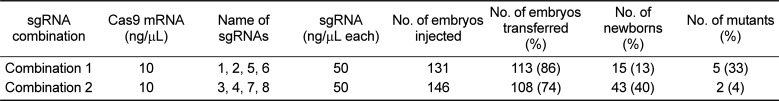
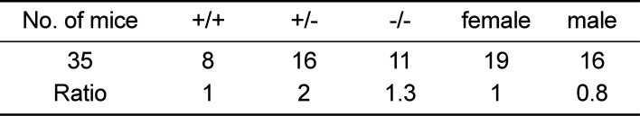
To generate Tff1-KO mice we deleted exon 1 using multiple sgRNAs targeting the promoter and intron 1 of Tff1. To minimize off-target effects and maximize on-target activity of CRISPR/Cas9, we designed eight different sgRNAs using CRISPR Design (http://crispr.mit.edu, Figure 1A). Two combinations consisting of four sgRNAs targeting either the promoter or intron 1 were used (Table 1, Figures 1A and B). The targeting efficiency of combination 1 (sgRNA 1, 2, 5, 6; 36%) in the embryos was higher than that of combination 2 (sgRNA 3, 4, 7, 8; 6%; Table 1, Figure 1B), resulting in seven candidates with large deletions (five in combination 1 and two in combination 2), as identified by PCR screening (Figure 1B). Each candidate was validated by sequencing analysis, which confirmed various large deletion mutations (800–1100 nucleotides), eliminating exon 1 of Tff1 (Figure 1C). Finally, we selected candidate #14, which carries an 856-bp deletion mutation, as the Tff1-KO mouse strain to use for further experiments (Figure 1C and D). Genotypes of KO mice were confirmed by PCR (Figure 1D). Homozygous KO mice generated from intercrosses between heterozygous mice were born at the expected Mendelian ratio (Table 2), indicating that Tff1 deficiency does not result in embryonic lethality.
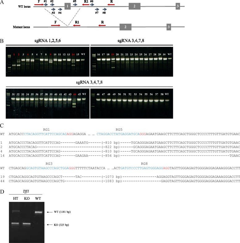 | Figure 1(A) CRISPR/Cas9 targeting strategy to generate Tff1-KO mice. Blue arrows #1–8 indicate the location and orientation of sgRNAs designed to target specific sites. Exons and introns are indicated with boxes and lines, respectively. PCR primers (F, forward; R, reverse) for genotyping are denoted by red arrows. (B) PCR screening results for newborns edited by Cas9 with sgRNAs 1, 2, 5, and 6 (combination 1) or 3, 4, 7, and 8 (combination 2). Mice carrying deletion mutations are highlighted in red. (C) Sequencing results for candidate newborns from combination 1 (upper panel) and 2 (bottom panel). The protospacer adjacent motif (PAM) and target sequences of sgRNAs are shown in red and blue, respectively. The symbol, “-”, indicates a single-nucleotide deletion. (D) PCR genotyping results for heterozygous (HT), homozygous (KO), and wild-type (WT) mice. Each arrow indicates WT-(1181 bp) and KO-specific (325 bp) bands.
|
Table 1
CRISPR/Cas9-mediated gene targeting
![]()
Body weight reduction in Tff1-KO male mice
To examine the phenotypic properties of Tff1-KO mice, we measured body weight weekly throughout the experimental period. Interestingly, the body weight of male Tff1-KO mice significantly decreased in comparison to wild-type (WT) mice after 4 weeks, whereas the body weight of female mice was not affected by Tff1 deficiency (Figures 2A and B). To further investigate the reason for the change in body weight, we measured the weight of each organ of male mice at the end stage of experiments when they were approximately 40 weeks old. Upon sacrifice, the weight of specific organs, including the heart, liver, kidney, lung, and spleen, were immediately measured using an electronic balance. At the end stage, the overall body weight loss was still observed in male Tff1-KO mice, with weight loss seen in the liver, heart, and spleen (Figure 2C). However, there was no significant difference between WT and Tff1-KO mice when organ weight was normalized to total body weight (data not shown). This result shows that Tff1 deficiency induces total body weight loss and organ weight loss in male mice.
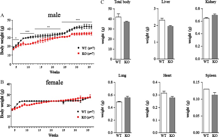 | Figure 2(A–B) Body weight changes in WT (black, n=7) and Tff1-KO (red, n=7) mice. Total body weights of male (A) and female (B) mice were measured once a week from 4 weeks to 36 weeks. Data are presented as mean±SEM (Student's t-test, *P<0.05, **P<0.01, ***P<0.001). (C) Mean weights (g) of total body, liver, kidney, lung, heart, and spleen in male WT and Tff1-KO mice after sacrifice. Data are presented as mean±SEM (WT, n=2; KO, n=7).
|
Loss of Tff1 promotes stomach lesions, especially in the antrum
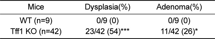
As presented in Figure 3A, macroscopic observations clearly showed an increase in the thickness of the antrum and mucosal projections in Tff1-KO mice (lower panel). Gastric lesions were not seen in WT mice (upper panel). However, morphological changes in the fundus were not observed in either mouse strain. To diagnose distinct morphological features, we performed histopathological analysis on 8- and 14-month-old Tff1-KO mice. In 8-month-old mice, Tff1 deficiency resulted in a conspicuous histological feature of antral dysplasia, characterized by irregular morphology of the gastric gland, occurrence of atypical cells, and an increase in infiltration of inflammatory cells when compared to WT mice with normal gland architecture (Figure 3Ba-d, Table 3). Adenoma was observed in 14-month-old Tff1-KO mice. Atypical epithelial gland architecture was observed in the cancerous region, with growth of the stromal stalk and marked inflammation (Figures 3Be and f). Some gland structures were observed in the adjacent region, but they were hard to distinguish from normal glandular architecture (Figures 3Be and f). Cystic dilatation was seen at the base mucosa in 14-month-old Tff1-KO mice (Figure 3Bg and h, Table 3). Indeed, all Tff1-KO mice showed precancerous or cancerous changes in the antrum, but no severe alterations were found in the fundus when compared with WT mice (data was not shown). Taken together, Tff1 deficiency promotes gastric tumorigenesis, especially in the antrum region, as previously reported [8].
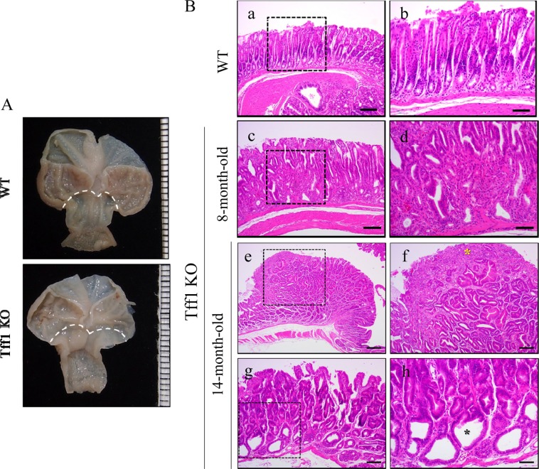 | Figure 3(A) Representative macroscopic images of WT (upper panel) and Tff1-KO mice (lower panel) at 8 months. The dotted line indicates the boundaries between the fundus and the antrum. (B) Representative H&E images of WT (a, b), 8-month-old (c, d) and 14-month-old Tff1-KO mice (e-h). Higher magnification views of images in the dotted boxes in the left panels are shown on the right. Hyperproliferation of the gastric epithelium and infiltration of immune cells are observed in the antrum of Tff1-KO mice (c, d). Adenoma is observed in the antrum region of Tff1-KO mice (e, f). Yellow asterisk indicates the tumor region (f). Mild lesions are observed in 14-month-old Tff1-KO mice (g, h). Black asterisk indicates cystic dilatation (h). Bars: 200 µm (e); 100 µm (a, c, f, g); 40 µm (b, d, h).
|
Go to :
Discussion
We report the generation of Tff1-KO mice using CRISPR/Cas9 and the analysis of the gastric phenotype of homozygous null mice to determine their utility as a stomach cancer model. We eliminated exon 1 of Tff1, including the initiation codon, using Cas9 with multiple sgRNAs. Consistent with previous results, [8] Tff1 deficiency in mice led to morphological changes in the antrum and the presence of atypical cells in the gastric glands. Although the antral and pyloric mucosa were thicker in Tff1-KO mice, the fundus did not show any obvious abnormality. Both sexes were equally affected and these phenotypes were seen in 54% of Tff1-KO mice examined. At 14 months, 26% of KO mice exhibited adenoma and many cancerous changes in the antrum region. These results are consistent with the lack of Tff1 expression observed in human gastric carcinomas [15161718]. Gastric cancer patients usually suffer from symptoms such as vomiting, stomach pain, and weight loss. Weight loss occurs in less than 40% of patients with early-stage gastric cancer and in approximately 60% of patients with advanced gastric cancer [1920]. Unlike previous embryonic-stem (ES)-cell-based Tff1-KO mice, which showed no weight loss compared to WT mice [21], significant weight loss was observed in male Tff1-KO mice in our study. Consistent with our results, weight loss and muscle wasting (cachexia) are more frequently observed in male than female cancer patients [22]. Although further studies regarding body weight are needed, these results suggest that the gender difference seen in gastric cancer patients may be influenced by Tff1 expression. In addition, the difference between our Tff1-KO mice and ES-cell-based Tff1-KO mice may be due to the differences in gene targeting processes used. The expression of NeoR, which is inserted in ES-cell-based KO mice to enable screening of correctly targeted clones [8], can affect gene expression and cell physiology [2324]. For example, the NeoR cassette can lead to an increase in the expression of the endogenous c-myc gene, which is commonly amplified or overexpressed in primary tumors [25]. On the other hand, our Tff1-KO mice were produced using the CRISPR/Cas9 system, without inserting exogenous antibiotic resistance genes. Therefore, these Tff1-KO mice will provide us a benefit to gain further insights into the more accurate genetic and environmental influences in gastric cancer.
Go to :
 XML Download