

 PDF
PDF ePub
ePub Citation
 Print
Print


Constipation is a complex affliction comprising of difficult defecation, hard stool, incomplete evacuation, sensation of blockage, anorectal obstruction and infrequent bowel movements [12]. Various types of chemical drugs including bulking agents, osmotic laxatives, stimulant laxatives, emollient laxatives and prokinetic agents are known to successfully treat constipated patients, although non-pharmacological treatments are the first approaches in the management of constipation [3]. Also, numerous medicinal plants and functional foods exhibiting laxative activity have received increased attention as novel therapeutic strategies for the treatment of constipation and its related diseases [4567].
The regulation of various gastrointestinal hormones has been investigated as one of the strategies for treating constipation. Zhizhuwan (ZZ) treatment induces an increase of substance P (SP) and decreases the vasoactive intestinal polypeptide (VIP), resulting in improved defecation and stool frequency [8]. Motilin (MTL) and ghrelin levels were enhanced after administering fat-free milk and Fuzhengliqi mixture along with acupuncture [910]. Furthermore, treatment with laxative mixtures such as lactobacillus fermentum Zhao [11], ultrafine powder of Dendrobium officinale [12], and Naringenin (NAR) [13] resulted in significant increase of several gastrointestinal metabolic components including MTL, gastrin (GAS), endothelin (ET), SP, VIP, and acetylcholinesterase (AChE), while levels of somatostatin (SS) were decreased under same conditions [1112]. However, although the laxative effects of GEGR have previously been studied [7], the regulation of gastrointestinal hormones during the laxative activity of GEGR have not yet been investigated in the Sprague Dawley (SD) rat constipation model induced by loperamide (Lop) injection.
The present study was therefore undertaken to examine the regulation of CCK, GAS, SS and MTL during the laxative activity of GEGR in a Lop-induced constipation model. Our results indicate that GEGR successfully improves the pathological features of constipation through the regulation of gastrointestinal hormones and their receptor signaling pathways in the constipated animal model.
Materials and Methods
Preparation of GEGR
GEGR were prepared as described in previous studies [1415]. Briefly, Galla Rhois harvested in the Hongcheon region of Korea in October 2013 was obtained from the Hongcheon National Agricultural Cooperation Federation (http://www.hcari.co.kr). Galla Rhois samples were dried in a hot-air drying machine (JSR, Seoul, Korea) at 60℃, and deposited as voucher specimens (WPC-14-001) at the Functional Materials Bank of the Wellbeing Regional Innovation System Center (WRISC) at Pusan National University (PNU). After powdering the dry samples, a circulating extraction equipment (IKA Labortechnik, Staufen, Germany) was used to obtain aqueous extract of Galla Rhois from the powders at 90℃ for 9 h in a fixed liquor ratio (solid Galla Rhois powder/water ratio, 1:10). These solutions were subsequently passed through a 0.4 µm filter, after which they were concentrated by vacuum evaporation and lyophilization using the circulating extraction equipment (IKA Labortechnik, Staufen, Germany).
Animal experiment
The animal protocol used in this study was reviewed and approved as per the ethical procedures for scientific care established by the PNU-Institutional Animal Care and Use Committee (PNU-IACUC; Approval Number PNU-2014-0572). Adult SD rats purchased from Samtako BioKorea Inc. (Osan, Korea) were handled at the PNU-Laboratory Animal Resources Center, accredited by the Korea Food and Drug Administration (FDA) (Accredited Unit Number-000231) and Association for Assessment and Accreditation of Laboratory Animal Care (AAALAC) International (Accredited Unit Number; 001525). During the experimental period, animals were provided ad libitum access to water and a standard irradiated chow diet (Samtako BioKorea Inc.) consisting of moisture (12.5%), crude protein (25.43%), crude fat (6.06%), crude fiber (3.9%), crude ash (5.31%), calcium (1.14%) and phosphorus (0.99%). The rats were maintained in a specific pathogen-free (SPF) state under a strict light cycle (lights on at 08:00 h and off at 20:00 h) at 23±2℃ and 50±10% relative humidity.
Constipation of SD rats was induced by Lop treatment as previously described [616]. Briefly, 8-week-old SD rats (n=18) were assigned to either a non-constipation group (No group, n=6) or a constipation group (n=12). Constipation was induced by subcutaneous injection of Lop (4 mg/kg weight) (Sigma-Aldrich, Saint Louis, MO, USA) prepared in 0.9% sodium chloride, twice a day for 3 days, whereas the non-constipation group was injected with 0.9% sodium chloride alone. The constipation group was further divided into a Lop+vehicle treated group (Lop+vehicle, n=6), and Lop+GEGR treated group (Lop+GEGR, n=6). The Lop+GEGR treated groups received 1,000 mg/kg body weight of GEGR, while the Lop+vehicle treated group received the same volume of dH2O, once after the induction of constipation. The pathological conditions of all animals in each group was regularly monitored, daily at 10 am, during the experimental period; no severely ill or dead animals were observed during the entire period. At 24 h after GEGR and vehicle administration, all animals were euthanized using CO2 gas, and transverse colons were acquired and stored in Eppendorf tubes at −70℃ until further assay.
Analysis of food intake and water consumption
Alterations in food intake and water consumption of SD rats treated with Lop+GEGR were measured daily at 10:00 am throughout the experimental period using an electrical balance and a measuring cylinder. All measurements were performed three times to ensure accuracy.
Measurement of stool parameters
To enable collection of pure stools and urine without any contamination, all SD rats were bred in metabolic cages (Daejong Instrument Industry Co., Ltd., Seoul, Korea) during the experimental period. The number of stools and their weight were measured as described in previous studies [616]. Stools excreted from each SD rat were collected at 10:00 am and weighed three times per sample using an electric balance. The number of stools were also tallied three times. The water content of each stool was analyzed using the following formula:
where A is the weight of fresh stools collected from SD rats of subset groups, and B is the weight of stools after drying at 60℃ for 12 h.
Histopathological analysis of transverse colon
Transverse colons collected from the subset constipation groups treated with vehicle and GEGR were fixed in 10% formalin for 24–48 h, embedded in paraffin wax, and sectioned into 4 µm thick slices that were stained with hematoxylin and eosin (H&E, Sigma-Aldrich Co., Saint Louis, MO, USA). Morphological features of these tissue sections were observed under light microscopy, after which the mucosa thickness, muscle thickness and flat luminal surface thickness were measured using the Leica Application Suite (Leica Microsystems, Heerbrugg, Switzerland).
Measurement of hormone concentrations
CCK, MTL, SS and GAS were quantified using ELISA kits (Cusabio Biotech Co., Ltd., Wuhan, China) according to the manufacturer's instructions. Briefly, the tissue of transverse colons (100 mg) were homogenized in ice-cold 1x PBS (pH 7.2–7.4) using a glass homogenizer (Sigma-Aldrich Co.). The tissue lysates were then centrifuged at 1,000 rpm for 5 min at 4℃, after which the supernatant was collected for analysis. After addition of the four specific hormone antibodies, separately in each well, the supernatant was incubated for 60 min at 37℃, following which HRP-Streptavidin solution was added and incubated for 60 min at 37℃. The TMP One-Step Substrate Reagent was then added, and the mixture was further incubated for 30 min at 37℃. The reaction was terminated following addition of the stop solution. Finally, the absorbance of the reaction mixture was read at 450 nm using a Molecular Devices VERSA max Plate reader (Sunnyvale, CA, USA).
Western blotting
Total proteins collected from the transverse colons of all subset groups (No, Lop+vehicle, Lop+GEGR) were separated by 4–20% sodium dodecyl sulfate-polyacrylamide gel electrophoresis (SDS-PAGE) for 3 h, after which the resolved proteins were transferred to nitrocellulose membranes for 2 h at 40 V. Each membrane was then incubated separately with the following respective primary antibody, overnight at 4℃: anti-PKC (Cell Signaling Technology Inc.), anti-p-PKC (Cell Signaling Technology Inc.), anti-Bax (Abcam, Cambridge, UK), anti-p53 (Abcam, Cambridge, UK), anti-JNK (Cell Signaling Technology Inc.), anti-p-JNK (Cell Signaling Technology Inc.) or anti-actin (Sigma-Aldrich Co.). Next, the membranes were washed with washing buffer (137 mM NaCl, 2.7 mM KCl, 10 mM Na2HPO4, 2 mM KH2PO4, and 0.05% Tween 20) and incubated with horseradish peroxidase-conjugated goat anti-rabbit IgG (Zymed Laboratories, South San Francisco, CA, USA) at a dilution of 1:1,000 for 2 h at room temperature. Finally, the membrane blots were developed using Chemiluminescence Reagent Plus kits (Pfizer, New York, NY, USA and Pharmacia, New York, NY, USA).
ELISA for inositol trisphosphate (IP3) concentration
The levels of IP3 were determined using an IP3 ELISA kit (Cusabio Biotech Co., Ltd., Wuhan, China) according to the manufacturer's instructions. Briefly, transverse colons (100mg) were washed and homogenized in ice-cold 1x PBS (pH 7.2–7.4) using a glass homogenizer (Sigma-Aldrich Co.). The tissue lysates were centrifuged at 1,000 rpm for 5 min at room temperature, after which the supernatant was collected for analysis. An anti-IP3 detection antibody was added and incubated for 60 min at 37℃, after which substrate solution was added and incubated for 15 min at 37℃. The reaction was terminated following the addition of stop solution, and the plates were read at an absorbance of 450 nm using a Molecular Devices VERSA max Plate reader (Sunnyvale, CA, USA).
Statistical analysis
Statistical significance was evaluated using one-way Analysis of Variance (ANOVA) (SPSS for Windows, Release 10.10, Standard Version, Chicago, IL, USA) followed by Tukey's post hoc t-test for multiple comparison. All data were expressed as the mean±standard deviation (SD). A P value less than 0.05 is considered to be statistically significant.
Results
Laxative effects of GEGR on stool parameters
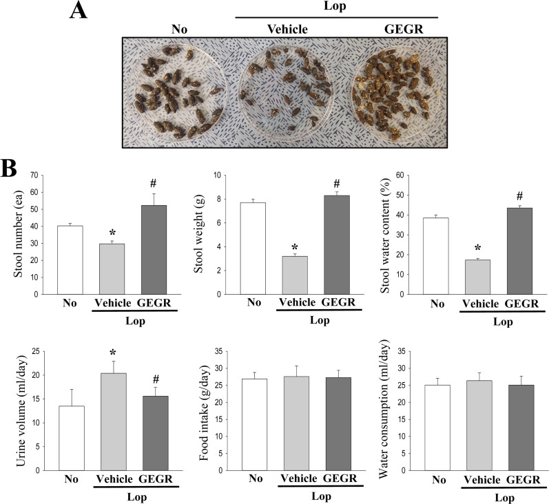
To investigate the effects of GEGR treatment on the feeding behavior and stool parameters of Lop-treated SD rats, we assessed the alteration in levels of food intake, water consumption, and stool-related factors in the constipated rat model after GEGR administration. Some significant alterations were observed in the stool parameters, whereas the food intake and water consumption did not differ significantly among groups. The number, weight and water contents of stools were lower in the Lop+vehicle treated group than the No treated group. However, levels were almost recovered in the Lop+GEGR treated groups when compared to the No treated group (Figures 1A and B). Furthermore, the enhanced urine volume observed in the Lop+vehicle treated group also recovered after GEGR treatment (Figure 1B). These results indicate that GEGR treatment induces laxative effects and results in the recovery of affected stool parameters and urine volume.
Laxative effect of GEGR on the histological structure of the transverse colon
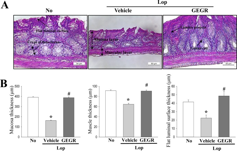
We next examined whether the laxative effects of GEGR on the stool parameters is accompanied with recovery of the histological structure of the transverse colon in Lop-induced constipation rats. Significant recovery was observed for the three parameters assessed for histopathological changes in the Lop+GEGR treated groups. The thickness of mucosa, muscle and flat luminal surface was significantly decreased in the Lop+vehicle treated group as compared to the No treated group. These levels remarkably recovered by 166.7, 38.5, and 100.3% following Lop+GEGR treatment (Figure 2). Taken together, these results indicate that the laxative effect of GEGR is accompanied with a recovery of the histopathological structure of the transverse colon in the Lop-induced constipation model.
Effect of GEGR on the secretion of gastrointestinal hormones and the activation of their receptors
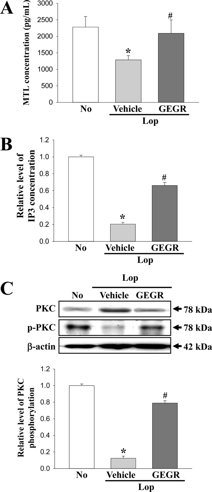
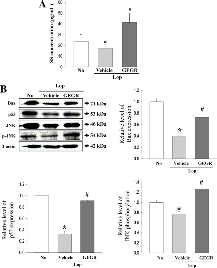
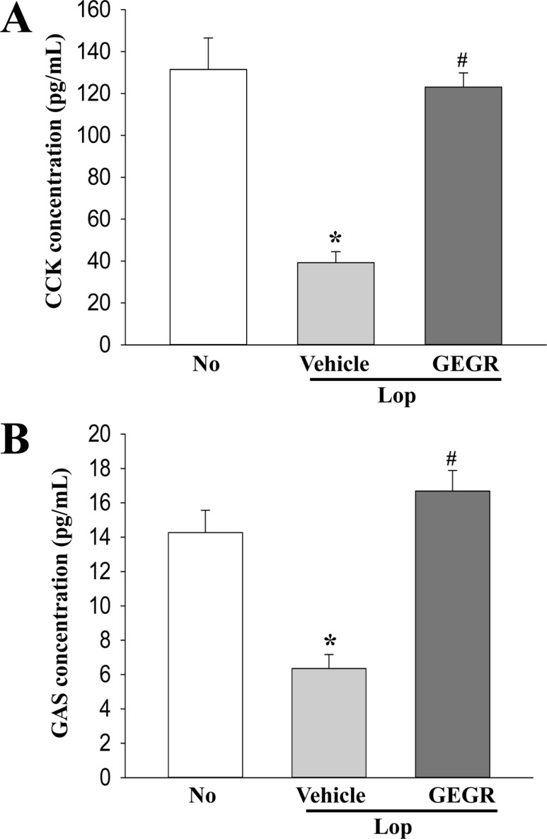
To investigate the effects of GEGR treatment on the ability to secrete gastrointestinal hormones and to regulate the downstream signaling pathway of their receptors, the levels of four gastrointestinal hormones, namely CCK, GAS, SS and MTL, as well as the expression of their signaling molecules were measured in the transverse colon of the subset group. A similar regulation pattern was observed for the levels of all four hormones. The level of MTL was lower in Lop+vehicle treated group than No treated group, which was observed to recover dramatically in the Lop+GEGR treated group as compared to the Lop+vehicle treated group (Figure 3A). Furthermore, the levels of IP3 and PKC phosphorylation (members of downstream signaling pathway of the MTL receptor) were remarkably enhanced after GEGR treatment (Figures 3B and C). The levels of SS were similarly increased in the Lop+GEGR treated group as compared to the Lop+vehicle treated group (Figure 4A). This hormone level primarily reflects the expression levels of downstream members of SS receptors. The levels of Bax, p53 expression and JNK phosphorylation were recovered in the Lop+GEGR treated group, which were observed to have decreased in the Lop+vehicle treated group (Figure 4B). Furthermore, a similar recovery was measured for CCK and GAS concentrations. The levels of both hormones were significantly enhanced in the Lop+GEGR treated group as compared to the Lop+ vehicle treated group (Figure 5). These results indicate that the laxative effects of GEGR correlate with the control of gastrointestinal hormones and regulation of the downstream signaling pathways of their receptors in transverse colons of constipated SD rats.
Discussion
To date, several studies have investigated gastrointestinal hormones in their quest to develop them as therapeutic and diagnostic strategies for the treatment of various chronic diseases. Secretin is applied to evaluate pancreatic secretion, while CCK estimates the gallbladder ejection fraction [17]. Also, a number of gastrointestinal hormones are clinically used to treat diarrhea [18], diabetes mellitus [19], gastrointestinal bleeding [20], obesity [21] and Crohn's disease [22]. In an effort to verify the role of gastrointestinal hormones in the laxative effects of GEGR, we investigated the levels of four hormones and activation of their receptors in Lopinduced constipated rats after GEGR administration. Our results demonstrate that the laxative effects of GEGR are associated with the functional recovery of gastrointestinal hormones in the Lop-induced constipation model. Moreover, these results provide additional evidences that gastrointestinal hormones can be considered as key markers to evaluate the laxative effects of chemical drugs and natural products.
Currently, some studies have reported the importance of regulation of gastrointestinal hormones during the laxative effects of natural products. Few herbal plants such as Zhizhuwan, fat-free milk, Fuzhengliqi mixture, lactobacillus fermentum, Dendrobium officiate and Naringenin are known to alleviate the chronic symptoms of constipation [8910111213]. During their activity, the serum level of many gastrointestinal metabolic components change remarkably. Especially, a significant enhancement has been detected in the levels of MTL, GAS, ET, SP, AChE and VIP, whereas SS levels were observed to decrease [8910111213]. Based on these results, we hypothesized that GEGR would regulate gastrointestinal hormones when exerting its laxative effects on the constipation induced by Lop treatment in SD rats. Especially, we investigated the effect of GEGR on the levels of four hormones (CCK, GAS, SS and MTL), and the activation of their respective receptors. In the current study, we detected similar alterations in the levels of all four gastrointestinal hormones. As presented in Figures 3, 4 and 5, the total level for all four hormones decreased in the Lop+vehicle treated group, but recovered in the Lop+GEGR treated group. These results indicate that regulation of gastrointestinal hormones is an important mechanism for improving the symptoms of constipation in Lop-induced animal models after GEGR administration.
Taken together, the results of the present study reveals that one of the mechanisms involved in exerting the laxative effect of GEGR is through the regulation of gastrointestinal hormones. In addition, these results successfully demonstrate that CCK, GAS, SS and MTL may contribute towards relief from constipation in Lop-induced constipated SD rats after administration of GEGR. However, it should be noted that the present study has limitations, in that levels of only four hormones are measured in the animal model for constipation.
 XML Download