Journal List > Lab Anim Res > v.34(4) > 1111355

Park, Lee, and Lee: Mouse models of breast cancer in preclinical research

Abstract

Breast cancer remains the second leading cause of cancer death among woman, worldwide, despite advances in identifying novel targeted therapies and the development of treating strategies. Classification of clinical subtypes (ER+, PR+, HER2+, and TNBC (Triple-negative)) increases the complexity of breast cancers, which thus necessitates further investigation. Mouse models used in breast cancer research provide an essential approach to examine the mechanisms and genetic pathway in cancer progression and metastasis and to develop and evaluate clinical therapeutics. In this review, we summarize tumor transplantation models and genetically engineered mouse models (GEMMs) of breast cancer and their applications in the field of human breast cancer research and anti-cancer drug development. These models may help to improve the knowledge of underlying mechanisms and genetic pathways, as well as creating approaches for modeling clinical tumor subtypes, and developing innovative cancer therapy.

Breast cancer is the second most common cancer in Korean women [1], with 19,219 cases diagnosed in Korea in 2015 [2]; it is also the second leading cause of cancer-related death among women worldwide [3]. Developing and applying mouse models, using both human tumor xenograft models and genetic modifications, have led to our current understanding of molecular mechanisms that operate during breast cancer progression and metastasis [4]. Breast cancers are classified into five intrinsic subtypes based on hormonal and Human Epidermal growth factor Receptor-2 (HER2) receptor status, namely luminal A (Estrogen Receptor (ER)/Progesterone Receptor (PR)-positive major problem), luminal B (ER/PR-positive, HER-negative, high Ki-67, higher histological grade than luminal A), ER-negative/HER2-positive, HER2-positive, and basal-like triple-negative (ER/PR/HER2-negative) [5]. Treatment of human breast cancer patients is based on the hormone receptor status, specifically ER, PR, and HER2 [67], and these hormonal therapies are effective for most patients with hormonal receptor-positive breast cancer; for example, tamoxifen for ER-positive breast cancer and Pertuzumab (Perjeta), Trastuzumab (Herceptin), and Docetaxel (Taxotere) for HER2-positive breast cancer [891011]. However, primary and acquired resistance to hormonal treatments remains to be resolved [101213]. Xenograft models are used to elucidate the underlying mechanisms of resistance, and genetically engineered mouse models (GEMMs) are also useful to understand the mechanisms involved in the pathogenesis and molecular processes of breast cancer and metastasis. In this review, we summarize several mouse models used in breast cancer research and drug development, and their contribution to understanding the molecular pathways in tumorigenesis and metastasis, providing us invaluable insights into possible developments of innovative cancer therapy [14].

Tumor transplantation models

Cell-derived xenografts (CDX), patient-derived xenografts (PDX), and a syngeneic model are well-established tools for evaluating therapeutic efficacy and toxicity and for applying to preclinical assessment (Table 1). CDX transplantation models are useful for investigating breast cancer and metastatic progress [15]. These models are also convenient for investigating genetic alterations that are associated with tumor initiation and growth, but unfortunately show poor clinical predictability.
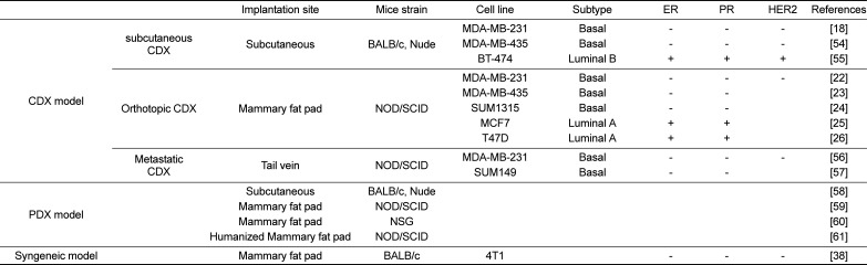
CDX transplantation models, in which tumor cells are subcutaneously transplanted in nude mice, facilitate straightforward monitoring of tumor growth. Cancer cells in these models show primary tumor growth but do not metastasize [16]. For example, models using BT474 cells (luminal B) [17], as well as MDA-MB-231 [1819] and MDA-MB-435 cells (triple negative) are well-established [20]. Orthotopic CDX transplantation models in which tumor cells are transplanted to a mammary fat pad of NOD/SCID mice are suitable for studying metastatic functions of genes of interest [21] and to investigate malignant and metastatic phenotypes in mice [4]. Several cancer cell lines, such as MDA-MB-231, MDA-MB-435, and SUM1315 (triple negative), as well as MCF7 and T47D cells (luminal A), are used to generate spontaneous metastasis models of breast cancer through orthotopic injection [2223242526]. Metastatic CDX transplantation models in which cancer cells, such as MDA-MB-231 and SUM149 cells, are injected into mouse tail veins are suitable for monitoring experimental metastasis. Taken together, these CDX models allow validation of target genes of interest, and facilitate evaluation of a candidate anti-cancer drug and therapeutics for breast cancer.
In PDX transplantation models, primary human breast carcinomas or tumor fragments are implanted into immune-deficient mice (e.g., nude, NOD/SCID, or NOD/SCID/IL2-receptor null (NSG) mice) subcutaneously or orthotopically [27]. Resulting tumors have characteristics that are similar to those of the original patients' tumors with respect to histology, genomic signature, and heterogeneity, as well as high predictive drug response [142829303132]. These models are used to identify biomarkers for personalized drug selection, and to overcome the limitation of CDX transplantation in clinical therapies [33]. While subcutaneous PDX transplantation models have been used in studies to measure primary tumor growth, orthotopic PDX transplantation models are suitable for mechanistic studies of metastasis and therapeutic resistance [34].
Syngeneic mouse models in which murine cancer cells, such as 4T1, are injected into immune-competent mice (e.g. BALB/c) show more effective metastasis, with characteristics similar to those of breast cancer patients. Advantages of these models over CDX transplantation models include the use of immune-competent mice with normal immune cells and immune system, enabling investigation and development of various immune therapies, for example, with anti-PD-1/PD-L1. These models are also useful to investigate the anti-tumor and anti-metastatic effects of multiple drugs due to the high invasiveness of murine cancer cells [3536373839]. While CDX transplantation models, in which human cancer cells are injected into immunocompromised mice, are well-established in the study of tumor growth and metastasis and the validation related gene profiles, human cancer cells in these models poorly metastasize and show poorly predictable metastatic characteristics.
Collectively, CDX, PDX, and syngeneic models are well-established for evaluating therapeutic efficacy and toxicity and for applying to preclinical assessment. These models could be critical tools for understanding breast cancer progression, and for evaluating responses to targeted therapeutics, in order to predict therapeutic outcomes for breast cancer patient.

Genetically engineered mouse models

GEMMs of breast cancer have greatly contributed to cancer research, improving understanding and validating human cancer genes, genetic pathways, and therapeutic approaches for cancer, as well as investigating cancer progression and metastasis [40]. They are also essential tools for gene expression profiling in breast cancer progression and metastasis. Histopathological features in over 25 different murine GEMMs of breast cancer have been reported [41]. Promoters frequently used in GEMMs of breast cancer are the mouse mammary tumor virus-long terminal repeat (MMTV-LTR), C3(1), and the whey acidic protein (WAP) promoter, which are used to drive mammary expression of oncogenes, such as neu/ErbB2, cyclin D1, Ras, Myc, and Wnt1 [42]. Promoters, origins, activations, active proteins, incidences, latencies, and pathology subtypes of GEMMs of breast cancer are summarized in Table 2.
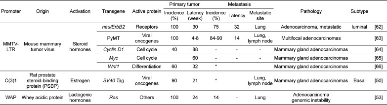
ErbB2 (neu/HER2) is the EGFR family of receptor tyrosine kinases (RTKs) [43], which form homo- and hetero-dimers in response to ligand stimulation, leading to proliferation, differentiation, cell survival, and apoptosis. ErbB2 functions as a prognostic marker, especially in tumors from patients with lymph node metastasis, and predicts poor survival and early relapse in patients [4445]. Transgenic mice develop multifocal adenocarcinomas by 30 weeks of age, which metastasize to the lungs [4647].
Mammary gland-specific expression of the polyoma middle T antigen (PyMT) -under the control of the MMTV promoter- exhibits multifocal adenocarcinomas with a short median latency, high penetrance, and metastasis to the lungs and lymph nodes. These transgenic mice develop palpable tumors at 4–8 weeks of age, and 84–90% of them show pulmonary metastasis by 14 weeks of age [46]. Pathology of these mice is very similar to that of human breast cancer, regarding hyperplasia, adenoma, and early or late carcinoma [46].
Cyclin D1 has been known to play a critical role in the development of normal alveolar mammary gland tumors induced by c-neu, v-Ha-ras, and other oncogenes. As the expression of Cyclin D1 gene is associated with poor prognosis of estrogen receptor (ER) positive patients, which is supported by relapse-free and overall survival, tumor growth in MMTV-Cyclin D1 models is expected to be ER-positive and estrogen-dependent [4849]. In MMTV-Cyclin D1 mice, mammary adenocarcinoma develops by 22 months of age, which is observed in 40% of the transgenic mice. As Cyclin D1 is a weak oncogene, its expression causes long latency and low incidence of breast cancer in the transgenic mice, and co-expression with potent oncogenes is required for carcinogenesis.
Wnt signaling in human breast cancer has been known to be related to the overexpression of β-catenin. It was also reported that human breast cancers highly express β-catenin which is correlated with poor prognosis of breast cancer patients. In MMTV-Wnt1 transgenic mice, mammary adenocarcinoma develops by 32 weeks of age, which is observed in 60% of the mice, and metastasis to lymph nodes and lungs is observed.
In C3(1)/Tag mice, the SV40 large T-antigen (Tag) is expressed by the regulatory control of the rat prostatic steroid binding protein C3(1) gene, leading to development of prostate tumors in male mice or mammary gland adenocarcinomas in female mice [50]. Characteristics of these models include short latency (~21 weeks) and high penetrance (>90%) (Table 2), which is useful for colony management, as well as nonclinical therapeutic trials. In the transgenic mice, mammary adenocarcinoma develops by 21 weeks of age, which is observed in 90% of the mice. Identification of conserved gene expression features and DNA somatic alterations between the C3(1)/Tag models and human breast tumors suggests that this transgenic model recapitulates human Basal-like cancer (BLBC) [51].
WAP (whey acidic protein) promoter is activated by lactogenic hormones in mammary tumors of mice. Ras oncogene has been known to contribute to human cancer development, and the expression of H-ras oncogene driven by the WAP promoter causes genomic instability, adenocarcinoma, and pulmonary metastasis in mice [5253]. In the transgenic mice, mammary gland tumors develop at 24 weeks of age (Table 2).
Taken together, GEMMs of breast cancer are essential nonclinical models to understand the expression profile of target genes and to validate novel therapeutic strategies.

Conclusion

Breast cancer is the most common type of cancer in females. Despite recent advances in its diagnosis and effective therapeutic strategies, further investigations into tumorigenesis, metastasis, and resistance are urgently required. In this review, we provide an overview of tumor transplantation models and GEMMs in order to understand the molecular mechanisms underlying breast cancer progression and metastasis, and to validate the association of human breast cancer with clinical therapeutic trials. Technological advances in order to develop novel mouse models would give us new insights for developing innovative breast cancer therapeutics.

Acknowledgments

This work was supported by the National Cancer Center Grant (NCC-1810042) funded by the Korean government.

Notes

Conflict of interests: The authors declare that there is no financial conflict of interests to publish these results.

References

1. Kweon SS. Updates on Cancer Epidemiology in Korea, 2018. Chonnam Med J. 2018; 54(2):90–100. PMID: 29854674.
crossref
2. Jung KW, Won YJ, Kong HJ, Lee ES. Community of Population-Based Regional Cancer Registries. Cancer Statistics in Korea: Incidence, Mortality, Survival, and Prevalence in 2015. Cancer Res Treat. 2018; 50(2):303–316. PMID: 29566481.
crossref
3. Althuis MD, Dozier JM, Anderson WF, Devesa SS, Brinton LA. Global trends in breast cancer incidence and mortality 1973-1997. Int J Epidemiol. 2005; 34(2):405–412. PMID: 15737977.
crossref
4. Kim IS, Baek SH. Mouse models for breast cancer metastasis. Biochem Biophys Res Commun. 2010; 394(3):443–447. PMID: 20230796.
crossref
5. Rees CA, Pollack JR, Ross DT, Johnsen H, Akslen LA, Fluge O, Pergamenschikov A, Williams C, Zhu SX, Lønning PE, Børresen-Dale AL, Brown PO, Botstein D. Molecular portraits of human breast tumours. Nature. 2000; 406(6797):747–752. PMID: 10963602.
crossref
6. Cardiff RD, Kenney N. A compendium of the mouse mammary tumor biologist: from the initial observations in the house mouse to the development of genetically engineered mice. Cold Spring Harb Perspect Biol. 2011; 3(6):
crossref
7. Sørlie T, Perou CM, Tibshirani R, Aas T, Geisler S, Johnsen H, Hastie T, Eisen MB, van de Rijn M, Jeffrey SS, Thorsen T, Quist H, Matese JC, Brown PO, Botstein D, Lønning PE, Børresen-Dale AL. Gene expression patterns of breast carcinomas distinguish tumor subclasses with clinical implications. Proc Natl Acad Sci U S A. 2001; 98(19):10869–10874. PMID: 11553815.
8. Osborne CK, Schiff R. Mechanisms of endocrine resistance in breast cancer. Annu Rev Med. 2011; 62:233–247. PMID: 20887199.
crossref
9. Palmieri C, Patten DK, Januszewski A, Zucchini G, Howell SJ. Breast cancer: current and future endocrine therapies. Mol Cell Endocrinol. 2014; 382(1):695–723. PMID: 23933149.
crossref
10. Luque-Cabal M, García-Teijido P, Fernández-Pérez Y, Sánchez-Lorenzo L, Palacio-Vázquez I. Mechanisms Behind the Resistance to Trastuzumab in HER2-Amplified Breast Cancer and Strategies to Overcome It. Clin Med Insights Oncol. 2016; 10(Suppl 1):21–30. PMID: 27042153.
crossref
11. Swain SM, Baselga J, Kim SB, Ro J, Semiglazov V, Campone M, Ciruelos E, Ferrero JM, Schneeweiss A, Heeson S, Clark E, Ross G, Benyunes MC, Cortés J. CLEOPATRA Study Group. Pertuzumab, trastuzumab, and docetaxel in HER2-positive metastatic breast cancer. N Engl J Med. 2015; 372(8):724–734. PMID: 25693012.
crossref
12. Fan W, Chang J, Fu P. Endocrine therapy resistance in breast cancer: current status, possible mechanisms and overcoming strategies. Future Med Chem. 2015; 7(12):1511–1519. PMID: 26306654.
crossref
13. Palomeras S, Ruiz-Martínez S, Puig T. Targeting Breast Cancer Stem Cells to Overcome Treatment Resistance. Molecules. 2018; 23(9):
crossref
14. Cho SY, Kang W, Han JY, Min S, Kang J, Lee A, Kwon JY, Lee C, Park H. An Integrative Approach to Precision Cancer Medicine Using Patient-Derived Xenografts. Mol Cells Mol Cells Mol Cells. 2016; 39(2):77–86. PMID: 26831452.
15. Rygaard J, Povlsen CO. Heterotransplantation of a human malignant tumour to “Nude” mice. Acta Pathol Microbiol Scand. 1969; 77(4):758–760. PMID: 5383844.
crossref
16. Zhang Y, Zhang GL, Sun X, Cao KX, Ma C, Nan N, Yang GW, Yu MW, Wang XM. Establishment of a murine breast tumor model by subcutaneous or orthotopic implantation. Oncol Lett. 2018; 15(5):6233–6240. PMID: 29616105.
crossref
17. Ding H, Quan H, Yan W, Han J. Silencing of SOX12 by shRNA suppresses migration, invasion and proliferation of breast cancer cells. Biosci Rep. 2016.
crossref
18. Xiao X, Chen B, Liu X, Liu P, Zheng G, Ye F, Tang H, Xie X. Diallyl disulfide suppresses SRC/Ras/ERK signaling-mediated proliferation and metastasis in human breast cancer by up-regulating miR-34a. PLoS One. 2014; 9(11):e112720. PMID: 25396727.
crossref
19. Tang H, Liu P, Yang L, Xie X, Ye F, Wu M, Liu X, Chen B, Zhang L, Xie X. miR-185 suppresses tumor proliferation by directly targeting E2F6 and DNMT1 and indirectly upregulating BRCA1 in triple-negative breast cancer. Mol Cancer Ther. 2014; 13(12):3185–3197. PMID: 25319390.
crossref
20. Holliday DL, Speirs V. Choosing the right cell line for breast cancer research. Breast Cancer Res. 2011; 13(4):215. PMID: 21884641.
crossref
21. Hoffman RM. Orthotopic metastatic mouse models for anticancer drug discovery and evaluation: a bridge to the clinic. Invest New Drugs. 1999; 17(4):343–359. PMID: 10759402.
22. Borges S, Perez EA, Thompson EA, Radisky DC, Geiger XJ, Storz P. Effective Targeting of Estrogen Receptor-Negative Breast Cancers with the Protein Kinase D Inhibitor CRT0066101. Mol Cancer Ther. 2015; 14(6):1306–1316. PMID: 25852060.
crossref
23. Zhang C, Yan Z, Arango ME, Painter CL, Anderes K. Advancing bioluminescence imaging technology for the evaluation of anticancer agents in the MDA-MB-435-HAL-Luc mammary fat pad and subrenal capsule tumor models. Clin Cancer Res. 2009; 15(1):238–246. PMID: 19118051.
crossref
24. Aslakson CJ, Miller FR. Selective events in the metastatic process defined by analysis of the sequential dissemination of subpopulations of a mouse mammary tumor. Cancer Res. 1992; 52(6):1399–1405. PMID: 1540948.
25. Cochrane DR, Bernales S, Jacobsen BM, Cittelly DM, Howe EN, D'Amato NC, Spoelstra NS, Edgerton SM, Jean A, Guerrero J, Gómez F, Medicherla S, Alfaro IE, McCullagh E, Jedlicka P, Torkko KC, Thor AD, Elias AD, Protter AA, Richer JK. Role of the androgen receptor in breast cancer and preclinical analysis of enzalutamide. Breast Cancer Res. 2014; 16(1):R7. PMID: 24451109.
crossref
26. Cerliani JP, Guillardoy T, Giulianelli S, Vaque JP, Gutkind JS, Vanzulli SI, Martins R, Zeitlin E, Lamb CA, Lanari C. Interaction between FGFR-2, STAT5, and progesterone receptors in breast cancer. Cancer Res. 2011; 71(10):3720–3731. PMID: 21464042.
crossref
27. Whittle JR, Lewis MT, Lindeman GJ, Visvader JE. Patient-derived xenograft models of breast cancer and their predictive power. Breast Cancer Res. 2015; 17:17. PMID: 25849559.
crossref
28. Hoffman RM. Patient-derived orthotopic xenografts: better mimic of metastasis than subcutaneous xenografts. Nat Rev Cancer. 2015; 15(8):451–452. PMID: 26422835.
crossref
29. Hidalgo M, Amant F, Biankin AV, Budinská E, Byrne AT, Caldas C, Clarke RB, de Jong S, Jonkers J, Mælandsmo GM, Roman-Roman S, Seoane J, Trusolino L, Villanueva A. Patient-derived xenograft models: an emerging platform for translational cancer research. Cancer Discov. 2014; 4(9):998–1013. PMID: 25185190.
crossref
30. Gao H, Korn JM, Ferretti S, Monahan JE, Wang Y, Singh M, Zhang C, Schnell C, Yang G, Zhang Y, Balbin OA, Barbe S, Cai H, Casey F, Chatterjee S, Chiang DY, Chuai S, Cogan SM, Collins SD, Dammassa E, Ebel N, Embry M, Green J, Kauffmann A, Kowal C, Leary RJ, Lehar J, Liang Y, Loo A, Lorenzana E, Robert McDonald, McLaughlin ME, Merkin J, Meyer R, Naylor TL, Patawaran M, Reddy A, Röelli C, Ruddy DA, Salangsang F, Santacroce F, Singh AP, Tang Y, Tinetto W, Tobler S, Velazquez R, Venkatesan K, Von Arx F, Wang HQ, Wang Z, Wiesmann M, Wyss D, Xu F, Bitter H, Atadja P, Lees E, Hofmann F, Li E, Keen N, Cozens R, Jensen MR, Pryer NK, Williams JA, Sellers WR. High-throughput screening using patient-derived tumor xenografts to predict clinical trial drug response. Nat Med. 2015; 21(11):1318–1325. PMID: 26479923.
crossref
31. Kopetz S, Lemos R, Powis G. The promise of patient-derived xenografts: the best laid plans of mice and men. Clin Cancer Res. 2012; 18(19):5160–5162. PMID: 22912394.
32. Rosfjord E, Lucas J, Li G, Gerber HP. Advances in patient-derived tumor xenografts: from target identification to predicting clinical response rates in oncology. Biochem Pharmacol. 2014; 91(2):135–143. PMID: 24950467.
crossref
33. Pillai SG, Li S, Siddappa CM, Ellis MJ, Watson MA, Aft R. Identifying biomarkers of breast cancer micrometastatic disease in bone marrow using a patient-derived xenograft mouse model. Breast Cancer Res. 2018; 20(1):2. PMID: 29291741.
crossref
34. Garrido-Laguna I, Uson M, Rajeshkumar NV, Tan AC, de Oliveira E, Karikari C, Villaroel MC, Salomon A, Taylor G, Sharma R, Hruban RH, Maitra A, Laheru D, Rubio-Viqueira B, Jimeno A, Hidalgo M. Tumor engraftment in nude mice and enrichment in stroma- related gene pathways predict poor survival and resistance to gemcitabine in patients with pancreatic cancer. Clin Cancer Res. 2011; 17(17):5793–5800. PMID: 21742805.
crossref
35. Rashid OM, Takabe K. Animal models for exploring the pharmacokinetics of breast cancer therapies. Expert Opin Drug Metab Toxicol. 2015; 11(2):221–230. PMID: 25416501.
crossref
36. Singh M, Ramos I, Asafu-Adjei D, Quispe-Tintaya W, Chandra D, Jahangir A, Zang X, Aggarwal BB, Gravekamp C. Curcumin improves the therapeutic efficacy of Listeria(at)-Mage-b vaccine in correlation with improved T-cell responses in blood of a triple-negative breast cancer model 4T1. Cancer Med. 2013; 2(4):571–582. PMID: 24156030.
37. Takahashi K, Nagai N, Ogura K, Tsuneyama K, Saiki I, Irimura T, Hayakawa Y. Mammary tissue microenvironment determines T cell-dependent breast cancer-associated inflammation. Cancer Sci. 2015; 106(7):867–874. PMID: 25940224.
38. Tao K, Fang M, Alroy J, Sahagian GG. Imagable 4T1 model for the study of late stage breast cancer. BMC Cancer. 2008; 8:228. PMID: 18691423.
crossref
39. Zhou H, Roy S, Cochran E, Zouaoui R, Chu CL, Duffner J, Zhao G, Smith S, Galcheva-Gargova Z, Karlgren J, Dussault N, Kwan RY, Moy E, Barnes M, Long A, Honan C, Qi YW, Shriver Z, Ganguly T, Schultes B, Venkataraman G, Kishimoto TK. M402, a novel heparan sulfate mimetic, targets multiple pathways implicated in tumor progression and metastasis. PLoS One. 2011; 6(6):e21106. PMID: 21698156.
crossref
40. Hanahan D, Wagner EF, Palmiter RD. The origins of oncomice: a history of the first transgenic mice genetically engineered to develop cancer. Genes Dev. 2007; 21(18):2258–2270. PMID: 17875663.
crossref
41. Cardiff RD, Anver MR, Gusterson BA, Hennighausen L, Jensen RA, Merino MJ, Rehm S, Russo J, Tavassoli FA, Wakefield LM, Ward JM, Green JE. The mammary pathology of genetically engineered mice: the consensus report and recommendations from the Annapolis meeting. Oncogene. 2000; 19(8):968–988. PMID: 10713680.
crossref
42. Taneja P, Frazier DP, Kendig RD, Maglic D, Sugiyama T, Kai F, Taneja NK, Inoue K. MMTV mouse models and the diagnostic values of MMTV-like sequences in human breast cancer. Expert Rev Mol Diagn. 2009; 9(5):423–440. PMID: 19580428.
crossref
43. Hynes NE, MacDonald G. ErbB receptors and signaling pathways in cancer. Curr Opin Cell Biol. 2009; 21(2):177–184. PMID: 19208461.
crossref
44. Park JW, Neve RM, Szollosi J, Benz CC. Unraveling the biologic and clinical complexities of HER2. Clin Breast Cancer. 2008; 8(5):392–401. PMID: 18952552.
crossref
45. Allred DC, Clark GM, Molina R, Tandon AK, Schnitt SJ, Gilchrist KW, Osborne CK, Tormey DC, McGuire WL. Overexpression of HER-2/neu and its relationship with other prognostic factors change during the progression of in situ to invasive breast cancer. Hum Pathol. 1992; 23(9):974–979. PMID: 1355464.
crossref
46. Guy CT, Cardiff RD, Muller WJ. Induction of mammary tumors by expression of polyomavirus middle T oncogene: a transgenic mouse model for metastatic disease. Mol Cell Biol. 1992; 12(3):954–961. PMID: 1312220.
crossref
47. Guy CT, Cardiff RD, Muller WJ. Activated neu induces rapid tumor progression. J Biol Chem. 1996; 271(13):7673–7678. PMID: 8631805.
crossref
48. Hwang TS, Han HS, Hong YC, Lee HJ, Paik NS. Prognostic value of combined analysis of cyclin D1 and estrogen receptor status in breast cancer patients. Pathol Int. 2003; 53(2):74–80. PMID: 12588434.
crossref
49. Sutherland RL, Musgrove EA. Cyclins and breast cancer. J Mammary Gland Biol Neoplasia. 2004; 9(1):95–104. PMID: 15082921.
crossref
50. Maroulakou IG, Anver M, Garrett L, Green JE. Prostate and mammary adenocarcinoma in transgenic mice carrying a rat C3(1) simian virus 40 large tumor antigen fusion gene. Proc Natl Acad Sci U S A. 1994; 91(23):11236–11240. PMID: 7972041.
crossref
51. Herschkowitz JI, Simin K, Weigman VJ, Mikaelian I, Usary J, Hu Z, Rasmussen KE, Jones LP, Assefnia S, Chandrasekharan S, Backlund MG, Yin Y, Khramtsov AI, Bastein R, Quackenbush J, Glazer RI, Brown PH, Green JE, Kopelovich L, Furth PA, Palazzo JP, Olopade OI, Bernard PS, Churchill GA, Van Dyke T, Perou CM. Identification of conserved gene expression features between murine mammary carcinoma models and human breast tumors. Genome Biol. 2007; 8(5):R76. PMID: 17493263.
crossref
52. Oztürk-Winder F, Renner M, Klein D, Müller M, Salmons B, Günzburg WH. The murine whey acidic protein promoter directs expression to human mammary tumors after retroviral transduction. Cancer Gene Ther. 2002; 9(5):421–431. PMID: 11961665.
crossref
53. Nielsen LL, Discafani CM, Gurnani M, Tyler RD. Histopathology of salivary and mammary gland tumors in transgenic mice expressing a human Ha-ras oncogene. Cancer Res. 1991; 51(14):3762–3767. PMID: 2065330.
54. Liby K, Neltner B, Mohamet L, Menchen L, Ben-Jonathan N. Prolactin overexpression by MDA-MB-435 human breast cancer cells accelerates tumor growth. Breast Cancer Res Treat. 2003; 79(2):241–252. PMID: 12825859.
crossref
55. Jensen MR, Schoepfer J, Radimerski T, Massey A, Guy CT, Brueggen J, Quadt C, Buckler A, Cozens R, Drysdale MJ, Garcia-Echeverria C, Chène P. NVP-AUY922: a small molecule HSP90 inhibitor with potent antitumor activity in preclinical breast cancer models. Breast Cancer Res. 2008; 10(2):R33. PMID: 18430202.
crossref
56. Zhang T, Chen Y, Li J, Yang F, Wu H, Dai F, Hu M, Lu X, Peng Y, Liu M, Zhao Y, Yi Z. Antitumor action of a novel histone deacetylase inhibitor, YF479, in breast cancer. Neoplasia. 2014; 16(8):665–677. PMID: 25220594.
crossref
57. Kuperwasser C, Dessain S, Bierbaum BE, Garnet D, Sperandio K, Gauvin GP, Naber SP, Weinberg RA, Rosenblatt M. A mouse model of human breast cancer metastasis to human bone. Cancer Res. 2005; 65(14):6130–6138. PMID: 16024614.
crossref
58. Marangoni E, Vincent-Salomon A, Auger N, Degeorges A, Assayag F, de Cremoux P, de Plater L, Guyader C, De Pinieux G, Judde JG, Rebucci M, Tran-Perennou C, Sastre-Garau X, Sigal-Zafrani B, Delattre O, Diéras V, Poupon MF. A new model of patient tumor-derived breast cancer xenografts for preclinical assays. Clin Cancer Res. 2007; 13(13):3989–3998. PMID: 17606733.
crossref
59. DeRose YS, Wang G, Lin YC, Bernard PS, Buys SS, Ebbert MT, Factor R, Matsen C, Milash BA, Nelson E, Neumayer L, Randall RL, Stijleman IJ, Welm BE, Welm AL. Tumor grafts derived from women with breast cancer authentically reflect tumor pathology, growth, metastasis and disease outcomes. Nat Med. 2011; 17(11):1514–1520. PMID: 22019887.
crossref
60. Zhang X, Claerhout S, Prat A, Dobrolecki LE, Petrovic I, Lai Q, Landis MD, Wiechmann L, Schiff R, Giuliano M, Wong H, Fuqua SW, Contreras A, Gutierrez C, Huang J, Mao S, Pavlick AC, Froehlich AM, Wu MF, Tsimelzon A, Hilsenbeck SG, Chen ES, Zuloaga P, Shaw CA, Rimawi MF, Perou CM, Mills GB, Chang JC, Lewis MT. A renewable tissue resource of phenotypically stable, biologically and ethnically diverse, patient-derived human breast cancer xenograft models. Cancer Res. 2013; 73(15):4885–4897. PMID: 23737486.
crossref
61. Charafe-Jauffret E, Ginestier C, Bertucci F, Cabaud O, Wicinski J, Finetti P, Josselin E, Adelaide J, Nguyen TT, Monville F, Jacquemier J, Thomassin-Piana J, Pinna G, Jalaguier A, Lambaudie E, Houvenaeghel G, Xerri L, Harel-Bellan A, Chaffanet M, Viens P, Birnbaum D. ALDH1-positive cancer stem cells predict engraftment of primary breast tumors and are governed by a common stem cell program. Cancer Res. 2013; 73(24):7290–7300. PMID: 24142344.
crossref
62. Muller WJ, Sinn E, Pattengale PK, Wallace R, Leder P. Single-step induction of mammary adenocarcinoma in transgenic mice bearing the activated c-neu oncogene. Cell. 1988; 54(1):105–115. PMID: 2898299.
crossref
63. Almholt K, Lund LR, Rygaard J, Nielsen BS, Danø K, Rømer J, Johnsen M. Reduced metastasis of transgenic mammary cancer in urokinase-deficient mice. Int J Cancer. 2005; 113(4):525–532. PMID: 15472905.
crossref
64. Wang TC, Cardiff RD, Zukerberg L, Lees E, Arnold A, Schmidt EV. Mammary hyperplasia and carcinoma in MMTV-cyclin D1 transgenic mice. Nature. 1994; 369(6482):669–671. PMID: 8208295.
crossref
65. Stewart TA, Pattengale PK, Leder P. Spontaneous mammary adenocarcinomas in transgenic mice that carry and express MTV/ myc fusion genes. Cell. 1984; 38(3):627–637. PMID: 6488314.
66. Li Y, Hively WP, Varmus HE. Use of MMTV-Wnt-1 transgenic mice for studying the genetic basis of breast cancer. Oncogene. 2000; 19(8):1002–1009. PMID: 10713683.
crossref
Table 1

Brief overview of breast cancer cell line-derived xenograft (CDX), patient-derived xenograft (PDX) and syngeneic mouse models

lar-34-160-i001
Implantation site Mice strain Cell line Subtype ER PR HER2 References
CDX model subcutaneous CDX Subcutaneous BALB/c, Nude MDA-MB-231 Basal [18]
MDA-MB-435 Basal [54]
BT-474 Luminal B + + + [55]
Orthotopic CDX Mammary fat pad NOD/SCID MDA-MB-231 Basal [22]
MDA-MB-435 Basal [23]
SUM1315 Basal [24]
MCF7 Luminal A + + [25]
T47D Luminal A + + [26]
Metastatic CDX Tail vein NOD/SCID MDA-MB-231 Basal [56]
SUM149 Basal [57]
PDX model Subcutaneous BALB/c, Nude [58]
Mammary fat pad NOD/SCID [59]
Mammary fat pad NSG [60]
Humanized Mammary fat pad NOD/SCID [61]
Syngeneic model Mammary fat pad BALB/c 4T1 - - - [38]
Table 2

Examples of genetically engineered animal tumor models in breast cancer

lar-34-160-i002
Promoter Origin Activation Transgene Active protein Primary tumor Metastasis Pathology Subtype
Incidence (%) Latency (week) Incidence (%) Latency Metastatic site
MMTV-LTR Mouse mammary tumor virus Steroid hormones neu/ErbB2 Receptors 100 30 75 32 Lung Adenocarcinoma, metastatic luminal [62]
PyMT Viral oncogenes 100 4-8 84-90 14 Lung, lymph node Multifocal adenocarcinomas [63]
Cyclin D1 Cell cycle 40 88 - - - Mammary gland adenocarcinomas [64]
Myc Cell cycle 60 - - - Mammary gland adenocarcinomas [65]
Wnt1 Differentiation 60 32 * Mammary gland adenocarcinomas [66]
C(3)1 Rat prostate steroid-binding protein (PSBP) Estrogen SV40 Tag Viral oncogenes 90 21 * Lung, lymph node Mammary gland adenocarcinomas Basal [50]
WAP Whey acidic protein Lactogenic hormones Ras Others 100 24 14 - Lung Adenocarcinoma genomic instability [53]

*Metastasis/tumor appearance but not incidence was reported.

TOOLS
Similar articles