

 PDF
PDF ePub
ePub Citation
 Print
Print


INTRODUCTION
Abernethy malformation is a rare condition, which was first described in 1793 as a congenital extrahepatic porto-systemic shunt (CEPS) directing splanchnic blood flow directly into the inferior vena cava [1]. Clinical manifestations, especially in children, are extremely variable, involving hepatic, neurological, pulmonary, metabolic, and other systems. Morgan and Superina classified CEPS into two types [2] (Table 1). In a type 1 shunt, portal blood is diverted completely into the inferior vena cava, with absence of the portal vein. Type 1 Abernethy malformation can be further divided into subtype 1a and 1b. In subtype 1a, the superior mesenteric vein and the splenic vein do not connect and thus there is no anatomic portal vein, whereas in subtype 1b, the superior mesenteric vein and splenic vein connect to form a portal vein, which then drains into a systemic vein [3]. In type 2 Abernethy malformation, the portal vein is intact, but a side-to-side anastomosis with the inferior vena cava leads to shunting. Therefore, structurally, type 2 shunt is not characterized by true absence of the portal vein. A type 2 shunt is more common in males and is rarely associated with other malformations [4].
In type 1 CEPS, which is characterized by absence of intrahepatic portal vein, the portal blood is completely drained into the systemic venous circulation. In type 2 CEPS, the portal blood is partially drained into the systemic venous circulation, and a hypoplastic intrahepatic portal vein is present [5].
Eighty cases have been published so far that reported extrahepatic portosystemic shunts [6]; while in Korea, very few cases have been reported as CEPS. Through this study, we present 6 cases of the patients diagnosed with CEPS at Samsung Medical Center while comparing these with the other cases published in France and China.
MATERIALS AND METHODS
From June 2004 to April 2017, 6 children were investigated for CEPS, treated, and followed-up at the Samsung Medical Center. The clinical sources of all CEPS cases were reviewed, including electronic medical records, histopathologic material (if available), and imaging and laboratory findings. The presence of shunt was confirmed by abdominal ultrasonography (US), computed tomography (CT), magnetic resonance imaging, or a combination of these techniques. Investigations for complications of the shunt and associated anomalies included liver function tests; estimation of the plasma ammonia levels and serum alpha-fetoprotein levels; coagulation studies; echocardiography; imaging studies of liver tumors and liver histology. The portosystemic shunts were classified anatomically based on the results of the imaging studies. The follow-up investigations consisted of clinical, biochemical, and US examinations.
RESULTS
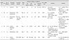
The main findings are summarized in Table 2, including the clinical presentation, type of shunt, associated anomaly, associated abnormalities, treatment and outcome. In the review, 6 patients were identified with a mean age of 1.5 years at the initial diagnosis (range, 11 days to 4 years). There were two male and four female children in our study.
Of six patients, one presented with Abernethy malformation type 1a, two with type 1b, and three with type 2. Four of the six patients presented with CEPS-related complications; whereas, two patients were asymptomatic. One patient presented with cholestatic jaundice and large hepatic mass at 2 months after birth. The investigations for other causes of cholestasis showed negative results. The abdominal US revealed a single shunt and an intrahepatic mass, which manifested as focal nodular hyperplasia on biopsy. Three elder children, aging from 1 to 4 years, presented with an initial symptom of gastrointestinal bleeding (hematemesis, melena, hematochezia). In these patients, the imaging studies with abdominal CT revealed esophageal varices or varicose veins in the perirectal region. In the remaining 2 children who presented with urinary tract infection and congenital hypertrophic cardiomyopathy associated with Noonan syndrome, the portosystemic shunts were found in abdominal US incidentally.
Liver function tests showed elevated serum aspartate aminotransferase and alanine aminotransferase in 3 patients, abnormal prothrombin time in 5 patients, and elevated blood ammonia in 3 patients. At presentation, elevated alpha-fetoprotein levels and liver mass were reported in only one patient, who underwent right lobectomy for focal nodular hyperplasia. In addition, 3 children presented with benign liver mass, which were suspected as focal nodular hyperplasia, with diameter ranging from 1.0 to 8.5 cm.
Among three patients with type 1 CEPS, two patients are waiting for liver transplantation because of increasing the number of liver tumors and one patient is in stable clinical situation with regular screening tests for associated morbidities.
In patients with type 2 CEPS, one patient was planned on surgical shunting vessel ligation, but the patient suddenly developed hepatic encephalopathy, and died of acute hepatic failure, although he underwent a liver transplantation [7]. Another patient diagnosed with CEPS type 2 underwent shunting vessel ligation allowing for age, 2 years-old, and anatomy of the shunt. While the third patient had been in stable clinical situation but was lost to follow-up.
DISCUSSION
In embryonic period, epithelial and mesenchymal interactions occur in the network of early embryonic and then fetal vessels, which involute both inside and outside the liver. Abnormal vascular communications can occur between any vein of the portal system and any branch of the inferior vena cava because of the incomplete involution of one or several of these primordial vessels [89]. These abnormal structures, like extrahepatic portal veins and ductus venosus, can persist throughout life and give rise to risks of complications. Severe complications are neonatal cholestasis; hyperammonemia and encephalopathy (35%); benign and malignant liver tumors (26%); hepatopulmonary syndrome and porto-pulmonary hypertension (18%) [1011].
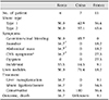
Since the initial description about Abernethy malformation, formerly called CEPS, only about 80 cases have been reported, with most of the patients under the age of 18 years [12]. As reported earlier, type 1 malformations are predominantly found in females (74%), and are more associated with other congenital defects than type 2 CEPS, such as congenital heart disease, genitourinary disease, skeletal abnormalities, etc. Our study reported more number of female patients (66.7%) who presented with type 1 CEPS, and all those patients also presented with other associated congenital defects (Table 2).
We compared our cases with other such cases reported in France and China (Table 3) [578]. In our study, there was no remarkable difference in the incidence of type 1 and 2 CEPS; on the other hand, in China and France, the incidence of type 2 malformation was higher. At presentation, the most common initial symptom was gastrointestinal bleeding followed by central nervous system (CNS) symptoms, which included mental retardation, drowsiness, and portal systemic encephalopathy. Abdominal mass, which was confirmed by imaging studies, jaundice, and dyspnea were also frequently found as initial presenting symptoms. Since numerous developing collaterals, such as the superior hemorrhoidal plexus and varicose veins in the perirectal region may cause hemorrhoidal bleeding, gastrointestinal bleeding is speculated as the most common symptom of CEPS [3]. In China, the most common presenting symptom was also gastrointestinal bleeding, but in France, the most frequent complications were dyspnea and CNS symptoms.
In our study, one patient (16.7%) with type 2 CEPS was treated by liver transplantation, and another patient (16.7%) with type 2 CEPS was treated by shunting vessel ligation, while rest of the patients were regularly followed-up with conservative treatment. In China, all patients were managed by conservative treatment; while in France, 6 patients (54.5%) were treated by shunt ligation or closure, 1 patient (9.1%) was treated by liver transplantation, and 4 patients (36.4%) were treated by conservative treatment.
The most notable differentiating point is that, the proportion of active treatment varies from country to country. One of three patients with type 2 CEPS in Korea (33.3%), six of seven patients with type 2 CEPS (85.7%) in France, none in China (0%), were treated by shunt ligation or closure. However, since long-term and large-sample studies have not yet been published, we do not know the difference of life-long outcome among the methods of treating CEPS.
Many other studies have reported that, early recognition is important because liver nodules may develop into hepatic neoplasms [13], and hepatic encephalopathy may occur suddenly [8], as in one of type 2 CEPS patients in our case. There are no guidelines yet for advising earlier treatment of CEPS to prevent the development of complications [6]. However, a few literatures about Abernethy malformation emphasize that when CEPS is suspected, the diagnosis should be confirmed immediately, and complications should be investigated to start early treatment for restoring portal flow and preventing complications [14]. The treatment options for CEPS depend on the type of shunts, the anatomic situation of the intrahepatic portal vein, condition of the patient, and local expertise [1516]. In type 1 CEPS, the only effective treatment option is liver transplantation, because occlusion of the shunt will obstruct the only drainage route of the mesenteric venous blood [817]; however, transplantation should be performed only in cases with exceptionally complex anatomy where closure of the shunt is not possible. Patients with type 2 CEPS with serious symptoms such as hepatic encephalopathy, may benefit from early shunt closure, in one or two steps, depending on the level of portal pressure assessed by an occlusion test [13161819]. Owing to the severity of complications and their potential reversibility after suppression of the shunts, closure or ligation of a shunt in type 2 CEPS is necessary [7]. It is performed either surgically or with percutaneous transcatheter embolization [20]. Various studies have strongly recommended that, when a shunt is detected with no clinically significant complications and is confirmed to be permanent, it should be closed before a significant complication occurs if a closure can be performed satisfactorily [821].
Since CEPS is a rare disease, and only small-sample studies have been reported, the disease often remains underestimated. In patients with unexplained signs and symptoms of gastrointestinal bleeding, encephalopathy, liver mass, porto-pulmonary hypertension, or hepatopulmonary syndrome, and especially in cases with chromosomal syndromes such as Noonan's syndrome, the associated presence of CEPS needs further consideration. Finally, clinical diagnosis must be carried out by radiologic evaluation, which is of primary importance to confirm the diagnosis and to plan management, thus avoiding the most severe consequences of this malformation.
The present study has a few limitations. Our study was limited to a relatively small number of patients in a single center, and therefore, the study population may not be representative of all pediatric patients in Korea. Furthermore, comparative groups of China and France were also small sized cohorts, hence these studies may also not be representative of all pediatric patients in each of these countries. Large-sample studies including CEPS are needed to evaluate the characteristics of patients with CEPS furthermore and determine the treatment guidelines for CEPS.

 XML Download