

 PDF
PDF ePub
ePub Citation
 Print
Print


INTRODUCTION
The number of patients with heart failure (HF) has been on the rise as a consequence of the aging of society and the improvement in screening and diagnostic techniques. Therefore, there is an increasing need for developing guidelines for the diagnosis and treatment of HF. Although guidelines for HF management have already been issued by American and European associations, many aspects of such guidelines do not reflect the domestic reality in Korea due to the social and anthropological characteristics of the Korean population. Thus, to help clinicians establish the best treatment plan for Korean patients with HF, it is necessary to develop specific guidelines that reflect the clinical situation in Korea. In 2012, the Korean Society of Heart Failure (KSHF) established the Guideline Writing Committee to develop the treatment guidelines for HF. The Korean guidelines for the diagnosis and management of chronic heart failure (CHF) were introduced in March 2016.1) However, CHF and acute heart failure (AHF) are distinct disease entities, warranting different approaches for diagnosis and treatment. Here, we introduce the Korean guidelines for the management of AHF with reduced or preserved ejection fraction (HFrEF and HFpEF, respectively). In Part I of this guideline, we discuss the definition, epidemiology, and diagnosis of AHF.
This guideline was developed based on previously issued international guidelines and amended to reflect the clinical situation in Korea. A committee of KSHF members decided on the format of the guideline, the selection of topics addressed, and the composition of the Writing Committee. To develop the guidelines, we considered all clinical studies and evidence included in the international guidelines, as well as domestic research conducted in Korean patients with HF. Members of the Writing Committee first drafted thematic manuscripts, which were then assembled and arranged according to the evidence-based scales. To ensure transparency and facilitate future revision of the guidelines, we documented the process followed for issuing a recommendation for each topic based on the gathered evidence. The guideline was drafted, reviewed by the advisory committee, and finalized after receiving endorsement from the Korean Society of Cardiology, Korean Society of Hypertension, Korean Society of Interventional Cardiology, Korean Society of Echocardiography, and Korean Society of Lipid and Atherosclerosis. While working on this guideline, the members of the Writing Committee were not affected by external influences and made every effort to exclude conflicts of interests.
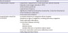
The Writing Committee issued the level of recommendation upon a comprehensive analysis of evidence from studies describing real clinical experience, from surveys, from epidemiologic, observational, and randomized clinical studies, and from meta-analyses. The level of evidence and class of recommendation were defined so as to have a clear formulation, provide straightforward instructions, and be easily adopted in daily clinical practice (Tables 1 and 2).
Table 1
Criteria used to judge the level of evidence and establish the class of recommendation for AHF
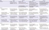
Table 2
Formulations typically used with each class of recommendation

This guideline is intended to help improve clinical practice by providing recommendations based on clinical evidence. As such, the guideline does not serve as a basis for clinical judgement. The final decision in the treatment of each patient should be made by the treating physician according to their personal opinion and judgment, while using the guideline to support these decisions.
GENERAL CONCEPTS OF ACUTE HEART FAILURE
Definition and classification of acute heart failure
Definition of acute heart failure
Patients with AHF are usually hospitalized for prompt treatment because the symptoms manifest and worsen rapidly. “Compensated HF” is usually defined as HF that responds to treatment, with symptoms stable for at least 1 month. “Acute decompensated HF” is defined as a sudden deterioration of symptoms or signs in patients with compensated HF, whereas acute presentation of HF in patients with no previous symptoms is referred to as “Acute de novo HF.” Acute myocardial infarction (AMI) is the most representative manifestation of AHF, though some patients remain asymptomatic despite decreased cardiac function or may exhibit gradual symptoms as decompensation develops.
Classification of acute heart failure
Although there are several ways to classify AHF, the classification based on the clinical condition at the time of admission is most useful from a practical perspective because it facilitates identification of high-risk patients and initiation of treatment according to a pre-established treat-to-target strategy. In patients with AHF, systolic blood pressure is usually preserved (90–140 mmHg) or elevated (>140 mmHg; hypertensive AHF) but may also be decreased (<90 mmHg; hypotensive AHF). Hypotensive AHF generally has poor prognosis, especially if accompanied by decreased perfusion.
Regarding causes and aggravating factors of AHF, five clinical conditions require immediate treatment: acute coronary syndrome, hypertensive emergencies, tachycardia/severe bradycardia/conduction disorders, structural heart damage, and acute pulmonary embolism.
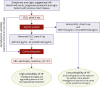
The clinical condition is defined based on the findings of the bedside physical examination, focused mainly on clarifying the presence, nature, and degree of congestion (“wet” or “dry”) and/or tissue perfusion (“warm” or “cold”) according to the Forrester classification (Figure 1). AHF patients can be classified into four groups with increasingly poor prognosis: group A, with “warm” and “dry” HF (compensated, well perfused, without congestion); group B, with “warm” and “wet” HF (well perfused but congested); group C, “cold” and “dry” HF (hypoperfused but without congestion); and group D, “cold” and “wet” HF (hypoperfused and congested). To confirm the presence and degree of congestion, it is necessary to assess jugular venous pressure and check for ascites or peripheral edema, rales or abnormal heart sound on chest auscultation, and pulmonary edema on chest radiography. Decreased tissue perfusion can be confirmed based on decreased skin temperature, reduced urine output, and altered mentality. Pulmonary capillary wedge pressure is the best indicator of congestion, while cardiac index is the best marker of perfusion.
Figure 1
Clinical classification of acute heart failure.
CI = cardiac index; PCWP = pulmonary capillary wedge pressure.
In patients with AMI, AHF can be classified according to the Killip classification: class I, no clinical signs of HF; class II, HF with rales in half of the lung field and with S3 gallop; class III, frank acute pulmonary edema; and class IV, cardiogenic shock, hypotension, and evidence of peripheral vasoconstriction such as cyanosis, oliguria, and diaphoresis.
Aggravating factors, risk factors, and causes of acute heart failure
Various aggravating factors are known to induce HF decompensation (see section 2.2. Aggravating factors). Risk factors include concomitant diseases leading to structural heart disease, including hypertension, diabetes, metabolic syndrome, and atherosclerotic disease. Risk factors of AHF comprise myocardial disorders (coronary artery disease, hypertension, myocarditis, cardiomyopathy), valvular heart diseases, pericardial diseases, endocarditis, congenital heart disease, arrhythmia, conduction disturbance, high cardiac output state (anemia, sepsis, arteriovenous fistula), and right HF.
Epidemiology
The prevalence of AHF has been increasing in parallel with the rates of cardiovascular disease, reflecting the aging of society and widespread adoption of a westernized lifestyle. AHF is a major cause of hospitalization as well as death during hospital admission and readmission, especially among individuals aged >65 years. The prevalence of AHF continues to increase despite recent advances in medical technology and reduced in-hospital mortality rates. According to epidemiological studies conducted in the United States, more than one million hospitalizations for AHF occur annually, and hospitalization rates are expected to continue to increase over the next 20 years.2) Approximately 15.5 million emergency room visits due to AHF were recorded between 1992 and 2001, with an average increase of 18,500 cases per year.3) It is estimated that, by 2050, 20% of the population of the United States will consist of individuals aged >65 years, approximately 80% of whom will likely require hospitalization for AHF.4) While no epidemiological studies on this topic have been conducted in Korea, the prevalence of AHF is expected to increase in the future due to the recent increase in the prevalence of risk factors for cardiovascular disease, reflecting the westernization of eating habits, lack of physical activity, and rapid increase in the proportion of elderly people. AHF is also associated with a heavy socioeconomic burden. In the United States, the current annual cost for hospitalizations due to AHF is approximately $34 billion, which is similar to the figures reported in Europe.5) In Korea, AHF-related expenses associated with an 8-day hospital stay amount to approximately 7.7 million won (about $7,000).6)
In a Korean AHF cohort, 52% of patients were identified as having de novo HF, while the remaining 48% had decompensated HF.6) The same study concluded that, at the time of AHF diagnosis, Korean patients tended to be younger (mean age, 69 years) than those in Japan, USA, and Europe; the proportion of men (55%) was slightly higher among Korean AHF patients than among patients from AHF registries in other countries. In this Korean AHF cohort, 59% of patients had hypertension, 35% had diabetes, and 28% had atrial fibrillation. The major cause of AHF was ischemic heart disease (37%), followed by cardiomyopathies (21%), valvular heart disease (14%), and tachycardia-induced cardiomyopathy (11%). The prognosis of AHF is reportedly very poor. A study conducted in the United States reported a 5-year survival rate of less than 1 in 3 patients hospitalized for AHF.7) Studies conducted in Korea reported similarly poor outcomes: in-hospital mortality, 6.1%; short-term all-cause mortality, 1.2% and 9.2% at 1 and 6 months, respectively;6) long-term mortality, 15%, 21%, 26%, and 30% at 1, 2, 3, and 4 years, respectively8); re-admission rate, 6.4% within 1 month and 24% within 6 months.6)
INITIAL ASSESSMENT
Symptoms and signs
The traditional clinical approach involves identifying symptoms and signs by taking a clinical history and performing a physical examination in all patients who visit the emergency room with acute symptoms suggestive of HF (Class of Recommendation, I; Level of Evidence, C). The diagnosis can also be made based on objective evidence of pulmonary edema or cardiac dysfunction in patients with signs and symptoms of AHF.9)10)11)
In general, symptoms and signs specific to HF can be divided into two categories: congestion and decreased perfusion of the peripheral tissues. AHF symptoms include orthopnea and paroxysmal nocturnal dyspnea as typical symptoms of congestion, as well as fatigue and decreased exercise capacity associated with decreased perfusion of peripheral tissues. AHF signs include pulmonary edema, jugular venous engorgement, hepatic-jugular reflux reflex, third heart sound (S3), left downward deviation of the apical pulse, peripheral edema, and decreased urine output. However, it is difficult to discriminate the cause of such symptoms and signs, especially in patients with obesity, advanced age, and chronic lung disease.9-13) Therefore, it is recommended that the following medical history be recorded and physical examination be conducted in patients with AHF.
Orthopnea
At the bedside, the presence and severity of orthopnea can be easily evaluated if sufficient pillow height is used to achieve a position in which the patient can breathe comfortably. Thus, orthopnea can be defined as: mild or non-existent if the patient can breathe comfortably without a pillow or with minimal elevation of the neck: moderate if the patient can breathe comfortably only when using at least one pillow, with neck elevation of up to 10 cm; and severe if the patient can breathe comfortably only when using at least two pillows.
Jugular venous distention
To evaluate jugular venous distention, the patient must sit in a relaxed posture, with the back at an angle of 30°–45° from the horizontal. The neck and chest should be exposed from the middle of the sternum to the antihelix of the ear. Upon turning the patient's neck to one side, jugular venous distention can be measured vertically with illumination. If it is difficult to distinguish the jugular venous pulse from the internal carotid pulse, the hepatojugular reflux can be measured by applying pressure to the right upper quadrant for about 10 seconds and then measuring the time it takes for jugular venous pressure to recover from the transient increase; if the elevated jugular venous pressure persists, right ventricular dysfunction may be suspected. Jugular venous distention, which is measured from the angle of Lewis to the highest point of internal jugular venous pulsation, is considered severe if it exceeds 15 cm.
Peripheral edema
Peripheral edema can be examined by applying pressure with the thumb for 5 seconds to the medial malleolus, posterior side of the tibia, or sacrum (in patients who cannot walk). Edema is confirmed if the skin takes longer than 10 seconds to return to the original shape, and is defined as mild edema if observed in only one lower limb or the sacrum, versus moderate edema if observed in the bilateral lower limbs. The patient is considered to have severe edema if a large amount of edema is noted in the lower portion of the calf, if edema extends from the leg to the sacrum (in immobile patients), if pitting edema develops easily and disappears after >30 seconds, or if acute/subacute skin changes including cutaneous distension or cleavage are noted.
Rales
Rales are graded as follows: grade 1, unilateral or bilateral rales across the lower third of the lung; grade 2, bilateral rales across the lower half or two thirds of the chest; and grade 3, bilateral rales throughout all lung fields. It is also necessary to distinguish rales from wheezing (stridor) or dry crackles.
Dyspnea
Dyspnea is graded according to the New York Heart Association functional classification (Table 3).
Table 3
New York Heart Association functional classification of dyspnea

Aggravating factors of acute heart failure
1. To improve symptoms in patients with AHF, the aggravating factors of AHF should be evaluated and treated (class of recommendation, I; level of evidence, B).
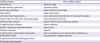
In the initial evaluation of patients with AHF, it is important to perform an accurate hemodynamic assessment and to identify the aggravating factors. Common causes of AHF include: acute coronary syndrome, inappropriate control of hypertension, high salt intake, arrhythmia (including atrial fibrillation), medications reducing cardiac contractility, renal impairment, pulmonary embolism, alcohol abuse, worsening of underlying thyroid disease, infection, and worsening of underlying cardiovascular disease (Table 4).14)
Table 4
Causes and aggravating factors of AHF
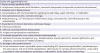
Ponikowski P, et al.; 2016 ESC Guidelines for the diagnosis and treatment of acute and chronic heart failure: The Task Force for the diagnosis and treatment of acute and chronic heart failure of the European Society of Cardiology (ESC) Developed with the special contribution of the Heart Failure Association (HFA) of the ESC, European Heart Journal 2016; 37 (27): 2129–2200, doi: 10.1093/eurheartj/ehw128. Reproduced by permission of Oxford University Press on behalf of the European Society of Cardiology. © European Society of Cardiology.
AHF = acute heart failure.
Because acute coronary syndrome is an important factor that can cause AHF, the patients should be carefully checked for chest pain, changes in the ST segment on electrocardiography, and elevation in blood troponin levels. If necessary, coronary angiography should be performed to exclude the possibility of coronary artery disease as a cause of worsening HF.15) Hypertension is a common cause of AHF exacerbation, especially in women and in patients with HFpEF. Patients with HF who are taking antihypertensive medications can also develop AHF if they suddenly stop their medication.16)17) The symptoms may worsen without adequate restriction of water or salt.18)19) Atrial fibrillation is common in patients with HF, and an excessively increased heart rate may be a cause of HF exacerbation due to increased left atrial pressure and decreased cardiac output.14) The use of medications such as calcium channel blockers, non-steroidal anti-inflammatory drugs, corticosteroids, and thiazolidinedione diabetic medications can decrease left ventricular contractility or worsen HF symptoms.14)
Excessive long-term alcohol consumption will deteriorate cardiac function and can deteriorate HF symptoms. Infections such as pneumonia and septicemia generally increase the metabolic demand or reduce myocardial contractility, which may exacerbate HF symptoms.14)20) An exacerbation of renal dysfunction or pulmonary embolism or abnormal production of thyroid hormones can exacerbate cardiac dysfunction. A worsening of pre-existing heart disease such as valvular heart disease would also cause HF symptoms.14) Therefore, aggravating factors should be assessed and treated in all AHF patients.
Noninvasive diagnostic tests
1. The initial assessment of patients with suspected AHF should include 12-lead electrocardiography, chest X-ray, and blood tests to evaluate the levels of blood urea nitrogen, creatinine, electrolytes, and serum glucose, as well as a complete blood count, a liver function test, and a thyroid function test (class of recommendation, I; level of evidence, C).
2. Measuring serum natriuretic peptide levels is useful for making the clinical diagnosis in the presence of signs and symptoms of AHF, and especially useful for the differential diagnosis of AHF in patients with idiopathic dyspnea (class of recommendation, I; level of evidence, A).
3. Echocardiography should be performed in patients with hemodynamic instability or suspected functional or structural heart disease (class of recommendation, I; level of evidence, C).
4. Determining initial serum natriuretic peptide levels is useful for predicting in-hospital mortality and pre-discharge follow up levels is useful for assessing prognosis. (class of recommendation, I; level of evidence, A).
5. Measuring the serum levels of troponin is helpful for predicting in-hospital mortality and for assessing prognosis (class of recommendation, I; level of evidence, A).
6. Measuring the serum levels of ST2, a biomarker of myocardial damage or fibrosis, may be helpful in predicting mortality risk in patients with acute decompensated HF (class of recommendation, IIb; level of evidence, A).
7. The usefulness of treatment based on serum natriuretic peptide levels has not been well established (class of recommendation, IIb; level of evidence, C).
Symptoms, physical findings, and laboratory findings in decompensated acute heart failure
The main symptoms of AHF include dyspnea, excessive body fluid retention, and fatigue, all of which are nonspecific findings that may occur in AHF or other cardiovascular diseases. The major symptoms and physical findings suggestive of AHF are summarized in Table 5. Because these symptoms and physical findings can be seen in other diseases, it is necessary to conduct differential diagnosis for pneumonia, pulmonary diseases (chronic obstructive pulmonary disease, pulmonary embolism, pulmonary arterial hypertension), renal impairment, severe infectious diseases, and acute coronary syndrome including AMI.
Table 5
Major symptoms and physical findings of patients with AHF
In patients with suspected AHF, the initial assessment should include general blood tests including biomarkers, electrocardiography, chest radiography, and echocardiography (Figure 2). Typical symptoms and signs of AHF in patients with suspected decompensated AHF include the characteristic findings of fluid overload (pulmonary edema, peripheral edema); less often, these findings are symptoms associated with reduced cardiac output and decreased peripheral circulation. Because the sensitivity and accuracy of these symptoms and physical findings are not high, the above-mentioned basic tests are often needed in addition to the clinical evaluation in order to discriminate the cause.
Baseline evaluation
1) Chest X-ray
Although pulmonary congestion, pleural effusion, and cardiomegaly are the most specific findings related to AHF, chest X-ray scans are normal in up to 20% of patients with AHF.21) Nevertheless, chest radiography can help detect non-cardiac diseases potentially responsible for the patients' symptoms.
2) Electrocardiography
Because patients with AHF rarely have normal findings on electrocardiography, this type of assessment is very helpful for identifying the underlying cardiac disease and potential triggering factors.22)
3) Echocardiography
An immediate echocardiographic examination is necessary only in patients with hemodynamic instability (cardiogenic shock), structural heart disease, or acute life-threatening structural or functional cardiac abnormalities (acute valvular regurgitation or acute aortic dissection). Early echocardiography should be considered in all patients with de novo AHF or for whom cardiac function information is lacking. It is common for patients to undergo echocardiography performed by a specialist within 48 hours of admission. However, the optimal timing of echocardiographic assessment in AHF remains to be established. Repeated echocardiographic examinations are usually not needed unless a change in clinical condition becomes evident.
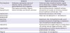
Laboratory tests
Recent routine blood tests reflect many aspects of the pathophysiology of AHF. Biomarkers for myocardial wall stress, hemodynamic abnormalities, inflammation, myocardial damage, neurohormone changes, myocardial remodeling, myocardial extracellular matrix changes, and myocardial fibrosis are known to provide powerful additional information for the standard diagnosis, treatment, and prognosis of AHF.
1) Natriuretic peptides: the brain natriuretic peptide and N-terminal pro-BNP
Sodium natriuretic peptides are used in the diagnosis or differential diagnosis of AHF, especially in patients with idiopathic dyspnea. The brain natriuretic peptide (BNP) and N-terminal pro-BNP (NT-proBNP) have high diagnostic accuracy and negative predictive value in patients admitted to the emergency room for acute dyspnea, and are not affected by ejection fraction (i.e., are useful in patients with HFrEF or HFpEF).23)24)25)26)27)28)29) BNP levels were shown to be moderately correlated with left ventricular end-diastolic pressure,30) and BNP levels at admission were direct predictors of in-hospital mortality, re-hospitalization within 30 days after discharge, and subsequent re-admission and mortality in patients with AHF.31)32)33)34)35)36) Importantly, BNP levels immediately before discharge after HF treatment were good predictors of re-admission and death.27)37)38) Increased levels of natriuretic peptides are very useful indicators of AHF in patients with AHF-related symptoms such as dyspnea (BNP >100 pg/mL or NT-proBNP >300 pg/mL). However, since various cardiac and non-cardiac conditions can result in elevated levels of natriuretic peptides, this criterion alone cannot be used to establish the diagnosis of AHF (Table 6). Thus, clinical characteristics should be considered, along with other laboratory findings.
Table 6
Cardiac and non-cardiac causes of elevated concentrations of natriuretic peptides
Ponikowski P, et al.; 2016 ESC Guidelines for the diagnosis and treatment of acute and chronic heart failure: The Task Force for the diagnosis and treatment of acute and chronic heart failure of the European Society of Cardiology (ESC) Developed with the special contribution of the Heart Failure Association (HFA) of the ESC, European Heart Journal 2016; 37 (27): 2129–2200, doi: 10.1093/eurheartj/ehw128. Reproduced by permission of Oxford University Press on behalf of the European Society of Cardiology. © European Society of Cardiology.
The levels of natriuretic peptides decrease under treatment for HF, and the magnitude of the decrease is reflected in the extent of clinical improvement.36)37)39)40) Some studies have focused on optimizing drug therapies to lower natriuretic peptide levels has been investigated whether it can improve clinical outcomes compared to those attainable with standard HF therapy, but the prospective randomized studies conducted to date have not reported consistent results. Therefore, further studies are warranted to clarify the usefulness of these therapeutic strategies.
2) Cardiac biomarkers: troponins
The levels of troponins, which are markers of myocardial necrosis, may be increased in patients with AHF in the absence of AMI or obstructive coronary artery disease,41) suggesting that myocardial damage or necrosis persists and accumulates. It has been reported that AHF patients with elevated troponin levels are at an increased risk of mortality during hospitalization and after discharge, and that prognosis is better in patients whose troponin levels decrease during treatment, reflecting treatment response.32)35)40)42)
3) Markers of myocardial fibrosis: soluble ST2 and galectin-3
In combination with natriuretic peptides, soluble ST2 and galectin-3, as biomarkers of myocardial fibrosis, help establish the diagnosis of HF and predict the risk of hospital admission and death. Soluble ST2 is a product of the ST2 gene, a member of the interleukin-1 receptor family. In animals with HF, increased ST2 transcription is associated with progressive myocardial fibrosis and hypertrophy induced by myocardial elongation. In clinical studies, soluble ST2 levels were significantly elevated in patients with AHF, representing a significant predictor of 1-year mortality.43)44) Galectin-3 plays an important role in fibroblast activation and fibrosis in animal cell models, and its elevation is associated with long-term survival in patients with HFrEF.45)
There is clinical evidence to support the future clinical use of biomarkers of myocardial damage and myocardial fibrosis in combination with already established markers such as natriuretic peptides. However, further studies are warranted to clarify the usefulness of such biomarkers for evaluating the prognosis of HF.
Invasive diagnostic methods
1. Invasive hemodynamic monitoring using a pulmonary artery catheter should be performed to determine the treatment direction when it is difficult to adequately assess left ventricular filling pressure in patients with dyspnea or hypoperfusion (class of recommendation, I; level of evidence, C).
2. Invasive hemodynamic monitoring can be used in the following cases where symptoms persist despite standard therapy (class of recommendation, IIa; level of evidence, C):
-
a. Fluid status, perfusion, and systemic or pulmonary vascular resistance are uncertain
b. Systolic blood pressure remains low despite initial treatmentc. Renal function decreases after the start of treatmentd. An intravenous injection is required to raise blood pressuree. Mechanical circulatory assist devices or heart transplantation are considered
3. Coronary angiography and intervention should be performed if ischemia is the suspected cause of HF (class of recommendation, IIa; level of evidence, C).
4. Endomyocardial biopsy may be useful in patients suspected of having a specific disease that may affect treatment decision (class of recommendation, IIa; level of evidence, C).
5. Invasive hemodynamic monitoring is not recommended if pulmonary congestion symptoms improve after diuretic and vasodilator administration in acute non-compensated HF patients with normal blood pressure (class of recommendation, III; level of evidence, B).
6. Routine endomyocardial biopsy is not recommended in patients with HF (class of recommendation, III; level of evidence, B).
Right heart catheterization
Hemodynamic monitoring is used when it is clinically impossible to assess the fluid status or in patients who are unresponsive to initial therapy, especially if left ventricular filling pressure and cardiac output are unclear. Patients with clinically severe hypotension (systolic blood pressure <90 mmHg or symptomatic hypotension) and patients with renal impairment during initial treatment also undergo invasive hemodynamic monitoring. Patients indicated for heart transplantation or insertion of a mechanical circulatory assist device are also required to undergo right heart catheterization including the measurement of pulmonary vascular resistance, an essential element for evaluating the candidacy of heart transplantation. Invasive hemodynamic monitoring should be used in the following cases: i) cardiogenic shock with an increased demand for vasopressors and a mechanical circulatory assist device; ii) clinically significant non-compensated HF states in which treatment is limited due to uncertainty regarding left ventricular filling pressure, perfusion status, and vascular tension; iii) clinically significant dependence with vasopressor use after the initial clinical improvement; and iv) persistence of severe symptoms after adequate use of standard therapy. On the other hand, routine invasive hemodynamic monitoring is not recommended in patients with decompensated AHF and normal blood pressure who experience symptomatic improvement with diuretics and vasodilators.
Left heart catheterization
Left heart catheterization may be useful in patients with left ventricular dysfunction.
Coronary angiography
Invasive coronary angiography should be performed in patients who may need coronary artery reperfusion. Coronary angiography is indicated in patients who were previously diagnosed with coronary artery disease and angina or those with ventricular dysfunction and severe coronary ischemic findings on electrocardiography or other noninvasive examinations. In patients without a previous diagnosis of obstructive coronary artery disease, the possibility of recurrent obstructive coronary artery disease should be considered if left ventricular dysfunction is present. In these patients, coronary angiography may help confirm the presence and location of coronary artery occlusion. If obstructive coronary artery disease has not been identified as a cause of left ventricular dysfunction, coronary angiography is generally unnecessary unless there is a change in clinical status suggestive of the progression of myocardial ischemia.
Endomyocardial biopsy
Endomyocardial biopsy is useful for diagnosing a specific disease that may affect treatment decision. Therefore, endomyocardial biopsy should be considered in patients with rapidly progressing HF or left ventricular function deterioration despite adequate medication. Endomyocardial biopsy should also be considered in patients with acute rejection after heart transplantation, infiltrative myocardial diseases (including primary amyloidosis), or acute myocarditis (especially if giant cell-myocarditis is suspected). However, due to the limited diagnostic rate and risk of procedural complications, routine endomyocardial biopsy is not recommended in HF.
Triage and hospitalization
AHF includes both acute de novo HF and acute exacerbations of compensated HF. In AHF, the mortality rate is high, and professional medical treatment such as mechanical ventilation is often required. Therefore, it is desirable that the patient be transferred to a specialized medical institution with an intensive care unit. Early diagnosis and appropriate treatment are critical for improving symptoms and stabilizing the patient's condition.46)47)
Classification of acute heart failure
The hemodynamic status of patients with AHF can be assessed via history taking and physical examination. The clinical condition is classified based on the findings of bedside physical examination, aiming mainly to detect clinical signs/symptoms of congestion (“wet” or “dry”) and/or tissue perfusion (“warm” or “cold”) according to the Forrester classification (Figure 1).48)49) Among these categories, “warm-dry” HF, which does not exhibit congestion and is characterized by good perfusion, corresponds to a stable compensated state. Patients with AHF most commonly exhibit congestion and good peripheral perfusion (“warm-wet” HF). Adequate classification of AHF at the initial evaluation is a key to accurately predicting prognosis and choosing the optimal treatment direction.
Initial assessment algorithm
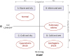
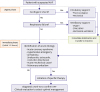
Continuous non-invasive monitoring of the patient's condition is required at the time of the initial evaluation and treatment. The patient's ventilatory status, peripheral perfusion status, and oxygen supply should be assessed through continuous monitoring of blood pressure, heart rate, oxygen saturation, and urine output.50) In hemodynamically unstable conditions, medication or mechanically assisted circulation therapy should be considered, while mechanical ventilation therapy is required if the respiratory disturbance persists despite appropriate oxygen therapy. To prevent deterioration of the patient's condition during the initial treatment, it is necessary to identify and treat the triggering factors of HF, including acute coronary syndrome, hypertensive emergency, arrhythmia, acute structural cardiac damage, and acute pulmonary embolism (Figure 3).
Figure 3
Initial management strategy for AHF.
Ponikowski P, et al.; 2016 ESC Guidelines for the diagnosis and treatment of acute and chronic heart failure: The Task Force for the diagnosis and treatment of acute and chronic heart failure of the European Society of Cardiology (ESC) Developed with the special contribution of the Heart Failure Association (HFA) of the ESC, European Heart Journal 2016; 37 (27): 2129–2200, doi: 10.1093/eurheartj/ehw128. Reproduced by permission of Oxford University Press on behalf of the European Society of Cardiology. © European Society of Cardiology.
AHF = acute heart failure; BiPAP = bilevel positive airway pressure; CCU = coronary care unit; CPAP = continuous positive airway pressure; ICU = intensive care unit.
*Acute mechanical cause: interventricular septal perforation, ventricular free wall rupture, and acute mitral regurgitation complicating acute coronary syndrome, ii) complications due to thoracic trauma, iii) acute native or prosthetic valve dysfunction secondary to endocarditis, and iv) complications due to aortic dissection or thrombosis.
1) Acute coronary syndrome
In patients with acute coronary syndrome, early diagnosis and treatment are important and can reduce the incidence of further HF.51)52)53)54)55)56)57)58)59) Patients with congestive HF and acute coronary syndrome are at high risk and require concomitant active reperfusion therapy for acute coronary syndrome.56)57)58)59)
2) Hypertensive emergencies
AHF can occur with a rapid and excessive increase in arterial blood pressure and mainly manifests as acute pulmonary edema. In the presence of hypertensive episodes, the treatment for HF and that for hypertension should be performed concomitantly. Blood pressure should be reduced aggressively by up to 25% during the first few hours, and the use of intravenous vasodilators with loop diuretics is recommended for this purpose.60)61)
3) Arrhythmia (rapid arrhythmia or severe bradycardia/conduction disturbance)
Arrhythmia associated with hemodynamic instability is common in AHF. If serious arrhythmias are noted in AHF patients, they should be stabilized early using medication, electrical cardioversion, or a temporary pacemaker. If the patient has hemodynamic instability due to atrial or ventricular arrhythmias, these arrhythmias should be terminated by electrical cardioversion. If ventricular arrhythmias occur repeatedly, appropriate treatment is required. If arrhythmia is caused by myocardial ischemia, immediate reperfusion therapy is necessary and radiofrequency catheter ablation may be considered in some cases.62)
4) Acute mechanical causes
Acute mechanical causes include i) interventricular septal perforation, ventricular free wall rupture, and acute mitral regurgitation resulting from ischemic heart disease, ii) complications due to thoracic trauma, iii) valvular regurgitation secondary to acute endocarditis, and iv) complications due to aortic dissection or thrombosis. Echocardiographic studies play an important role in diagnosis and treatment. In patients with acute structural damage, hemodynamic instability or cardiogenic shock occurs frequently and warrants the use of circulatory assist devices; in such patients, active surgical correction should be considered early.63)64)65)66)
5) Acute pulmonary embolism
Reperfusion therapy using a thrombolytic agent, percutaneous catheter removal of the thrombus, or surgical embolectomy is recommended in patients with hypotension or shock.67)68) Early detection of these triggering factors of HF is important, and treatment should be initiated as soon as possible (within the first 1–2 hours) (Figure 3).
Treatment plan
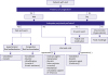
The initial treatment plan for patients with AHF is created according to the patient's condition, which is defined in terms of the degree of congestion and peripheral perfusion. The management strategy for AHF is illustrated in Figure 4.
Figure 4
Management strategy for AHF based on the degrees of congestion and peripheral perfusion.
Ponikowski P, et al.; 2016 ESC Guidelines for the diagnosis and treatment of acute and chronic heart failure: The Task Force for the diagnosis and treatment of acute and chronic heart failure of the European Society of Cardiology (ESC) Developed with the special contribution of the Heart Failure Association (HFA) of the ESC, European Heart Journal 2016; 37 (27): 2129–2200, doi: 10.1093/eurheartj/ehw128. Reproduced by permission of Oxford University Press on behalf of the European Society of Cardiology. © European Society of Cardiology.
AHF = acute heart failure.
Criteria for hospitalization
In patients with AHF, hospitalization should be considered if there are symptoms or signs of congestion. The indications of hospitalization are listed in Table 7. Few patients are discharged from the emergency room within a few hours due to good response to the initial diuretic therapy. The following criteria should be considered when considering discharge from the emergency room:
• Have the patient's symptoms improved sufficiently?
• Has the patient's heart rate stabilized?
• Is orthostatic hypotension absent at standing?
• Is urine output appropriate?
• Is there any deterioration of renal function?
• Is oxygen saturation maintained (>95%)?
Table 7
Criteria for hospitalization of patients with AHF
If there is persistent dyspnea or hemodynamic instability, it is necessary to observe AHF patients in a space where cardiopulmonary resuscitation is possible. Patients with severe respiratory distress, hemodynamic instability, recurrent arrhythmia, and acute coronary syndrome are considered high risk; it is thus recommended to have such patients monitored and treated in the intensive care unit. Patients who meet one or more of the following criteria are also candidates for intensive care monitoring50):
CONCLUSION
As the prevalence of AHF has been increasing, AHF is a major cause of hospitalization and mortality. In patients with symptoms suggesting AHF, initial assessment should include general blood tests including biomarkers, electrocardiography, chest radiography, and echocardiography. Initial identification and prompt treatment of aggravating factors are crucial in AHF management. The management strategy should be decided based on the patient's status of congestion and perfusion.
 XML Download