

 PDF
PDF ePub
ePub Citation
 Print
Print


Introduction
The prevalence of spontaneously conceived triplet pregnancies is 1 in 8,000–9,000 deliveries and that of monochorionic triplet pregnancies is approximately 1 in 45,500 deliveries.12 Although the occurrence of monozygotic twins is thought to be higher after the use of assisted reproductive technology (ART), including in vitro fertilization (IVF), extended embryo culture, and embryo transfer (ET) procedures, the rate of monochorionic and triamniotic triplet pregnancies remains very low. Selective reduction can be attempted to reduce the potential risk for monochorionic triplet pregnancy complications, such as abortion, stillbirth, extreme preterm birth, fetofetal transfusion syndrome (FFTS), and fetal anomaly; however, determining the timing and method of selective reduction and the number of fetuses to eliminate remains challenging.
We report a rare case of monochorionic triamniotic (MT) triplet pregnancy following IVF-ET and introduce a selective reductive technique with radiofrequency ablation (RFA) in early pregnancy.
Case
A 33-year-old woman (gravida 0, para 0) at 8+5 weeks' gestation was transferred to our obstetric unit for the evaluation and management of a high-order multiple pregnancy following IVF and two-blastocyst transfer. She had been diagnosed with unexplained infertility 7 months earlier without past or familial medical history of the condition. Medical records from the infertility clinic revealed that controlled ovarian hyperstimulation (COH) was performed using recombinant follicle stimulating hormone in a luteal down-regulation cycle with a gonadotropin releasing hormone agonist. Subcutaneous human chorionic gonadotropin was injected on day 10 after starting the gonadotropin agent. Eighteen oocytes were aspirated under ultrasound guidance, and 13 oocytes were fertilized and cultured. Intramuscular injection of progesterone in oil 50 mg for luteal support was initiated on the day after oocyte retrieval. Two fresh blastocysts without assisted hatching were transferred on postfertilization day 5. The remaining seven blastocysts were cryopreserved. A single intrauterine gestational sac was observed using transvaginal ultrasonography at 5+2 weeks' gestation. This gestational sac was suspected to have monochorionic twins at 6+2 weeks' gestation. At 8+2 weeks' gestation, three embryos were observed using transvaginal ultrasonography.
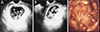
Precise transvaginal ultrasonography at our obstetric unit demonstrated a single intrauterine gestational sac with three separate embryos. Each of the embryos had an active heartbeat, slow body movement, and a yolk sac. Three amniotic sacs divided by thin membranes were clearly distinguishable from one another (Fig. 1A, 1B). MT triplet pregnancy was diagnosed using two-dimensional ultrasound at 8+5 weeks' gestation. Three-dimensional (3D) ultrasound at 12+1 weeks' gestation aided in evaluating the fetal structures and confirming the diagnosis by showing the three distinct fetuses within one chorionic sac (Fig. 1C).
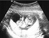
The increased maternal and fetal risks of continuing the pregnancy in this condition were comprehensively discussed with the couple. Three weeks after initial diagnosis of MT triplet pregnancy, they decided to attempt selective reduction of two fetuses and save one fetus. The fetuses located in the uppermost portion of the uterus were selected for abortion, and selective reduction via RFA of the heart was performed at 12+3 weeks' gestation (Fig. 2).
A single needle puncture for each fetus with lidocaine injection was performed with guidance of continuous ultrasound. A 17-gauge RFA (VRS01, VP01; STARmed Co., Goyang, Korea) needle was percutaneously inserted into each fetal heart through the uterus and amniotic membrane without penetrating the amniotic sac of the fetus designated to be the survivor. After a 5-min application of RFA energy increasing by 10 W every minute up to 40 W, cessation of cardiac activity was noted in the selected fetuses.
The survivor fetus and pregnant woman had an uneventful obstetric progression until she went to a local obstetric clinic at 20 weeks' gestation. Thereafter, a spontaneous preterm premature rupture of membrane (PPROM) was occurred at the period of periviability and delivery was inevitable. The patient was terminated without any complication.
Discussion
A maximum of two embryos after prolonged culture for 5–6 days have been transferred to reduce multiple pregnancies following ART. However, this effort can lower the occurrence of multifetal pregnancies in multizygotic twins, but not in monozygotic twins. After ART, 3.2% of all conceptions have a monochorionic component, and 9.8% of all multiple pregnancies are monochorionic twins.3 If a single embryo divides into twins and one of the twins consecutively splits between day 4 and day 8 after IVF, an MT triplet pregnancy occurs. Factors increasing the rate of monozygotic twins after ART include COH, prolonged culture for blastocyst transfer, and manipulation of the zona pellucida. Gonadotropin treatment during ovulation induction might be the etiology of the increased incidence of monozygotic twins after ART.4 Milki et al.5 reported that compared with cleavage-stage transfer, blastocyst transfer appeared to significantly increase the incidence of monozygotic twins. Ménézo and Sakkas6 suggested that the over-stimulation of apoptosis in media containing high glucose levels leads to the development of monozygotic twins. Ovarian stimulation and extended culture seem to be responsible for zygote splitting and changes in the structure of the zona pellucida.7 Tarlatzis et al.8 reported that the incidence of monozygotic twins was significantly associated with intracytoplasmic sperm injection among patients undergoing ET in the blastocyst stage. Artificial trauma to the zona pellucida is suspected to cause the splitting of the inner cell mass via trapping in the opening of the zona pellucida during the hatching process.9 However, no single risk factor has been suggested as the leading inducer of monozygotic multiple development.
Although there was a reported case of MT triplet pregnancy without obstetric complication except preterm birth at 33 weeks,10 the mortality rate of MT triplet pregnancy is usually high. Among triplet pregnancies, the perinatal mortality rate shows a positive correlation with the presence of a monochorionic component. Considering the perinatal death of triplet pregnancies according to chorionicity, the risk of intrauterine fetal death and neonatal death is higher in MT triplets compared with that in dichorionic or trichorionic triplets. The mortality rate (fetal death at >22 weeks of gestation; neonatal death) is reported to be 2.1% in trichorionic triamniotic triplet pregnancies, 3.2% in dichorionic triamniotic triplet pregnancies, and 5.3% in MT triplet pregnancies.11 Selective intrauterine growth restriction owing to uneven placental sharing and FFTS caused by placental vascular anastomoses are regarded as the main reasons for the high mortality rate in MT triplets. The incidence of FFTS in MT triplet pregnancies is reported to be approximately 8.0%, but this figure may be higher because triplet pregnancies at <22 weeks' gestation and cases of intrauterine fetal death in MT triplets were excluded in those studies.11 In a systemic review of FFTS in MT triplets treated using fetoscopic laser ablation, the FFTS procedure was performed between 15 and 23 weeks' gestation, and 84.6% of FFTS occurred at <22 weeks.12 Therefore, the majority of FFTS complications in MT triplet pregnancies usually develop early in the second trimester. The true incidence of FFTS in MT triplet pregnancies is thought to be much higher than that reported in the literature and must be accurately adjusted.
The determination of chorionicity and amnionicity for all multiple pregnancies is necessary for optimal obstetric management and improved pregnancy outcomes. Defining one chorionic cavity and the ipsilon zone of amniotic membranes using ultrasound is helpful in diagnosing MT triplet pregnancy.13 In the present case, the center of the triamniotic membranes assumed a triangle before forming the ipsilon zone via complete fusion of the three membranes. Because thin amniotic membranes are faint on 3D ultrasound, the differentiation of MT triplet and monochorionic monoamniotic triplet pregnancy with a single 3D ultrasound is difficult.
The effort to increase the overall success of ART by decreasing the rate of multifetal pregnancies is futile if monozygotic multiple pregnancies occur, even when the number of transferred embryos is restricted. Selective reduction has been an acceptable management approach to improve perinatal outcomes in triplet pregnancies. The risk for complications, such as abortion, stillbirth, extreme preterm birth, FFTS, and fetal anomaly, is significantly high when MT triplet pregnancy continues without fetal reduction. Therefore, obstetricians may feel less encumbered by the ethical issues surrounding selective reduction in MT triplet cases than in cases of dichorionic or trichorionic triplets.
It is difficult to determine the timing, method, and number of fetuses to be aborted for selective reduction. A gestational age of 10–13 weeks is considered to be a suitable timing for selective reduction because the odds of preterm delivery of the remaining twin increase with the death of one fetus during the second trimester and procedures performed too early can result in the loss of the entire pregnancy. The potential risk of twin to twin transfusion syndrome (TTTS) in remaining monochorionic diamniotic twins exists if the selective reduction of one fetus is performed in MT triplet pregnancy. Standard managements, such as amnioreduction, laser coagulation, and selective termination, for this condition should be applied if TTTS develops in monochorionic twins. The initial reduction of two fetuses in MT triplet pregnancy may be a painful decision that eliminates the chance of intact survival for two fetuses in favor of a successful outcome for one. The abortion of all fetuses should be considered only when selective reduction is unavailable.
When MT triplet pregnancy continues without intervention during the first trimester, it can be complicated by FFTS during the second trimester. In these cases, prognosis is related to Quintero stage and gestational age at presentation. Outcomes in FFTS identified at stage III or higher are poor, and the mortality rate is extremely high without intervention. Selective reduction using potassium chloride is contraindicated in monochorionic twins or triplets because agents administered to a selected fetus can enter into the circulation of the other fetus or fetuses through placental vascular anastomoses.14 A fetoscopic umbilical cord occlusive technique or ultrasound-directed bipolar cautery of the umbilical cord has been used for selective termination in monochorionic twins to avoid damaging the surviving fetus. These procedures are usually performed during mid-pregnancy, not at an early gestational age, and the risk for PPROM due to large-caliber puncture of the amniotic sac remains high.13 In the MT triplet pregnancy described herein, the couple requested that two fetuses be aborted because of the fear of TTTS in the remaining twins; therefore, we used RFA for selective reduction at 12+3 weeks' gestation. The period of gestation between 12 and 15 weeks may be the optimal timing for selective reduction with RFA in MT triplet pregnancy because FFTS is not usually established before this time.
RFA, one of the selective reduction methods for early pregnancy, is easier and safer than bipolar cord occlusion. Furthermore, this technique has many advantages such as smaller needle diameter, less maternal discomfort, and ability to perform the procedure under local anesthesia.15 The use of a small-diameter RFA needle and frequency of puncture is also correlation in reducing the risk of PPROM.
In summary, MT triplet pregnancies are very rare but occur owing to the increasing incidence of monochorionic components after ART. The appropriate managements for MT triplets have not been established. In this case, RFA was performed for selective reduction in early pregnancy and the pregnancy was maintained without complications for 2 months. Considering the serious obstetric complications and the high mortality rate in these pregnancies, RFA can be one of the options for selective reduction.
 XML Download