

 PDF
PDF ePub
ePub Citation
 Print
Print


INTRODUCTION
Pneumocystis jirovecii causes opportunistic respiratory infections in immunocompromised hosts(1). The importance of P. jirovecii pneumonia (PJP) in solid organ transplant patients has been increasingly recognized over the past three decades owing to its increasing incidence. Without prophylaxis, 4% to 16% of patients experience PJP after renal transplantation. Although the mortality rate has improved greatly from 50% in 1990s, about 14% of renal transplant patients die owing to PJP(2). Currently, universal prophylaxis is recommended after all solid organ transplantations, including renal transplantation(3).
The first-line agent for PJP prevention is trimethoprim-sulfamethoxazole (TMP-SMX). After solid organ transplantation, guidelines recommend administering 80/400 mg daily TMP-SMX or 160/800 mg thrice a week(345). Although TMP-SMX is highly effective in preventing PJP(6), it is far from ideal. Given the high rate of adverse reactions, TMP-SMX is frequently discontinued during therapy. Mitsides et al.(7) reported that 38% of renal transplant recipients experienced adverse reaction after TMP-SMX administration, leading to its discontinuation. Another controversial issue regarding PJP prophylaxis in renal allograft recipients is the therapy duration. While the European Renal Association recommends at least 4 months of prophylaxis after transplantation, 3 to 6 months are recommended in the Kidney Disease Improving Global Outcomes guideline(45). The latest guideline from the American Society of Transplantation recommends 6 to 12 months of prophylaxis for all solid organ transplant recipients(3). Recent recommendations of prolonged prophylaxis stem from data showing increased risk of infection after completing prophylaxis, especially within 2 years post-transplantation(8). PJP outbreaks even years after transplantation have also been increasingly reported worldwide(9). Hence, considering the high PJP-associated mortality and morbidity, some even advocate lifelong prophylaxis(10). However, considering the aforementioned adverse reactions of TMP-SMX and emergence of TMP-SMX-resistant PJP, an individualized risk-based prophylaxis may be a more rational approach.
Here, to establish a patient-centred PJP prophylaxis protocol based on individual risk after renal transplantation, we evaluated the risk factors of PJP development at different post-transplantation periods. Furthermore, to correctly evaluate the limitation of current PJP prophylaxis protocol, we investigated the incidence and risk factors of adverse reactions causing premature TMP-SMX discontinuation and proposed a selective protocol based on the risk factors of PJP at different time points.
MATERIALS AND METHODS
1. Study population and design
This single-centre study retrospectively evaluated the risk factors of PJP development during the first 6 months (early PJP) and afterwards (late PJP) in renal transplant recipients with (prophylaxis group) and without (no-prophylaxis group) 6 months of PJP prophylaxis. A total of 578 patients aged ≥18 years who underwent kidney transplantation at the Seoul National University Hospital from January 2011 to December 2015 were included. Patients who underwent simultaneous transplantation of the liver, heart, or pancreas and those with documented poor compliance with immunosuppressive therapy were excluded. The data collection and analysis of this study were approved by the Institutional Review Board (IRB) of the Seoul National University Hospital (IRB no.: H-1708-044-876). Informed consent was waived due to the retrospective non-interventional study design.
2. PJP prophylaxis and diagnosis
From 2011 to 2015, PJP prophylaxis for renal transplant patients was not a routine practice in our centre. Although routine prophylaxis after renal transplantation was recommended worldwide, agreement on its implementation was not reached in our centre due to the drug's frequent adverse reactions. During the study period, PJP prophylaxis was administered according to the preference of the primary physician. For those with prophylaxis, patients were started on daily low-dose oral TMP-SMX (80 mg TMP/400 mg SMX) within 1 month after kidney transplantation. The prophylaxis was continued for 6 months. Second line agents were not an option in our institution during the study period, because of its high cost (pentamidine) or unavailability (dapsone and meprone).
PJP diagnosis was based on clinical symptoms (fever, non-productive cough, shortness of breath, or night sweats), characteristic features on chest computed tomography (patchy ground glass opacity), and microbiologic confirmation. Microbiologic diagnosis was made by identifying the organism, P. jirovecii, in respiratory samples (sputum, trans-tracheal aspirate, or bronchioalveolar lavage fluid) through immunofluorescent staining or detection of P. jirovecii DNA through polymerase chain reaction.
3. Immunosuppressive regimen
During the study period, most of the recipients received basiliximab as the induction therapy. In selected immunologically high-risk patients with panel reactive antibody (PRA) over 80% or high mean fluorescence intensity (MFI) of donor specific antibody (DSA), antithymocyte globulin (ATG) was administered instead.
After renal transplantation, patients were maintained on triple immunosuppressive regimen consisting of tacrolimus, mycophenolate mofetil or mycophenolic acid, and steroids. Tacrolimus was administered twice a day, with an initial dose of 0.075 mg/kg and adjusted according to the daily serum concentration thereafter. The target trough level of tacrolimus was 10 to 12 ng/mL during the first 3 months post-operation, 8 to 10 ng/mL until 6 months, 6 to 8 ng/mL until 12 months, and 4 to 6 ng/mL thereafter. Mycophenolate mofetil or mycophenolic acid was administered at fixed dosages (500 or 360 mg, respectively; two times a day). After intraoperatively administering 500-mg methylprednisolone, steroid was rapidly tapered from 1 mg/kg/day methylprednisolone to 5 mg/day oral prednisone within 4 weeks.
Highly sensitized patients in the deceased donor waiting list (PRA >30%) or patient scheduled to receive graft from ABO incompatible (ABOi) or human leukocyte antigen (HLA) incompatible living donor underwent desensitization therapy prior to transplant. In living donor transplantation candidates, rituximab was administered in a single dose for 2 to 3 weeks before renal transplantation. The dose of rituximab was 200 mg in ABOi patients, and 375 mg/m2 if otherwise. Plasmapheresis was performed for three to six sessions until MFI of DSA dropped below 3,000 or until the isoagglutinin titre decreased to <1:16 in ABOi patients. Intravenous immunoglobulin (IVIG) was administered (0.1 g/kg) after each session of plasmapheresis.
Acute rejection episodes were treated with 500 mg/day of methylprednisolone for 3 to 6 days and tapered afterwards. In patients with acute T-cell mediated rejection where there was no clinical improvement with steroid therapy, 1.5 mg/kg/day of ATG was administered for up to 3 weeks. PJP prophylaxis was not administered during the steroid therapy, but was administered during ATG therapy.
4. Data collection
Clinical data, including demographics, past medical history, and transplantation characteristics (donor type, history of previous renal transplantation, degree of HLA mismatch, presence of DSA, ABOi, immunosuppressive regimen), were collected. Each patient's post-transplantation infection history, including PJP prophylaxis administration, PJP development and severity, and cytomegalovirus (CMV) infection, and graft outcomes including rejection and graft loss, were also documented. PJP was defined as severe if patient's blood gas examination revealed hypoxemia (PaO2 <60 mmHg while breathing room air) or a widened alveolar-to-arterial oxygen difference ([A-a] DO2 ≥45 mmHg). For those who received TMP-SMX for PJP prophylaxis, presence and type of adverse reaction were searched for and recorded. Estimated glomerular filtration rate (eGFR) was calculated by using Modification of Diet in Renal Disease (MDRD) equation: eGFR=186×(serum Cr)−1.154×(age)−0.203×1.212 (if patient is black)×0.742 (if patient is female).
5. Statistical analysis
Baseline demographic and transplant characteristics of the study population were analysed using descriptive statistics. Chi-square or Fisher's exact test was performed to compare dichotomous variables, and independent sample t-test or a Mann-Whitney U test was used for continuous variables. To evaluate the risk factors of PJP development and factors associated with occurrence of adverse reaction after TMP-SMX prophylaxis, multivariate binary logistic regression was performed with factors with P<0.10 in the univariate analysis. To examine whether various clinicopathologic factors were associated with late PJP development, univariate and multivariate analyses with Cox regression were used. Subsequently, we used acute rejection or CMV infection as a time-varying covariate to include only those episodes that precede PJP. To estimate the effect of several potential risk-based protocols, the following parameters were calculated assuming 100% efficacy of TMP-SMX prophylaxis as described by de Boer et al.(11): proportion of prevented PJP cases, residual incidence, proportion of patients treated unnecessarily, and number required to treat to prevent one case. Receiver operator curve (ROC) analysis was performed to determine the optimal cut-off value of the continuous variables for a simplified risk-based protocol. All statistical analyses were performed using SPSS ver. 22 (IBM Co., Armonk, NY, USA) and R 3.4.1 (http://www.r-project.org). P<0.05 was considered statistically significant.
RESULTS
1. Patient characteristics
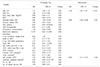
In total, 578 renal transplant recipients were included, of which 241 (41.7%) were started on TMP-SMX within 1 month post-transplant to prevent PJP and 171 (29.6%; prophylaxis group) completed the 6-month prophylaxis course. Patients who prematurely discontinued the drug (n=70, 12.1%) were included in the no-prophylaxis group with patients who were not administered prophylactic TMP-SMX (n=337, 58.3%). Baseline demographic characteristics of each group are shown in Table 1. The prophylaxis and no-prophylaxis group significantly differed in the proportion of male sex (50.9% vs. 67.8%), rate of ABOi transplantation (15.2% vs. 7.6%), preoperative desensitization status (22.8% vs. 12.3%), and rate of acute rejection (43.3% vs. 57.5%).
2. Incidence and outcomes of PJP
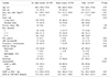
Overall, there were 39 cases of PJP (six and 33 cases in the prophylaxis and no-prophylaxis groups, respectively) during a median follow-up of 51 months (interquartile range, 35.0 to 66.0) (Fig. 1). Calculated incidence rate of PJP in patients with and without prophylaxis was 8.3 and 21.7 per 1,000 patient year, respectively. Among 39 patients with confirmed PJP, 18 (46.2%) was categorized as severe PJP based on its presentation, and nine (23.1%) required ventilator care (Table 2). In four patients, PJP was lethal; thus, PJP mortality rate was 10.3%. Of note, all mortality cases were from late PJP.
3. Incidence and risk factors of early PJP
The large difference in the incidence rate between the groups was due to the effective PJP prevention during the prophylaxis period in the prophylaxis group. Twenty-seven patients (6.6%) developed PJP within 6 months post-transplant in the no-prophylaxis group, whereas the prophylaxis group had none. The first case of early PJP documented occurred 50 days after transplant.
Factors associated with early PJP in the no-prophylaxis group were ABOi transplantation, preoperative desensitization, CMV infection within 6 months, and acute rejection (Table 3). To identify risk factors that may stratify patients' risk, thereby stratifying the individual need of PJP prophylaxis during the first 6 months, we performed a multivariate analysis including significant factors from univariate analysis that were identifiable at 1 month post-transplant (ABOi transplantation, preoperative desensitization, acute rejection within 1 month). Analysis revealed preoperative desensitization (odds ratio [OR], 4.59; 95% confidence interval [CI], 1.95 to 10.82; P=0.001) and acute rejection within 1 month (OR, 2.31; 95% CI, 1.03 to 5.17; P=0.04) as significant predictive factors of subsequent early PJP.
4. Estimated efficacy of the risk-based prophylaxis strategy to prevent early PJP
We simulated the efficacy of the risk-based strategy, which excludes early routine prophylaxis in patients with low risk, i.e., patients without preoperative desensitization and acute rejection within 1 month post-transplant (Supplementary Table 1). When simulated with the data from the no-prophylaxis group, the proposed strategy will reduce the number of patients with unnecessary prophylaxis (93% to 34%) compared to the universal prophylaxis strategy, but would result in 2.2% residual incidence of early PJP.
5. Incidence and risk factors of late PJP
Six patients each from the prophylaxis and no-prophylaxis groups developed PJP at 6 months post-transplant. The rate of late PJP was higher in the prophylaxis group (3.5% vs. 1.5%), but without statistical difference (Table 4). The earliest case of PJP in the prophylaxis group developed 51 days after completing prophylaxis. All late PJP cases developed within 2 years post-transplant, and none had recurrent PJP in our study population.
The two groups were combined in the subsequent analysis for risk factors of late PJP. Factors associated with late PJP were age, deceased donor transplant, steroid-resistant acute rejection requiring ATG, and number of biopsy proven acute rejection (Table 4). In multivariate Cox regression analysis, age (OR, 1.09; 95% CI, 1.03 to 1.19; P<0.01), deceased donor transplantation (OR, 4.89; 95% CI, 1.05 to 22.89; P=0.04), and steroid resistant acute rejection requiring ATG treatment (OR, 8.88; 95% CI, 1.11 to 71.07; P=0.04) were significantly associated with late PJP.
6. Estimated efficacy of the risk-based prophylaxis strategy to prevent late PJP
Several prophylaxis strategies to prevent late PJP are proposed based on risk factors (Supplementary Table 2). Theoretically, extended prophylaxis in patients aged ≥57 years and transplant from deceased donor would prevent 67% of the late PJP cases, resulting in the residual incidence of 0.7%. PJP risk in the proposed patient group for whom prophylaxis is indicated would be 10.3%, and the number required to treat to prevent one case would be 9.5. The cut-off age of 57 years was derived from the ROC curve analysis (Supplementary Fig. 1).
7. Adverse reaction related to TMP-SMX prophylaxis
TMP-SMX prophylaxis were prematurely discontinued in 29% (n=70) of 241 patients who had PJP prophylaxis offered initially. The main reasons for discontinuation were TMP-SMX-related adverse reaction including increased creatinine levels (n=59), hyperkalaemia (n=1), cytopenia (n=5), and nausea (n=1). Among the remaining seven patients, five had TMP-SMX discontinued due to increased creatinine levels, later revealed to be linked to other causes (rejection, n=1; bacterial infection, n=2; CMV infection, n=1; other viral infection, n=1), but the drug was not resumed. In two patients, the reason for discontinuation was not specified. Risk factors of increased creatinine levels causing premature drug discontinuation was male sex, high body surface area, concomitant use of azathioprine, and lower eGFR at TMP-SMX initiation in univariate analysis (Table 5). In multivariate analysis, the significant risk factors were male sex (OR, 4.24; 95% CI, 1.94 to 9.26; P<0.01), concomitant use of azathioprine (OR, 58.7; 95% CI, 2.78 to 1,240; P<0.01), and lower eGFR at TMP-SMX initiation (OR, 0.96; 95% CI, 0.94 to 0.98; P<0.01) (Supplementary Table 3).
DISCUSSION
To establish an effective and safe prophylaxis strategy, one should carefully balance the risks and benefits of a prophylaxis regimen. This study evaluated the effect of current PJP prophylaxis protocol on the rate of early and late PJP after renal transplantation and the adverse reactions associated with prophylactic TMP-SMX. Moreover, to propose a selective risk-based protocol, we investigated PJP risk factors at different time points post-transplantation.
Generally, universal prophylaxis is recommended in patient groups with PJP incidence of more than 3% to 5%(12). Cumulative rate of early PJP in our patients undergoing contemporary immunosuppressive therapy was 6.1% without prophylaxis. PJP prophylaxis with TMP-SMX was highly effective; thus, none had early infection in the prophylaxis group. However, we frequently encountered TMP-SMX-associated adverse reactions. Although adverse events associated with PJP prophylaxis in non-Human immunodeficiency virus (HIV) patients were rare in the meta-analysis by Stern et al.(6), 29% of our patients experienced adverse events, leading to early prophylaxis discontinuation. These frequent adverse reactions were the main reason for the delay in implementing universal PJP prophylaxis at our centre. Our adverse event rate is similar to those of Mitsides et al.(7) (38%) and Urbancic et al.(13) (35%).
The most frequent adverse reaction leading to premature prophylaxis termination was increased serum creatinine level, and factors associated with this were male sex, concomitant azathioprine use, and lower eGFR at TMP-SMX initiation. Increased serum creatinine levels are frequently attributed to the inhibition of tubular creatinine secretion by the TMP component of the drug and thus are suggested to occur without real change in glomerular filtration rate(14). However, not all increases in creatinine levels caused by TMP-SMX are benign, and tubulointerstitial nephritis, acute tubular necrosis, and acute kidney injury cases have been reported, especially when therapeutic doses are used(15). In the post-renal transplant setting, the increase in creatinine during TMP-SMX prophylaxis leads to difficulty in differentiating whether it is caused by inhibition of creatinine excretion by TMP, tubulointerstitial nephritis caused by SMX, or other problems unrelated to TMP-SMX, including graft rejection or infection. Hence, regardless of the reversibility and benign nature of majority of the cases with increased serum creatinine, it frequently necessitates drug discontinuation and subsequent evaluation for its cause. In contrast, mere observation without evaluation may mask rejection or infection episodes requiring urgent treatments.
Given the high rates of adverse reaction and aforementioned clinical complexity caused by TMP-SMX, a more selective risk-based approach in PJP prophylaxis may be more beneficial. If a patient group for whom routine prophylaxis can be safely avoided can be identified, it would lower the risk of adverse reaction, reduce cost, and reduce the development of microbial resistance in the long term. Here, we demonstrated that different patient groups were at risk of developing PJP at different time points after renal transplantation. The risk factors of PJP development during the first 6 months, which is the period of highest risks of PJP after renal transplant, were acute rejection within 30 days post-transplant and pre-transplant desensitization. Only the factors available at 30 days post-transplant were chosen because previous reports recommend prophylaxis to be started within 1 month post-transplant based on the finding that PJP are rare during the first month. Delaying the PJP prophylaxis 1 month post-transplant results in stable renal function, and thereby reducing the number of cases with renal toxicity resulting in premature discontinuation. Acute rejection has been frequently identified as a risk factor of early PJP in previous reports and is related to increased burden of immunosuppression during treatment of rejection. Additionally, our data show that preoperative desensitization confers additional risk for PJP. Preoperative desensitization regimen consists of rituximab in combination with plasmapheresis and/or IVIG. The association between T-cell immunosuppression and PJP is well known, whereas that between B-cell depletion therapy and PJP has only been described in a few recent reports(16). Although the mechanism is unclear, B-cells may be important in clearing pneumocystis infection by participating in the early priming of CD4+ T-cells(17).
There were 12 cases of PJP after 6 months post-transplant in our mixed study population of renal recipients with or without prophylaxis. All late PJP cases developed within 2 years post-transplant, and the rates were not affected by the previous prophylaxis. Iriart et al.(8) recently showed that 6-month prophylaxis prevented PJP in the first year and that the second year post-transplant was the period of highest risk. Our data also show that most of the late PJP cases occur within 2 years, but the rate of PJP was higher in 6 to 12 months post-transplant than in the second year (Fig. 1). The discrepancy may have been caused by the difference in transplant type (mixed solid organ transplant recipients vs. kidney transplant recipients), immunosuppression regimen, and possible regional difference in the risk of de novo infection. While more studies are needed to define the temporal change in the risk of PJP after prophylaxis, it is noteworthy that PJP can occur early (51 days in our study) after discontinuing PJP prophylaxis. Although documented severity of PJP was not different between early and late PJPs, all four mortality cases were from late PJPs, possibly indicating the clinical importance of preventing late PJPs.
Factors associated with late PJP were old age, transplant from deceased donor, and ATG treatment for steroid resistant acute rejection. Age and ATG were also suggested as risk factors for late pneumocystis infection in other recent studies that evaluated the risk factors of late PJP (Supplementary Table 4)(81819). By depleting T-cells, ATG increases host susceptibility to opportunistic infection, including PJP. Although the mechanism of how old age increases PJP risk is less clear, age-related immune dysfunctions, including thymic impairment, are possible underlying mechanisms(20). Other changes in T-cell function may also contribute to increased risks of infection in the elderly(21). In contrast, donor source is rarely linked to PJP. One explanation for the increased risk of late PJP in deceased donor transplant compared to living donor transplant would be the higher burden of cumulative immunosuppression due to higher number of acute rejection and delayed graft function; however, this needs to be evaluated in further studies. Additionally, the CMV infection is a factor that deserves mention. The association between CMV infection and PJP has been frequently demonstrated elsewhere(81819), suggesting that immune-modulating functions of CMV may have a role in PJP. We also observed a high rate of concurrent CMV and PJP infection that was frequently observed in our study population (Supplementary Fig. 2). The CMV infection was also associated with late PJP when a COX regression analysis was performed, with the CMV infection as a fixed covariate. However, the association was not significant when the CMV infection was treated as a time-dependent covariate. Our data suggest that CMV is not a risk factor for subsequent PJP, and thus does not necessarily predict subsequent PJP. It is more likely that both CMV and PJP reflect a high degree of immunosuppression.
We simulated the effectiveness of several prophylaxis strategies designed based on the risk factors of PJP identified in the present study. Estimates after simulation showed that while the strategy avoiding routine TMP-SMX prophylaxis within 6 months post-transplant in low risk patients would prevent two-thirds of the early PJP cases in our population, residual incidence of early PJP would not be negligible (2.2%). Thus we recommend universal PJP prophylaxis during the first 6 months after transplantation. Meanwhile, combination of age criteria and donor source seemed promising in discriminating patients with high risks of late PJP in which extended prophylaxis may be applied. Selective prophylaxis in patients aged ≥57 years and transplant from deceased donor until 2 years post-transplant would result in 0.7% of late PJP incidence while avoiding TMP-SMX use in 87% of the population. The selective prophylaxis we propose here are similar with the one of the scenarios proposed by de Boer et al.(11). By analysing the risk factor of overall PJP development in patients without PJP prophylaxis, de Boer et al.(11) proposed that 2 to 6 months universal prophylaxis and extended selective prophylaxis in >55 years of age or those with rejection as an optimal prophylaxis strategy. Our study differ in that we have separately evaluated the risk factors of PJP at different time points after transplantation, and in that we have incorporated those with PJP prophylaxis in the analysis for the late PJP. Regardless of the difference, de Boer et al.(11) and our study group have reached a similar prophylactic strategy.
While we started this study to propose a selective prophylaxis strategy during the first 6 months because of the high rate of adverse event, we were unable to define a group of patients whose risk of PJP was low enough that PJP prophylaxis during the first 6 months could be safely avoided. As PJP prophylaxis in the early transplant period seems unavoidable, future studies on less toxic prophylactic agent, as well as the accurate diagnosis and strategical management of TMP-SMX related adverse reaction are warranted.
The current study has several limitations, including the inherent limitation owing to its non-randomized retrospective single-centre design. The prophylaxis and no-prophylaxis groups were not randomly selected, and the two groups differed significantly in several baseline clinicopathologic properties. As prophylaxis virtually eliminated early PJP in the prophylaxis group, the risk factors of early PJP were evaluated only in the no-prophylaxis group. Although the incidence of early PJP was not associated with the factors that differed between the two groups, such difference should be taken into account when interpreting and applying our results. The overall incidence of PJP was high (6.6%) and generally, may not be applicable in centers with a lower incidence rate. While this was mainly due to the high rate of early PJP caused by the lack of prophylaxis, considering that the rate of late PJP in the prophylaxis group (2.2%) was higher compared with Western countries, who showed an incidence rate of 0.3% to 1.8% after prophylaxis(81922), there is also a possibility that the inherent rate of PJP in our center is high. Because the net effect of a prophylaxis strategy depends greatly on the incidence of infection, antimicrobial resistance profile of the organism, and drug-tolerability of the target population, local characteristics should be considered before generalizing the results of our study. Last, as we did not have a common protocol on the indication of prophylaxis and management of adverse events during the study period, the decision to start or stop prophylaxis was left to the treating physician, and thus, may have been biased by individual preferences. Without definite criteria of a renal adverse event (i.e., degree of rise in creatinine increase), and when to stop prophylaxis, we may have overestimated the rate of renal adverse events.
CONCLUSION
Different patient groups are at risk of developing PJP during early and late periods after transplantation. Adverse renal events were frequently encountered during TMP-SMX prophylaxis, leading to premature discontinuation. After modelling different prophylaxis scenarios, we proposed a universal prophylaxis of up to 6 months post-transplant combined with extended selective prophylaxis in patients aged ≥57 years and transplants from deceased donors as a possible strategy to effectively prevent PJP after renal transplantation.
 XML Download