

 PDF
PDF ePub
ePub Citation
 Print
Print


Abstract
Mucormycosis is an extremely rare but potentially life-threatening fungal infection. Gastrointestinal (GI) mucormycosis is very rare and occurs primarily in highly malnourished patients, especially in infants and children. A 55-year-old man with end-stage renal disease due to diabetic nephropathy, who had undergone deceased donor kidney transplantation 2 years prior, complained of abdominal pain and distension with a 3-day duration. Computed tomography revealed diffuse gastric wall thickening, and a huge amount of grey colored necrotic debris surrounded by erythematous erosive mucosa was observed at the antrum to upper body by GI endoscopy. The microscopic examination obtained from a GI endoscopic specimen demonstrated peptic detritus with numerous non-septate mucor hyphae in the mucosa and submucosa. Mucormycosis was diagnosed based on the clinical findings and morphological features. A total gastrectomy was performed and an antifungal agent was administered. A microscopic examination of the surgical specimen demonstrated invasive mucormycosis with numerous fungal hyphae with invasion into the mucosa to subserosa. The patient and graft were treated successfully by total gastrectomy and antifungal therapy.
REFERENCES
1). Neofytos D., Treadway S., Ostrander D., Alonso CD., Dierberg KL., Nussenblatt V, et al. Epidemiology, outcomes, and mortality predictors of invasive mold infections among transplant recipients: a 10-year, single-center experience. Transpl Infect Dis. 2013. 15:233–42.

2). Boelaert JR. Mucormycosis (zygomycosis): is there news for the clinician? J Infect. 1994. 28(Suppl 1):1–6.
3). Lehrer RI., Howard DH., Sypherd PS., Edwards JE., Segal GP., Winston DJ. Mucormycosis. Ann Intern Med. 1980. 93(1 Pt 1):93–108.
4). DeVane MS., Taskin V., Berty RM. Fatal sinonasal mucormycosis: case report and review. Infect Dis Clin Pract. 1997. 6:56–9.
5). Ingram CW., Sennesh J., Cooper JN., Perfect JR. Disseminated zygomycosis: report of four cases and review. Rev Infect Dis. 1989. 11:741–54.
6). Hameroff SB., Eckholdt JW., Lindenberg R. Cerebral phycomycosis in a heroin addict. Neurology. 1970. 20:261–5.
7). Dannheimer IP., Fouche W., Nel C. Gastric mucormycosis in a diabetic patient. S Afr Med J. 1974. 48:838–9.
8). Chkhotua A., Yussim A., Tovar A., Weinberger M., Sobolev V., Bar-Nathan N, et al. Mucormycosis of the renal allograft: case report and review of the literature. Transpl Int. 2001. 14:438–41.
9). Fogarty C., Regennitter F., Viozzi CF. Invasive fungal infection of the maxilla following dental extractions in a patient with chronic obstructive pulmonary disease. J Can Dent Assoc. 2006. 72:149–52.
10). Cherney CL., Chutuape A., Fikrig MK. Fatal invasive gastric mucormycosis occurring with emphysematous gastritis: case report and literature review. Am J Gastroenterol. 1999. 94:252–6.
11). Singh N., Gayowski T., Singh J., Yu VL. Invasive gastrointestinal zygomycosis in a liver transplant recipient: case report and review of zygomycosis in solid-organ transplant recipients. Clin Infect Dis. 1995. 20:617–20.
Fig. 1.
Gastrointestinal endoscopy showed a huge amount of grey colored elevated necrotic debris surrounded by erythematous erosive mucosa from antrum to upper body (arrows).
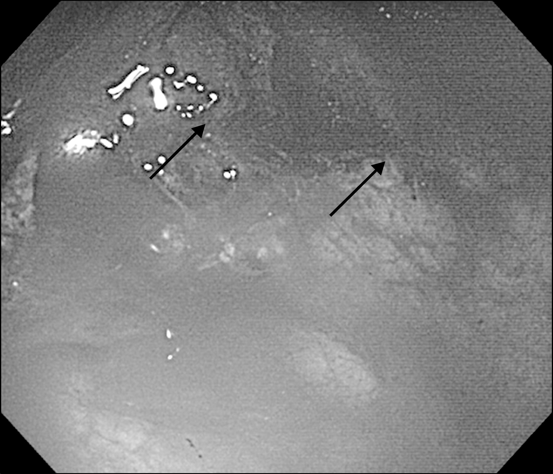
Fig. 2.
(A) Gross specimen of stomach. The black arrow indicates a perforation lesion. (B) Multifocal transluminal ulcer finding of stomach. (C) Necrotizing vasculitis finding of filled thrombi and vascular wall destruction. (D) Fungal ball (arrows). (E, F) PAS stain (E, ×400) and Gomori's Methenamine silver stain (F, ×400) sections showing broad, non-septated hyphae with right angled branches (B: HE stain, ×200; C, D: HE stain, ×400).

 XML Download