Abstract
Objectives
To document fistula formation between the disc and dura by an unrecognized dural tear after percutaneous endoscopic lumbar discectomy (PELD).
Summary of Literature Review
The risk of durotomy is relatively low with PELD, but cases of unrecognized durotomies have been reported. An effective diagnostic tool for such situations has not yet been identified.
Materials and Methods
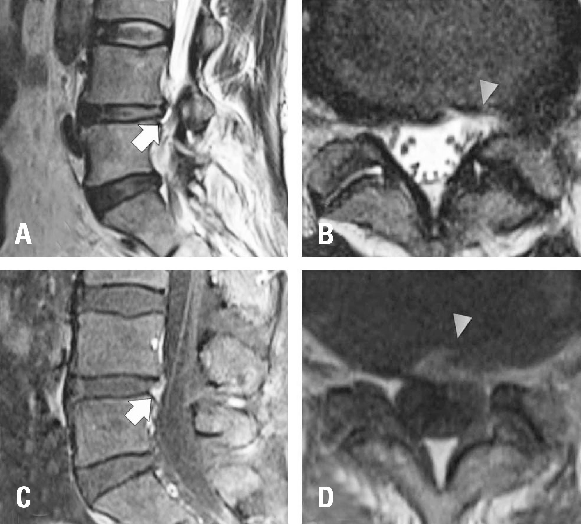
A patient twice underwent transforaminal PELD under the diagnosis of a herniated lumbar disc at L4-5. She still complained of intractable pain and motor weakness around the left lower extremity at 6 months postoperatively. Magnetic resonance imaging showed no specific findings suggestive of violation of the nerve root. However, L5 and S1 nerve root injury was noted on electromyography. An exploratory operation was planned to characterize damage to the neural structures.
Results
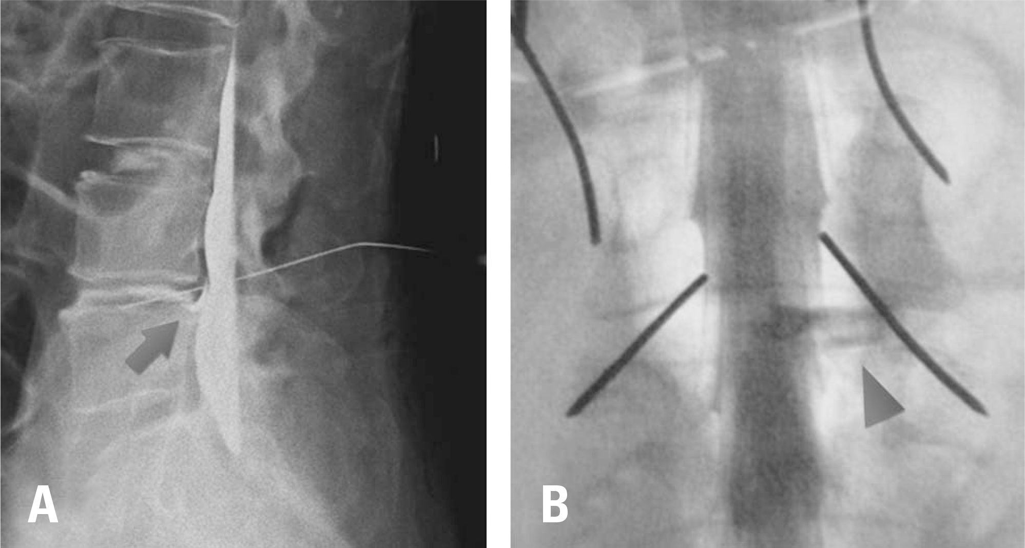
In the exploration, a dural tear was found at the previous operative site, along with a fistula between the disc and dura was also found at the dural tear site. The durotomy site was located on the ventrolateral side of the dura and measured approximately 5 mm. The durotomy site was repaired with Nylon 5-0 and adhesive sealants. The patient's preoperative symptoms diminished considerably.
REFERENCES
1. Smith JS, Ogden AT, Shafizadeh S, et al. Clinical outcomes after microendoscopic discectomy for recurrent lumbar disc herniation. J Spinal Disord Tech. 2010 Feb; 23(1):30–4. DOI: 10.1097/BSD.0b013e318193c16c.

2. Hermantin FU, Peters T, Quartararo L, et al. A prospective, randomized study comparing the results of open discectomy with those of video-assisted arthroscopic microdiscectomy. J Bone Joint Surg Am. 1999 Jul; 81(7):958–65. DOI: 10.2106/00004623-199907000-00008.

3. Kambin P, Brager MD. Percutaneous posterolateral discectomy. Anatomy and mechanism. Clin Orthop Relat Res. 1987 Oct; 223:145–54. DOI: 10.1097/00003086-198710000-00016.
4. Jang EC, Song KS, Kang KS, et al. Comparative evaluation of percutaneous endoscopic discectomy and microdiscectomy using tubular retractor system at L4-5 level. J Korean Soc Spine Surg. 2009 Sep; 16(3):186–93. DOI: 10.4184/jkss.2009.16.3.186.

5. Yeung AT, Tsou PM. Posterolateral endoscopic excision for lumbar disc herniation: surgical technique, outcome, and complications in 307 consecutive cases. Spine (Phila Pa 1976). 2002 Apr; 27(7):722–31. DOI: 10.1097/00007632-200204010-00009.
6. Ahn Y, Lee HY, Lee SH, et al. Dural tears in percutaneous endoscopic lumbar discectomy. Eur Spine J. 2011 Jan; 20(1):58–64. DOI: 10.1007/s00586-010-1493-8.

7. Hasegawa K, Yamamoto N. Nerve root herniation sec-ondary to lumbar puncture in the patient with lumbar canal stenosis. A case report. Spine (Phila Pa 1976). 1999 May; 24(9):915–7. DOI: 10.1097/00007632-199905010-00015.
8. Bosacco SJ, Gardner MJ, Guille JT. Evaluation and treatment of dural tears in lumbar spine surgery: a review. Clin Orthop Relat Res. 2001 Aug; 389:238–47. DOI: 10.1097/00003086-200108000-00033.
9. Khan MH, Rihn J, Steele G, et al. Postoperative management protocol for incidental dural tears during degenerative lumbar spine surgery: a review of 3,183 consecutive degenerative lumbar cases. Spine (Phila Pa 1976). 2006 Oct; 31(22):2609–13. DOI: 10.1097/01. brs.0000241066.55849.41.
10. Johnsin DB, Brennnan P, Toland J, et al. Magnetic resonance imaging in the evaluation of cerebrospinal fluid fistu-lae. Clin Radiol. 1996 Dec; 51(12):837–41. DOI: 10.1016/S0009-9260 (96)80079-1.
Fig. 1.
Magnetic resonance imaging (MRI) findings. (A, B) T2-weighted MRI images showing hyperintense signal changes in the annulus fibrosus at the previous discectomy site (arrow, sagittal view; arrowhead, axial view). (C, D) Gd-enhanced T1-weighted images showing mild enhancement of the annulus fibrosus at the previous discectomy site (arrow, sagittal view; arrowhead, axial view). Nerve root sleeve violation by the disc material was not observed.


 PDF
PDF Citation
 Print
Print


 XML Download