

 PDF
PDF Citation
 Print
Print


Abstract
Objectives
We report 3 cases of loss of disc height after spontaneous regression of a herniated lumbar disc.
Summary of Literature Review
Reports of spontaneous regression of a herniated lumbar disc were identified.
Materials and Methods
We conservatively treated 3 patients who were diagnosed with a herniated lumbar disc. During outpatient follow-up, radiating pain improved in all patients, but they complained of chronic lower back pain. Magnetic resonance imaging (MRI) was performed for diagnostic purposes.
Go to : 
REFERENCES
1. Boden SD, McCowin PR, Davis DO, et al. Abnormal mag-netic-resonance scans of the cervical spine in asymptomatic subjects. A prospective investigation. J Bone Joint Surg Am. 1990 Sep; 72(8):1178–84. DOI: 10.2106/00004623-199072080-00008.

2. Sabuncuoglu H, Ozdogan S, Timurkaynak E. Spontaneous regression of extruded lumbar disc herniation: report of two illustrative case and review of the literature. Turk Neurosurg. 2008 Oct; 18(4):392–6.
3. Guinto FC Jr., Hashim H, Stumer M. CT demonstration of disk regression after conservative therapy. AJNR Am J Neuroradiol. 1984 Sep-Oct; 5(5):632–3.
4. Benoist M. The natural history of lumbar disc herniation and radiculopathy. Joint Bone Spine. 2002 Mar; 69(2):155–60. DOI: 10.1016/S1297-319X(02)00385-8.
5. Komori H, Shinomiya K, Nakai O, et al. The natural history of herniated nucleus pulposus with radiculopathy. Spine (Phila Pa 1976). 1996 Jan; 21(2):225–9. DOI: 10.1097/00007632-199601150-00013.
6. Teplick JG, Haskin ME. Spontaneous regression of herniated nucleus pulposus. AJR Am J Roentgenol. 1985 Aug; 145(2):371–5. DOI: 10.2214/ajr.145.2.371.
7. Slavin KV, Raja A, Thornton J, et al. Spontaneous regression of a large lumbar disc herniation: report of an illustrative case. Surg Neurol. 2001 Nov; 56(5):333–6. DOI: 10.1016/S0090-3019 (01)00607-3.
8. Henmi T, Sairyo K, Nakano S, et al. Natural history of extruded lumbar intervertebral disc herniation. J Med Invest. 2002 Feb; 49(1-2):40–3.
9. Autio RA, Karppinen J, Niinimä ki J, et al. Determinants of spontaneous resorption of intervertebral disc herniations. Spine (Phila Pa 1976). 2006 May; 31(11):1247–52. DOI: 10.1097/01.brs.0000217681.83524.4a.
10. Barth M, Diepers M, Weiss C, et al. Two-year outcome after lumbar microdiscectomy versus microscopic seques-trectomy: part 2: radiographic evaluation and correlation with clinical outcome. Spine (Phila Pa 1976). 2008 Feb; 33(3):273–9. DOI: 10.1097/BRS.0b013e31816201a6.
Go to :
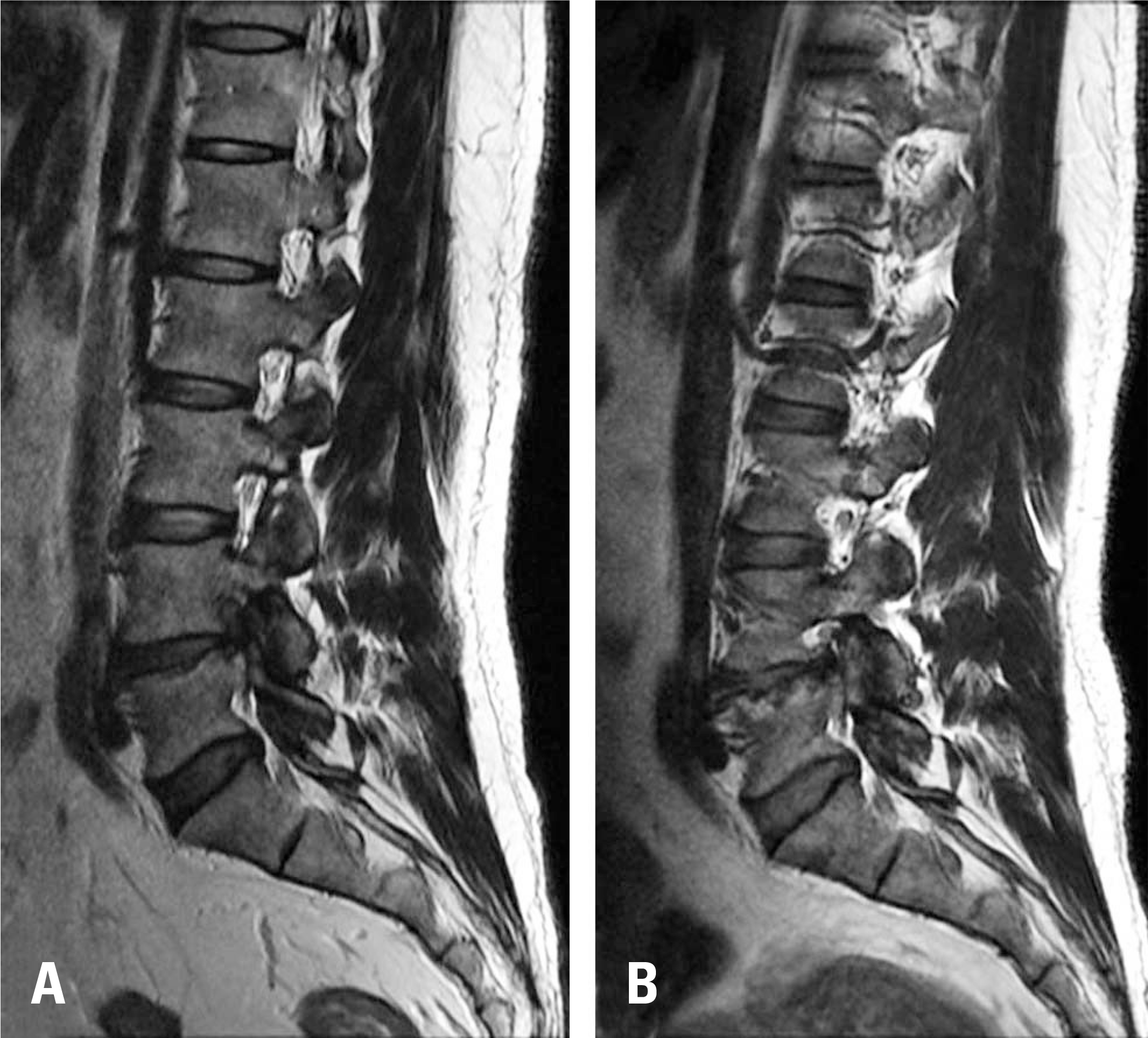
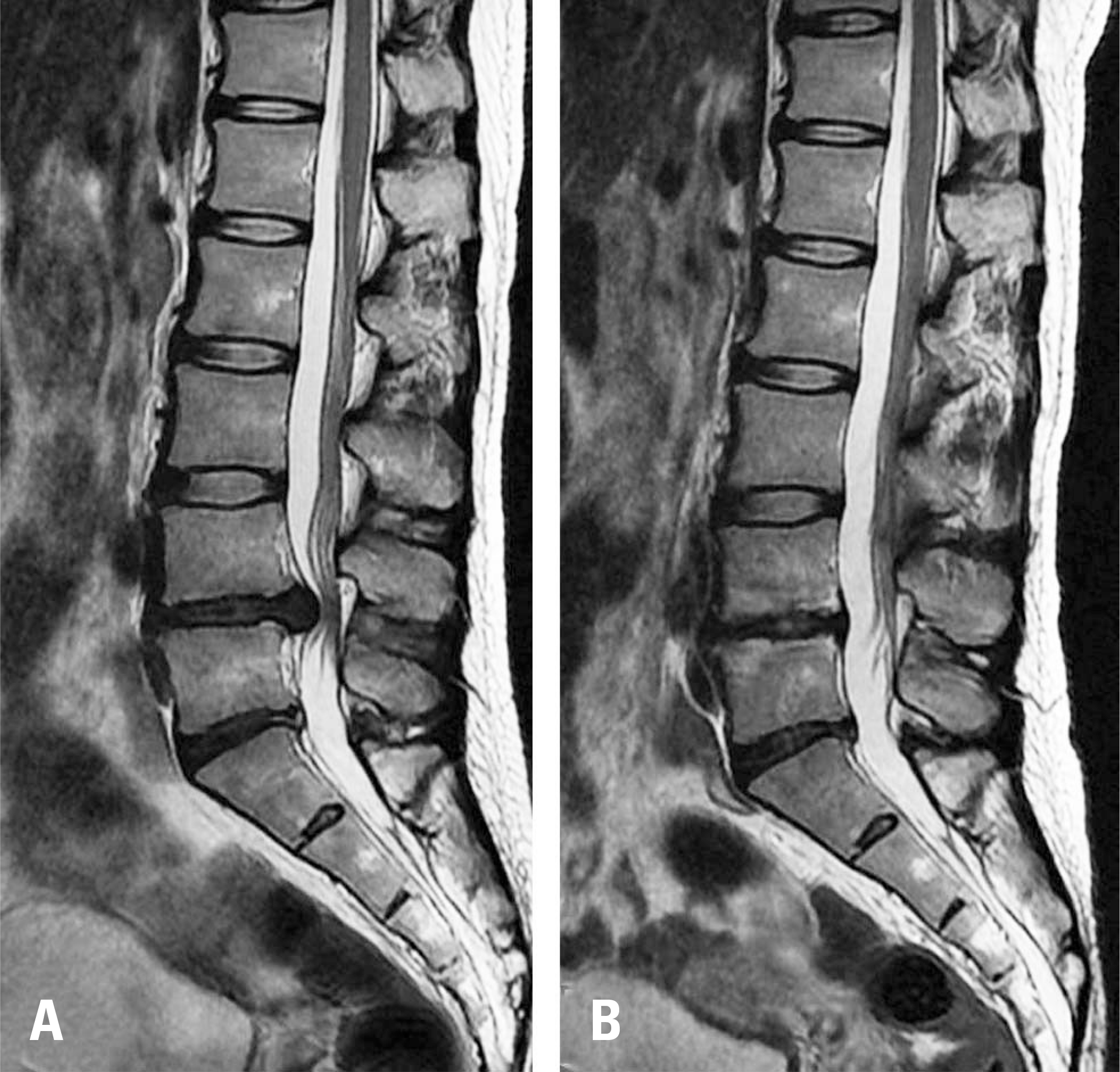 | Fig. 1.Magnetic resonance imaging (MRI) findings. (A) Initial MRI, showing a large area of disc extrusion at the L4/5 disc space. (B) MRI taken 2 years after the initial MRI, showing spontaneous regression of the herniated lumbar disc and loss of disc height. |

 XML Download