

 PDF
PDF ePub
ePub Citation
 Print
Print


Introduction
External iliac vein is used in catheterization procedures. Its variations might result in hazardous bleeding during the procedure. External iliac vein seldom shows variations. Very few cases have been reported about its variations. In rare cases, it may be absent [1]. It may undergo aplasia [2] or even be duplicated [3]. Its reported anomalies also include a prearterial external iliac vein [4] and a venous annulus formed around external iliac artery by it [5]. Drainage of left external iliac vein into left inferior vena cava and left internal iliac vein into the right common iliac vein is another reported variation about the iliac veins [6]. Presence of multiple communications between the iliac veins is a rare variation and due to its rarity, the report is worthwhile to update the clinicians about its occurrence.
Case Report
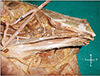
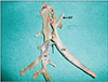
During dissection classes for medical undergraduates, four unusual communications between external and internal iliac veins of the left side were noted in a male cadaver aged approximately 70 years. The lowest communication was at the level of first sacral vertebra and the uppermost communication was at the level of intervertebral disc between fourth and fifth lumbar vertebrae. The lowest (first) communication was the narrowest measuring about 2 mm in diameter and 2 cm in length. The length and breadth of second communication was 0.6 cm each. The third communication was 1 cm long and 0.7 cm broad. The last communication (termination of the external iliac vein) measured 3 cm in length and 0.5 cm in breadth. The upper three communications were twisted in a helical manner. The internal jugular vein had its normal tributaries except that the iliolumbar vein drained into the external iliac vein at the level of the third communication. The external iliac vein was slightly dilated just below the level of lowest communication. The variations have been shown in Figs. 1 and 2. Since the variations were noted on a cadaver donated to the institution for research and teaching purpose, patient consent form was not taken.
Discussion
Variations in the internal iliac veins are observed in 20.9% of cases [7]. Shin et al. (2015) [7] have classified the variations into eight types as follows: type 1, normal type, which is found in 79.1% cases; type 2, higher union of internal iliac vein with ipsilateral external iliac vein in 8.7% cases; type 3, internal iliac vein ending in contralateral common iliac vein in 2.3% cases; type 4, internal iliac veins forming a common trunk in 0.9% of cases; type 5, presence of communicating vein connecting internal iliac vein with contralateral internal iliac or common iliac vein in 7.8% of cases; type 6, double internal iliac vein in 0.9% of cases; type 7, presence of left inferior vena cava in 0.1% cases; and type 8, fenestration of common iliac vein in 0.4% cases. The current variation being reported here, does not fall under any one of these categories. Thus it is unique and worth adding to the literature.
External iliac vein is exceedingly used in cardiac catheterizations for pacemaker [8] and defibrillator implantations [9]. The external iliac vein graft is also used in hepatic venous outflow reconstruction of hepatic veins for hepatic malignancies [10]. In all these procedures, the knowledge of variation of external iliac veins has to be kept in mind to avoid iatrogenic bleeding. The current case might complicate the catheterization procedure due to the twisted cross communicating channels and their narrower lumen size compared to the main vein. The lowest communication was the narrowest and it may not be visible in imaging technics as well. Hence its rupture may go unnoticed till late stage. Iliac vein stent placing is done in case of iliac vein disease [11].
The possible reason for the occurrence of peculiar twisted communications between the iliac veins in the current case was the failure of disappearance of some of the intercommunicating channels between the two iliac veins. Usually, during the embryonic development, the inferior vena cava and the pelvic vessels are derived from multiple venous channels. Inferior vena cava develops from the contribution from post cardinal veins, supracardinal veins, subcardinal veins, right hepatocardiac channel, etc. The lower limb and pelvis present a plexus of veins in the early development. Later on many of these veins disappear leaving behind one external iliac and one internal iliac veins which joint to form the common iliac vein. The left common iliac vein is longer and more oblique because it is derived mainly from an oblique anastomosis between right and left post cardinal veins. The possible reason for the twisted communications between the two iliac veins in the current case was failure of disappearance of all those channels which usually disappear leaving behind only external and internal iliac veins.
The variations like the one being reported here, can surely pose challenges in stent placing. The current case is unique due to the number of communications, the size of the communications and their twisted nature. This case could be the first report on such a variation and this could be of interest to cardiologists, orthopedic surgeons, gynecologists and radiologists.
 XML Download