

 PDF
PDF ePub
ePub Citation
 Print
Print


Introduction
The sternocleidomastoid muscle (SCM) is a superficial muscle of the neck, serving as an anatomical bridge between the anterior and lateral regions of the neck, and further triangular subdivisions. The SCM may be observed by the clinician as a muscular protrusion rising vertically and obliquely along the neck. The muscle itself is thick along its center, and thinner at its attachments [1]. In normal cases, there are two heads: one arising from the sternum and one from the clavicle. The sternal head arises from the anterior surface of the manubrium of sternum as a distinct rounded tendon of consistent width; the clavicular head spans the medial third of the clavicle and may vary in width, arising as a band-like tendon with musculofibrous elements [12].
Herein, we discovered bilateral variant SCMs send one tendon to the mastoid and six distinct tendons along the lateral superior nuchal line to the midline. In particular, this case highlights the unique morphological possibilities of the sternocleidomastoid, necessitating understanding of the clinician, as neighboring muscles, glandular structures, and cervical and upper brachial plexi may be covered by the supernumerary insertions along the superior nuchal line leading to possible complications, or confounding for the radiologist during imaging [1]. We present this case for the anestheologist, orthopedic surgeon, neurosurgeon, and head and neck surgeons as appropriate knowledge so that complications in such cases may be avoided.
Case Report
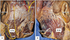
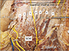
During routine dissection of an 81-year-old at death Caucasian male fresh-frozen cadaveric specimen, a unique variant of the SCM was discovered bilaterally (Fig. 1). Specifically, a wide belly was observed bilaterally. Upon approaching its attachment, a single, gradually thinning muscle and subsequent tendon was observed attaching onto the lateral aspect of the mastoid process. Six distinct tendons, with the transition of muscle-to-tendon highly discernable, were then sent attaching to the more medial occiput. The first of six of the tendons inserted onto the lateral aspect of the superior nuchal line, followed by a small space, then another inserted tendon. These tendons repeated this regular spacing between one another until the sixth tendon inserted at the midline (Fig. 2). The muscle belly as well as the tendon of the sixth insertion interfacing along the midline were both comparably thicker and wider than the other five tendons along the superior nuchal line. This SCM variant received innervation from the accessory nerve as well as the great auricular nerve formed by C2 and C3. The accessory nerve, in this case on each side, was traveling through the SCM belly. On both sides, the lesser occipital nerve ascended along the surface of the SCM. The seventh tendon of the SCM approached the greater occipital nerve (Fig. 2). As this dissection of the neck was separated from the clavicle and sternum, these attachments of the SCM were not observed.
Discussion
Different patterns of the origin of the variant SCM have been reported [23]. Mori [2] described five distinct portions of the SCM muscle belly: the superficial layer consisting of the superficial sternomastoid, sternooccipital, and cleidooccipital portions; and the deep layer consisting of the deep sternomastoid and cleidomastoid portions. The precise fusion of these parts varies greatly, but are generally separated into sternomastoid and cleidomastoid portions [3]. The two heads rise vertically, with the clavicular head passing beneath the sternal head into their fusion forming the noticeably thick belly midway along the neck. Normally, there are two distinct insertions: at the lateral surface of the mastoid process, and the lateral aspect of the superior nuchal line. Variants in which there are supernumerary origins, and or irregularities in the distinct anatomical location of the origins, are generally more common than aberrant or supernumerary insertions [456789].
In the present case, the several attachments along the superior nuchal line bilaterally appear to be uncommon but important for surgeons to be aware of during surgical dissections of the posterior head and neck. For example, muscle flaps of the SCM may be used in several types of corrective and reconstructive procedures and transmastoid approaches to the skull base commonly use the SCM attachment as a surgical landmark. A slip of the SCM may be harvested from its sternal or clavicular attachment with a segment of the clavicle or sternum as a pedicle flap for mandibular reconstruction, or may be transposed into the oral cavity following ablative surgery [10]. Interestingly, with the many insertions of the SCM as seen in our case, one variant slip of the SCM onto the occiput may be harvested with no significant muscular deficit due to the remaining insertions along the superior nuchal line. With this type of variation, we propose that, due to the supernumerary SCM insertions, a functional advantage may be observed in individuals harboring such a variant perhaps increasing strength of flexion to the contralateral side. The seventh tendon in this case approached the greater occipital nerve (Fig. 2), which might lead to nerve entrapment and result in pain syndromes such as occipital neuralgia [11]. There is a high degree of variability in the occipital cutaneous nerves; the greater and lesser occipital and great auricular nerves are all related to the SCM along their course in the superficial fascia, and may become entangled in the fascicles of a variant SCM [11].
Also intimate with the SCM is the great auricular nerve, arising from the ventral rami of the second and third cervical spinal nerves. It curves around the posterior aspect of the SCM, and crosses it to pass above the external jugular vein to provide sensory innervation for the skin over the surface of the ear. In this case, a direct branch of the cervical plexus was observed to pierce the belly of the SCM. A direct branch may be problematic as cervicogenic headache can arise from structures associated with upper cervical spinal nerves like the SCM [12]. Direct compression of a cervical spinal root in this SCM variant may thus result in increased surface area of pain for the patient. Irregularly wide or expansive SCM variants could perhaps compress the carotid sheath and its contents [1314]. The superior thyroid artery could also become occluded by an irregular SCM, as it emanates from under the anterior border of the muscle.
Developmentally, this variant may have occurred due to aberrancies in the signaling of the Hox gene in the development of the myotome that gives rise to the SCM and trapezius, both seen to fuse at their respective insertions, and on occasion make contact with one another along their medial parts [4715]. Embryologically, these structures arise together as an aggregation of cells readily observed at Carnegie stage 15 (CS15), and are referred to as the trapezius/SCM muscle complex. At CS17, the trapezius and SCM may be viewed as separate muscles, and at CS23, they are fully defined at their skeletal attachments [16]. Anomalous signaling in this developmental interval may result in deviations from the commonly observed morphology and attachments of the SCM.
Variations of the SCM might mislead surgeons during surgical dissection. The supernumerary attachments as seen in the present case might confuse the operator during posterior surgical approaches to the neck and occiput and should therefore be kept in mind.
 XML Download