Journal List > Anat Cell Biol > v.51(4) > 1110751

Baxla, Kumari, and Kaler: Bilateral thyrolinguofacial trunk: unusual and rare branching pattern of external carotid artery

Abstract

Prior knowledge of arterial supply to the head and neck is of substantial importance for well-planned surgeries involving the concerned region. We are reporting an unusual and rare variation in the branching pattern of external carotid artery in a 60-year-old female cadaver. A common trunk known as thyrolinguofacial trunk, originating from the anterior surface of the external carotid artery (right and left) giving of superior thyroid artery and a linguofacial trunk during a routine neck dissection. The linguofacial trunk then divided into a lingual and a facial artery. Vascular abnormalities are usually detected either on the dissection table or by the radiologists during imaging or accidently during surgeries leading to serious consequences.

Introduction

Our face and neck has a rich source of blood supply which predominantly comes from the branches given off by the external carotid artery. The external carotid artery gives rise to superior thyroid, ascending pharyngeal, lingual, facial, occipital, posterior auricular and two terminal branches, i.e., maxillary and superficial temporal arteries. The first three branches that is superior thyroid, lingual and the facial artery take independent origin from the anterior aspect of external carotid artery. Any variations in the branching pattern of the external carotid artery are important for the surgeons as planning and execution of surgery is purely based on the anatomical arrangement of the structures in the neck region. So, this study highlights the variation in the branching pattern of the external carotid artery.

Case Report

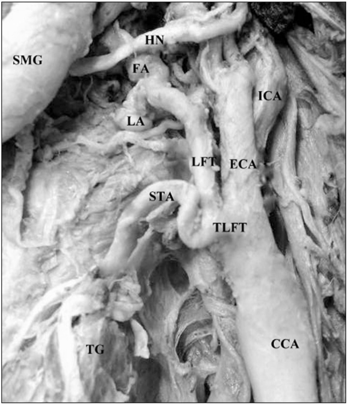
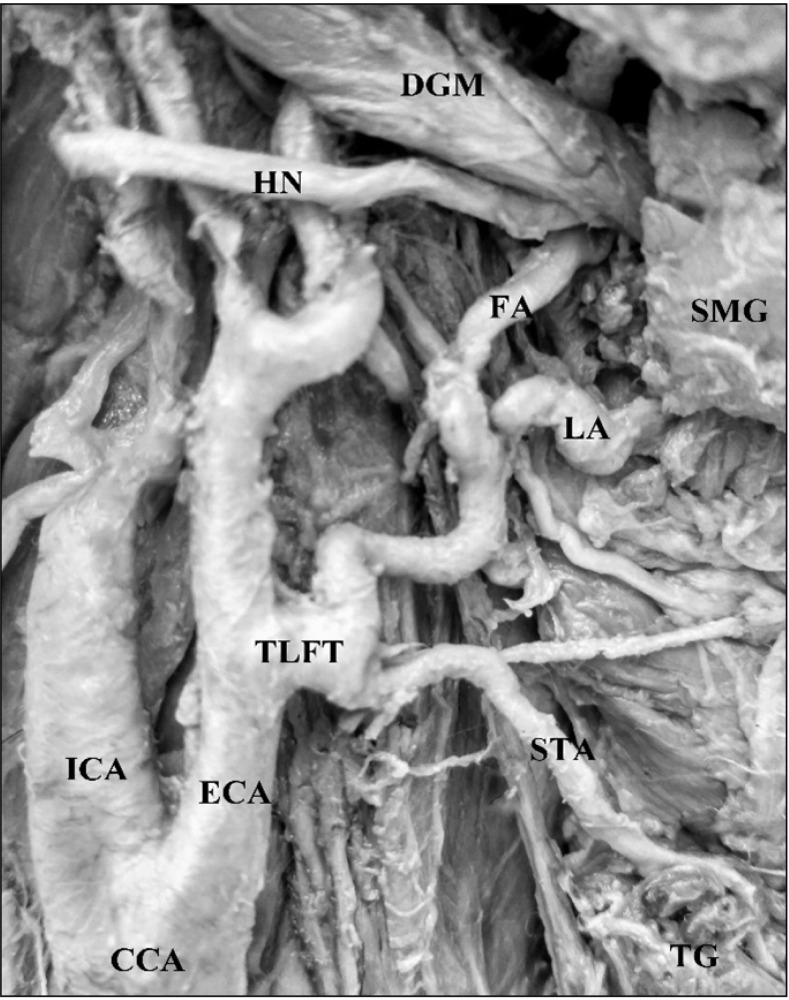
The present case was observed during a routine dissection for M.B.B.S. undergraduates in the Department of Anatomy, All India Institute of Medical Sciences, New Delhi. The cadaver was of a 60-year-old female; embalmed with formalin and had been donated to the department of Anatomy, All India Institute of Medical Sciences, New Delhi, India for study and research purposes. In this cadaveric specimen we observed that the first branch originating anteromedially from the external carotid artery was a common trunk (thyrolinguofacial trunk) on both the sides (right and left) of the neck region (Figs. 1, 2). This common trunk was 0.5 cm in length on the right side and about 0.3 cm on the left which soon divided to give of a superior thyroid artery and a linguofacial trunk (common trunk for lingual and facial artery). The superior thyroid artery coursed downwards to reach the thyroid gland and the linguofacial trunk after running upwards for about 1.8 cm divided into lingual and facial artery. The lingual artery travelled upwards to supply the tongue musculature. The facial artery travelled towards the face to reach the lower border of mandible. Since it's a bilateral presentation, we looked into all other possible variations in the body but there was none.

Discussion

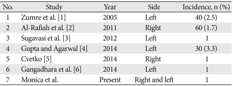
There are numerous variations of the great vessels of head and neck which needs to be explored as accurate knowledge of vascular patterns is necessary for avoiding any risk of iatrogenic bleeding. Thyrolinguofacial trunk is rather an uncommon variation though some of the authors have reported of this variation. Till now unilateral thyrolinguofacial trunk had been observed but in the present study we have observed a bilateral presence of the trunk. As we can see from the Table 1 that the incidence is very low.
In conclusion, this variation is surgically relevant as it is liable to get injured during head and neck surgeries such as thyroidectomy, reconstruction of an aneurysm hence important for clinical, radiological and surgical practices. Analysing the great vessels of head and neck through radiological interventions before a surgery is a must for diagnosing such variations which are very rare.

References

1. Zumre O, Salbacak A, Ciçekcibşi AE, Tuncer I, Seker M. Investigation of the bifurcation level of the common carotid artery and variations of the branches of the external carotid artery in human fetuses. Ann Anat. 2005; 187:361–369. PMID: 16163849.
2. Al-Rafiah A, El-Haggagy AA, Aal IH, Zaki AI. Anatomical study of the carotid bifurcation and origin variations of the ascending pharyngeal and superior thyroid arteries. Folia Morphol (Warsz). 2011; 70:47–55. PMID: 21604253.
3. Sugavasi R, Sujatha M, Indira Devi B, Sirisha B, Kanchana L. Variant origin of thyro linguo facial trunk from left common carotid artery: a case report. Int J Health Sci Res. 2012; 2:92–95.
4. Gupta V, Agarwal R. Anomalous branching pattern of the external carotid artery in cadavers. Int J Sci Stud. 2014; 2:28–31.
5. Cvetko E. Thyrolinguofacial trunk arising from the carotid bifurcation determined by cadaver dissection. Anat Sci Int. 2014; 89:246–249. PMID: 24307453.
6. Gangadhara , Shakuntala Rao N, Manivannan K, Krishna Rao HR. Unusual branching pattern of external carotid artery in the left carotid triangle. Int J Res Med Sci. 2014; 2:1743–1745.
Fig. 1

Photographic illustration of left thyrolinguofacial trunk (TLFT) originating from left external carotid artery (ECA). This common trunk divided to give origin to superior thyroid artery (STA) and linguofacial trunk (LFT). This trunk further gave rise to lingual artery (LA) and facial artery (FA). CCA, common carotid artery; HN, hypoglossal nerve; ICA, internal carotid artery; SMG, submandibular gland; TG, thyroid gland.

acb-51-302-g001
Fig. 2

Photographic illustration of right thyrolinguofacial trunk (TLFT) originating from the right external carotid artery (ECA). This common trunk (TLFT) is divided into in superior thyroid artery (STA) and linguofacial trunk which further gave off lingual artery (LA) and facial artery (FA). CCA, common carotid artery; DGM, digastric muscle; HN, hypoglossal nerve; ICA, internal carotid artery; LA, lingual artery; SMG, submandibular gland; TG, thyroid gland.

acb-51-302-g002
Table 1

Showing findings of previous and the present study

acb-51-302-i001
No. Study Year Side Incidence, n (%)
1 Zumre et al. [1] 2005 Left 40 (2.5)
2 Al-Rafiah et al. [2] 2011 Right 60 (1.7)
3 Sugavasi et al. [3] 2012 Left 1
4 Gupta and Agarwal [4] 2014 Left 30 (3.3)
5 Cvetko [5] 2014 Right 1
6 Gangadhara et al. [6] 2014 Left 1
7 Monica et al. Present Right and left 1
TOOLS
Similar articles