

 PDF
PDF ePub
ePub Citation
 Print
Print


INTRODUCTION
Early effective risk stratification is essential for the management of patients with acute coronary syndrome.1 Unlike patients with ST-elevation myocardial infarction (STEMI), Patients with non-STEMI have a different time limitation in performing intervention.2 So, in a treatment of non-STEMI patients, early prediction of prognosis makes decisions regarding pharmacologic and interventional treatments, allocation of clinical resources, and triage among alternative levels of hospital care.
Blood tests were conducted in all patients including complete blood count (CBC) test on admission. CBC is the most widely, easily, fast available laboratory datum on admission, especially in the first 30 minutes. Even among patients with non-STEMI, for whom initial therapeutic options are well-defined, patient risk assessments have an impact on early therapeutic decision making.3 Furthermore, if primary percutaneous coronary intervention (PCI) is done as soon as possible after admission, the CBC may be the only available laboratory data before, or at, the procedure. Previous study reported that high neutrophil to lymphocyte ratio (N/L)45 and anemia67 are independent prognostic predictors of mortality in patients with STEMI.8 The aim of the present study was to assess the value of early risk stratification using readily available CBC data in patients with non-STEMI.
MATERIALS AND METHODS
1. Study population
The study population consisted of 6,191 consecutive non-STEMI patients enrolled in the Korea Acute Myocardial Infarction Registry-National Institute of Health (KAMIR-NIH) between November 2011 and March 2015. Patients with no laboratory data or no vital sign were excluded, and 6,157 patients (65±12.4 years, male 69%) were included in the final analysis (Fig. 1).
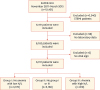 | Fig. 1Flow chart of the study population.KAMIR-NIH; Korea Acute Myocardial Infarction Registry-National Institute of Health, STEMI; ST-elevation myocardial infarction, N/L; neutrophil to lymphocyte ratio.
|
All patients were categorized into 3 groups by using the median value of N/L (4.42) and the presence of anemia: group I, low N/L & no anemia (n=3,170); group II, no group I or III (n=2,168); group III, high N/L & anemia (n=819).
The KAMIR-NIH was a Korean prospective multicenter data collection registry reflecting real-world treatment practices and outcomes in Asian patients diagnosed with acute myocardial infarction (AMI). The registry includes data from 20 hospitals with facilities for primary PCI and on-site cardiac surgery. Data were collected by a trained study coordinator using a standardized case report from and protocol. All management decisions were at the discretion of the treating cardiologists. Before the initiation or the KAMIR-NIH study, several investigator meetings were held, and a steering committee was selected to standardize the care given in clinical practice to minimize the differences in medical care among the hospitals. The ethics committee at each participating institution approved the study.
Eligible patients were on admission for and non-STEMI, and had ≥1 of the following: electrocardiographic changes consistent with an non-STEMI (defined as ST-segment depression and T-wave inversion of 0.2 mV in 2 contiguous leads), serial increases in serum biochemical markers of myocardial necrosis, and documentation of coronary artery disease (CAD) by coronary angiogram (CAG).2 Patients with ST-segment elevation or presumed new left bundle branch block were excluded, and ST-segment depression being defined as new horizontal or downsloping depression 0.05 mV in 2 contiguous leads. Cut-off value of cardiac troponin was defined as the lowest concentration with a value of coefficient of variation<10%.
2. Complete blood cell count
CBC values were obtained just after admission, before the patient's transfer to the catheter laboratory. It was possible to collect the data of 99% of all patients. Automated analyzers were used for hematologic measurements. Anemia was defined according to the World Health Organization definition as admission hemoglobin (Hb) level <13 mg/dL in men and <12 mg/dL in women.9
3. Coronary angiographic procedure and post-intervention management
All patients received aspirin ≥100 mg and a loading dose of clopidogrel 300 to 600 mg and unfractionated heparin 50 to 70 U/kg to maintain an activated clotting time of >250 to 300 seconds before or during the procedure. Primary PCI was performed using standard techniques if the coronary anatomy was suitable for angioplasty. Successful PCI was defined as thrombolysis in myocardial infarction (TIMI) grade 3 flow with residual stenosis ≤50% in the infarct-related artery. Multivessel coronary disease was defined as >50% diameter stenosis by quantitative coronary angiography in ≥2 coronary arteries or a left main coronary artery lesion. Coronary artery lesion type was determined according to American College of Cardiology and American Heart Association (ACC/AHA) classification.10 Platelet glycoprotein IIb/IIIa receptor inhibition was at the discretion of the operator. During the in-hospital period and at discharge, patients received essential medical treatment that included β-blockers, angiotensin-converting enzyme inhibitors, angiotensin II receptor blockers, or statins. All patients were prescribed lifelong aspirin and clopidogrel (75 mg/day) for 3 to 12 months, depending on stent type. The left ventricular ejection fraction (LVEF) was determined by echocardiography. Impaired renal function was defined as an estimate of the glomerular filtration rate of <60 mL/min/1.73 m2, as calculated using Cockcroft vault equation, including age, race, gender, and serum creatinine. Global Registry of Acute Coronary Events (GRACE) risk scores were calculated in all patients.11 Age, initial vital signs (systolic blood pressure [BP] and heart rate [HR]), and Killip classification on admission, ST-segment depression on electrocardiogram, and blood chemistry (baseline serum creatinine) were implemented. Major adverse cardiac events (MACEs) included death, myocardial infarction, repeat PCI and coronary artery bypass grafting.
4. Statistical analysis
All statistical analyses were performed using SPSS version 20.0 (SPSS, Inc., Chicago, IL, USA). Categorical variables were presented as number of cases and percentage and continuous variables as means±standard deviations. The relation between 2 continuous variables was assessed using bivariate correlation analysis. Comparative analysis was performed using Student's t-test or analysis of variance for continuous variables and Pearson χ2 test or Fisher's exact test for categorical variables. We used TIMI risk score, which is a well-known useful tool for early risk stratification in patients with non-STEMI undergoing primary PCI,12 to assess the prognostic discriminatory capacity of the combined use of Hb level and N/L in patients having higher mortality risk based on the risk score. Also, we used the GRACE score to estimate risk in individual patients. The risk factors included in the TIMI risk score and GRACE score were considered as covariates on logistic regression analysis.
The independent discriminatory capacity of the combined use of Hb level and N/L on short-term mortality was assessed by logistic regression models. Candidate covariates for logistic regression analysis were chosen based on previous medical knowledge and independent of their p value, namely age, gender, diabetes mellitus (DM), hypertension (HTN), previous CAD, systolic BP, HR, Killip class, LVEF, creatinine clearance, multivessel coronary disease, ACC/AHA class. A 2-sided p-value of <0.05 was considered statistically significant for all analyses.
RESULTS
1. Basal clinical characteristics
Mean and median values of N/L in all patients were 4.42±5.24 and 2.96, respectively. Mean and median values of Hb in all patients were 13.4±2.25 and 13.7 mg/dL, respectively. Of the 6,157 patients, anemia was found in 1,853 patients (30.1%). The correlation between values of N/L and age was positive but weak. Hb level was correlated moderately with age and decreased with increasing age (Fig. 2).
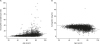 | Fig. 2Simple correlation analysis between age and (A) N/L (r=0.305; p<0.001) and (B) Hb level (r=−0.356; p<0.001).N/L; neutrophil to lymphocyte ratio, Hb; hemoglobin.
|
Baseline characteristics of the patients' groups are listed in Table 1. Group III were older and more frequently female, and had lower body weight, lower body mass index, lower admission blood pressure, more increased HR, higher rates of HTN, lower rates of DM and dyslipidemia, worse LVEF, worse renal function, and more multivessel coronary disease. Group III showed higher GRACE score and Killip class than other groups. Other significant differences included lower rates of history of smoking and lower low-density lipoprotein cholesterol levels.
Table 1
Baseline characteristics according to 3 groups based on Hb level and N/L
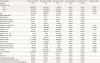
Data presented as mean±standard deviation or number (percentage). Group I, low N/L & no anemia; group II, no group I or III; group III, high N/L & anemia.
Hb; hemoglobin, N/L; neutrophil to lymphocyte ratio, BMI; body mass index, BP; blood pressure, HR; heart rate, GRACE; Global Registry of Acute Coronary Events, CAD; coronary artery disease, HTN; hypertension, DM; diabetes mellitus, LVEF; left ventricular ejection fraction.
*Statistical significance for linear by linear association between categorical variables, calculated using χ2 test for trend.
![]()
2. Coronary angiographic and procedural findings
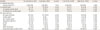
The baseline coronary angiographic characteristics are shown in Table 2. There are more patients with multivessel disease in group III. Patients in group III less underwent CAG and PCI. In lesion type, there are no significant differences between groups.
Table 2
Baseline coronary angiographic variables according to 3 groups based on Hb level and N/L

Data presented as number (percentage). Group I, low N/L & no anemia; group II, no group I or III; group III, high N/L & anemia.
Hb; hemoglobin, N/L; neutrophil to lymphocyte ratio, PCI; percutaneous coronary intervention, CAG; coronary angiogram.
*Statistical significance for linear by linear association between categorical variables, calculated using χ2 test for trend.
![]()
3. Clinical outcomes
During 180-day follow-up, there were 124 in-hospital cardiac death (2.0%), 40 in-hospital non-cardiac death (0.6%) and 87 cardiac deaths (1.4%), 52 non-cardiac death (0.8%) at 180-day. Table 3 showed clinical outcomes in-hospital and 180-day after PCI according to either the presence of anemia or N/L. Patients with anemia or high N/L had worse clinical outcomes for MACEs and deaths in-hospital or at 180-day. When patients were stratified by Hb level and N/L, there was a stepwise association with 180-day mortality (Fig. 3A) and MACEs (Fig. 3B).
Table 3
Clinical outcomes in the hospital period and follow-up at 180-day according to either the presence of anemia or N/L
Data presented as number (percentage).
N/L; neutrophil to lymphocyte ratio, PCI; percutaneous coronary intervention, CABG; coronary artery bypass surgery.
![]()
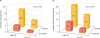 | Fig. 3(A) The 180-day mortality and (B) MACEs stratified by Hb level and N/L: no anemia & low N/L (n=3,170); no anemia & high N/L (n=1,134); anemia & low N/L (n=1,034); and anemia & high N/L (n=819).MACE; major adverse cardiac event, Hb; hemoglobin, N/L; neutrophil to lymphocyte ratio.
|
Clinical outcomes in-hospital and 180-day after PCI according to 3 groups based on Hb level and N/L are listed in Table 4. There were significant differences on clinical outcomes among 3 groups. The 180-day mortality rate and MACEs were respectively 10-fold, 6-fold higher in group III compared with group I.
Table 4
Clinical outcomes in the hospital period and follow-up at 180-day according to 3 groups based on Hb level and N/L

Data presented as number (percentage). Group I, low N/L & no anemia; group II, no group I or III; group III, high N/L & anemia.
Hb; hemoglobin, N/L; neutrophil to lymphocyte ratio, PCI; percutaneous coronary intervention, CABG; coronary artery bypass surgery.
*Statistical significance for linear by linear association between categorical variables, calculated using χ2 test for trend.
![]()
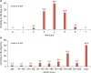
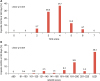
The 180-day mortality rates according to 3 groups in patients having higher mortality risk based on TIMI risk score and GRACE score are presented in Fig. 4. The TIMI risk score showed peak mortality in patients with TIMI score 3, 4 (Fig. 4A). GRACE score showed a strong association with mortality in all patients except score 201–220 (Fig. 4B). In MACEs, similar pattern was observed (Fig. 5). The TIMI risk score showed peak MACEs in patients with TIMI score 3, 4 (Fig. 5A). GRACE score showed a strong association with MACEs in all patients except score 141–160, 201–220 (Fig. 5B).
4. Multivariate analysis of predictors of 180-day mortality
Logistic regression analysis showed the independent prognostic value of the combined use of Hb level and N/L for MACEs (Table 5) and 180-day mortality (Table 6). After adjusting for multiple covariates, the coexistence of pre-procedural anemia and high N/L was still a significant predictor of MACEs after PCI in patients with non-STEMI. In 180-day mortality, after adjustment of age and sex, the coexistence of pre-procedural anemia and high N/L was still a significant predictor of mortality. But, after adjusting for multiple covariates, there was no significant difference among groups to predict mortality in patients with non-STMI undergoing PCI.
Table 5
Prognostic value of the combined use of Hb level and N/L for major cardiac adverse events(logistic regression)

Group I, low N/L & no anemia; group II, no group I or III; group III, high N/L & anemia.
Hb; hemoglobin, N/L; neutrophil to lymphocyte ratio.
*Adjusted for age and gender; †Adjusted for age, gender, diabetes, hypertension, previous coronary artery disease, systolic blood pressure, heart rate, Killip classification, left ventricular ejection fraction, creatinine clearance, multivessel disease, American College of Cardiology/American Heart Association class; ‡Reference group.
![]()
Table 6
Prognostic value of the combined use of Hb level and N/L for 180-day mortality (logistic regression)

Group I, low N/L & no anemia; group II, no group I or III; group III, high N/L & anemia.
Hb; hemoglobin, N/L; neutrophil to lymphocyte ratio.
*Adjusted for age and gender; †Adjusted for age, gender, diabetes, hypertension, previous coronary artery disease, systolic blood pressure, heart rate, Killip classification, left ventricular ejection fraction, creatinine clearance, multivessel disease, American College of Cardiology/American Heart Association class; ‡Reference group.
![]()
DISCUSSION
This study aimed at stratification of risk and prognosis with non-STEMI patients by simple Lab data, such as CBC. We investigated the clinical utility of the combined use of Hb level and N/L, which are readily available CBC data in the early in-hospital period, for predicting risk in patients with non-STEMI. We found that the combined use of Hb level and N/L had a significant discriminatory capacity for predicting short-term clinical outcome in non-STEMI patients. The combined use of Hb level and N/L were independent predictors of MACEs after adjusting for multiple covariates.
Cho et al.8 reported that the combined use of Hb level and N/L had a significant discriminatory capacity for predicting short-term clinical outcome in STEMI patients. They showed that patients who had high N/L and anemia had higher mortality at 180-day compared to patients who had low N/L and no anemia. Cho et al.8 suggested that combined use of Hb level and N/L provides valuable timely information for early risk stratification in patients with STEMI undergoing primary PCI.
Unlike previous study by Cho et al.8, this study have 2 differences. First, whereas previous study showed TIMI risk score had a strong association with mortality in all patients, the TIMI risk score showed peak mortality in patients with TIMI 3, 4 in this study. Second, Cho et al.8 reported that combined use of Hb and N/L was a predictor of 180-day mortality in patients with STEMI undergoing primary PCI. Although we found that combined use of Hb and N/L was not a predictor of 180-day mortality in patients with non-STEMI undergoing primary PCI, we showed that combined use of Hb and N/L was a predictor of MACEs in patients with non-STEMI undergoing primary PCI.
Our study revealed that patients with versus without anemia had higher incidence of MACEs at 180-day. Many patients with AMI usually have an anemia. And anemia increased adverse outcomes in patients with AMI.71314 Some studies reported that there are older and more frequently have co-morbidities that can affect mortality, including DM and renal insufficiency in patients with anemia,1516 and they are less likely to receive life saving medications such as β-blockers.17 Nonetheless, anemia showed discriminatory prognostic capacity of MACEs after adjusting for these variables. As described previously,8 several mechanisms have been suggested to be associated with risk of anemia on clinical outcome in patients with AMI undergoing PCI. In patients with AMI, anemia can directly exacerbate ischemia and provoke arrhythmia by decreasing blood oxygen carrying capacity and inadequate tissue-oxygen delivery.18 Also, myocardium demands more oxygen in anemia by stimulating greater cardiac output.19 Recently, relationship cardiovascular disease and anemia is considered to develop due to a complex interaction of iron deficiency, cytokine production and impaired renal function, although other factors such as blood loss may also contribute.20 This results in ventricular remodeling and ventricular hypertrophy long-term.21 Therefore, anemia may represent worse health status, larger extent of ischemic myocardium, and cardiac remodeling long-term.
Patients with high N/L showed higher incidence of MACEs at 180-day compared to those with low N/L in our study. There are major roles of white blood cells (WBC) in the inflammatory processes of atherogenesis.22 Moreover, previous studies demonstrated that an elevated leukocyte increase risk for ischemic events and death in patients with AMI.23 A study showed that, among WBC counts and its subtypes, increased N/L can predict cardiovascular risk in patients with CAD or at high-risk for CAD.4 Moreover, Duffy et al.24 reported that a positive and independent association between N/L and long-term mortality in patients undergoing PCI. Other study showed that there are increased neutrophil aggregation and oxidase activity in the coronary sinus of patients with CAD.25 And, Madjid et al.26 suggested that, during reperfusion of ischemic myocardium, neutrophil can plug capillaries in the coronary microcirculation, aggravating ischemia and extending the infarct area. Stress response can make lymphopenia27 and lymphopenia has a good discriminatory ability for diagnosis of AMI.28 Therefore, N/L can represent a combined assessment of the severity of an acute ischemic event and extent of myocardium at risk.
Taken together, combined use of Hb level and N/L may represent a biochemical integrated assessment of health status, severity and extent of myocardial ischemia, and cardiac remodeling long-term. The present study demonstrated that combined use of Hb level and N/L had a significant discriminatory capacity for predicting short-term clinical outcome in patients with non-STEMI, including high-risk subgroups. Prognostic discriminatory capacity remained significant after adjusting for multiple covariates including age, sex, comorbidities, renal function, and surrogates of the extension of myocardial ischemia including systolic BP, HR, LVEF, and Killip classification. Our study suggests that combined use of Hb level and N/L, which are readily available CBC data in the early in-hospital period, provides valuable timely information for early risk stratification in patients with non-STEMI.
There are several limitations to this study. First, our study was retrospective and nonrandomized and thus subject to selection bias. Second, our study lacked different causes of anemia that likely had different prognostic effects in patients with non-STEMI. We focused on clinical utility of combined use of Hb level and N/L for early risk stratification in patients with non-STEMI in the real-world. Third, the possibility of residual confounding from unmeasured or unselected covariates cannot be excluded, although we have adjusted 12 risk factors.
 XML Download