

 PDF
PDF ePub
ePub Citation
 Print
Print


INTRODUCTION
Globally, cardiovascular disease (CVD) is one of the most common causes of human morbidity and mortality.1 CVD includes coronary artery disease (CAD), which is the progressive, atherosclerotic occlusion of the arteries that supply oxygenated blood to the heart muscle. A challenge in the management of CAD lies in the selection of patients who require coronary intervention, and this process can be aided by the identification of biomarkers to predict the onset and severity of the disease. Many biochemical markers, such as cytokines and proinflammatory factors, are being investigated for potential targeting in the treatment and primary prevention of CVD.2 Among these markers, fibroblast growth factor (FGF) 21 has received attention for its proposed involvement in metabolic diseases, including CVD.3
FGF21 is a member of an endocrine subfamily of FGFs that function as hormones.4 FGF21 is expressed in the liver, pancreas, adipose tissue, and skeletal muscle, and is mainly secreted from the liver.5 Physiologically, endogenous induction of FGF21 during fasting or starvation conditions mediates fatty acid oxidation and ketogenesis.6 Metabolically, FGF21 has systemic beneficial effects in target tissues such as liver, pancreas, and adipose tissues.7 Pharmacologically, a therapeutic role for FGF21 in obesity and type 2 diabetes mellitus (T2DM) has been demonstrated in humans as well as in animal models.3 Exogenous administration of recombinant FGF21 in a diet-induced animal model of obesity decreases levels of serum triglycerides and glucose and causes weight loss by increasing energy expenditure.8
In addition to its therapeutic potential, the serum concentration of FGF21 has been investigated as a putative marker in metabolic diseases.2 However, in contrast to its favorable metabolic function, serum levels of FGF21 are elevated in individuals with obesity, T2DM, or nonalcoholic fatty liver disease,9 and are significantly correlated with multiple metabolic parameters, including levels of blood glucose, lipids, and glycosylated hemoglobin.2 Moreover, serum FGF21 concentrations are associated with established cardiovascular risk factors, and are higher in patients with CAD than in those without CAD.510 However, the precise nature of the association between serum FGF21 concentrations and CAD has yet to be confirmed.41112
In this study, we compared serum FGF21 concentrations in individuals with or without coronary artery narrowing, as identified by coronary angiography (CAG), and determined whether circulating FGF21 concentrations are associated with the severity of CAD.
MATERIALS AND METHODS
1. Study population
This cross-sectional study included 444 consecutive subjects with suspected acute coronary syndrome (ACS) who attended Kyungpook National University Hospital from December 2009 to July 2012. Subjects were screened for their eligibility to participate in the study, resulting in the enrolment of 137 men who underwent CAG. Exclusion criteria were subjects who had lipid-lowering agents within 3 months, infectious or systemic inflammatory diseases, history of hematologic disorder, thyroid dysfunction and severe liver disease (serum aspartate transaminase (AST) and alanine transaminase (ALT) levels >5 times upper limits of normal) and renal dysfunction (serum creatinine level >3.0 mg/dL). Among these participants, 112 (mean age, 61.2 years) had one or more narrowing lesions of the coronary arteries on CAG, and 25 (the control group; mean age, 55.5 years) had no such lesions. The participants were assessed for the presence of underlying metabolic diseases, including T2DM, hypertension, dyslipidemia, and ischemic heart disease.
2. Anthropometric and biochemical parameters
Body weight and height were measured at the hospital visit, and body mass index (BMI) was calculated as weight divided by squared height (kg/m2). Blood pressure (BP) was measured with a sphygmomanometer. Serum levels of N-terminal pro-B type natriuretic peptide (NT-proBNP) were measured with an electrochemiluminescence immunoassay method (Modular Analytics E170; Roche Diagnostics, Mannheim, Germany). Other biochemical variables were measured by standard laboratory techniques. Levels of serum total cholesterol (TC), high-density lipoprotein cholesterol (HDL-C), low-density lipoprotein cholesterol (LDL-C), triglyceride, C-reactive protein (CRP), creatinine, white blood cells (WBCs), AST, and ALT were determined from samples collected after an overnight fast (of ≥8 hours).
3. Measurement of levels of FGF19 and FGF21
Serum samples for measurement of FGF19 and FGF21 were collected prior to CAG procedures. Samples were centrifuged (13,800 ×g for 20 minutes at 4°C) immediately after collection, and the resulting serum samples were kept at −80°C until they were assayed. Serum FGF19 and FGF21 concentrations were assayed with a commercial ELISA kit (R&D Systems, Minneapolis, MN, USA) in accordance with the manufacturer's protocol. Blood samples were diluted 1:1 with dilution buffer before the assays. The upper and lower detection limits for FGF21 were 600 and 50 pg/mL. All samples were assayed in duplicate.
4. Assessment of the severity of coronary artery occlusion
The severity of coronary artery occlusion was evaluated by determination of the SYNergy between percutaneous coronary intervention with (paclitaxel-eluting) TAXus stent and cardiac surgery (SYNTAX) score and the Global Registry of Acute Coronary Events (GRACE) score. The detailed methodologies for calculating the SYNTAX and GRACE scores have been described elsewhere.1314
5. SYNTAX score
The SYNTAX score is an anatomically based scoring system that quantitatively characterizes coronary vessels with respect to the number, complexity, location, and functional aspect of angiographically obstructive lesions. Through CAG, each lesion with a stenosis diameter ≥50% in vessels ≥1.5 mm is scored. Each coronary segment is assigned a weight factor according to the location and severity of the associated lesions. The characteristics of the lesions, including total occlusion, trifurcation, bifurcation, calcification, tortuosity, length >20 mm, thrombus, and diffuse or small-vessel disease, are summed up with the internet-based SYNTAX calculator (www.syntaxscore.com) to derive the final score.1315 This score is an objective angiographic tool to grade the complexity of CAD and characterize its severity.
6. GRACE risk score
The GRACE score is used for risk assessment in ACS, which includes non-ST-elevation myocardial infarction (STEMI), STEMI, and unstable angina. The score reflects the prognoses of patients with ACS, and is calculated on the basis of age, Killip class, systolic BP, ST-segment deviation, cardiac arrest during presentation, serum creatinine level, heart rate, and elevated cardiac enzyme or marker levels.16 GRACE scores are calculated with an online risk calculator (http://www.outcomes-umassmed.org/grace).
7. Statistical analysis
The data are presented as the mean±standard deviation. Statistical analysis was performed using SPSS 18.0 software (SPSS Inc., Chicago, IL, USA), with calculation of Pearson's correlation coefficient (r) for determination of the association between quantitative variables. Analysis of covariance was applied for adjustment of established CVD risk factors including age, BMI, TC, LDL-C, and smoking status. Student's t-test and the χ2 test were performed to compare continuous and categorical variables between the 2 groups. Statistical significance was defined by p-values ≤0.05.
RESULTS
Baseline characteristics of the participants are shown in Table 1. Those with lesions identified by CAG had significantly higher mean age (p=0.01), WBC count (p<0.01), TC (p=0.03), LDL-C (p=0.028), and AST (p=0.01) than those without lesions. Left ventricular ejection fraction (p<0.01) and serum levels of cardiac enzyme (creatine kinase-MB, troponin I, p<0.01), NT-proBNP (p=0.003), and CRP (p=0.008) were significantly higher in patients with lesions identified by CAG than in those without such lesions.
Table 1
Baseline clinical characteristics of study participants with and without lesions identified by CAG
Data are expressed as the mean±standard deviation or number (percentage). ‘No lesion’ indicates that no coronary arterial lesion was identified by CAG. ‘Lesion’ indicates that one or more coronary arterial lesions were identified.
CAG; coronary angiography, BMI; body mass index, BP; blood pressure, WBC; white blood cell, TC; total cholesterol, HDL-C; high-density lipoprotein cholesterol, LDL-C; low-density lipoprotein cholesterol, AST; aspartate transaminase, ALT; alanine transaminase, eGFR; estimated glomerular filtration rate, LVEF; left ventricular ejection fraction, CK-MB; creatine kinase-MB, TnI; troponin I, NT-proBNP; N-terminal pro-B type natriuretic peptide, CRP; C-reactive protein.
![]()
The mean serum FGF21 concentration was significantly higher in individuals with CAG-identified lesions than in those without such lesions (370.9±196.9 pg/mL vs. 282.1±215.4 pg/mL; p=0.048, Table 2). The optimal cut-off value of serum FGF21 was 181.87 pg/mL determined through cox regression analysis. After adjustment for established CVD risk factors including age, BMI, TC, LDL-C, and smoking, the difference between the mean serum FGF21 concentrations in these groups was slightly greater (376.6±20.3 pg/mL vs. 269.4±44.0 pg/mL; p=0.031; Table 3). Because FGF21 was suggested as a putative marker of metabolic disease including diabetes,6 we further analyzed the difference of FGF21 concentrations according to the presence of diabetes. Although mean serum FGF21 concentrations were higher in diabetic subjects than non-diabetic subjects regardless of coronary arterial lesion, there was no statistically significant difference between 2 groups (307.6±248.4 pg/mL vs. 277.2±251.1 pg/mL, T2DM vs. no T2DM in no lesion group; p=0.803 and 404.3±188.9 pg/mL vs. 358.4±199.4 pg/mL, T2DM vs. no T2DM in lesion group; p=0.276, respectively; Table 4). Mean serum FGF19 concentrations were not significantly different in the 2 groups (Tables 2 and 3).
Table 2
Difference of serum FGF21 and FGF19 concentrations according to the presence or absence of lesions identified by CAG

| Biomarkers | No lesion (n=25) | Lesion (n=112) | p-value |
|---|---|---|---|
| FGF21 (pg/mL) | 282.1±215.4 | 370.9±196.9 | 0.048 |
| FGF19 (pg/mL) | 572.0±196.8 | 629.2±245.8 | 0.27 |
Values shown are means±standard deviation. ‘No lesion’ indicates that no coronary arterial lesion was identified by CAG. ‘Lesion’ indicates that one or more coronary arterial lesions were identified.
FGF; fibroblast growth factor, CAG; coronary angiography.
![]()
Table 3
Difference of serum FGF21 and FGF19 concentrations (after adjustment for established CVD risk factors) according to the presence or absence of lesions identified by CAG

| Biomarkers | No lesion (n=25) | Lesion (n=112) | p-value |
|---|---|---|---|
| FGF21 (pg/mL) | 269.4±44.0 | 376.6±20.3 | 0.031 |
| FGF19 (pg/mL) | 564.2±49.0 | 642.4±22.7 | 0.313 |
Values shown are means±standard error. Values were adjusted for established CVD risk factors including age, BMI, TC, and LDL-C and further adjusted for smoking. ‘No lesion’ indicates that no coronary arterial lesion was identified by CAG. ‘Lesion’ indicates that one or more coronary arterial lesions were identified.
FGF; fibroblast growth factor, CVD; cardiovascular disease, CAG; coronary angiography, BMI; body mass index, TC; total cholesterol, LDL-C; low-density lipoprotein cholesterol.
![]()
Table 4
Serum FGF21 concentrations in subjects according to presence or absence of lesions identified by CAG and T2DM

| Biomarker | No lesion (n=25) | p-value | Lesion (n=112) | p-value | ||
|---|---|---|---|---|---|---|
| No T2DM (n=21) | T2DM (n=4) | No T2DM (n=82) | T2DM (n=30) | |||
| FGF21 (pg/mL) | 277.2±251.1 | 307.6±248.4 | 0.803 | 358.4±199.4 | 404.3±188.9 | 0.276 |
Values shown are means±standard deviation. ‘No lesion’ indicates that no coronary arterial lesion was identified by CAG. ‘Lesion’ indicates that one or more coronary arterial lesions were identified.
FGF; fibroblast growth factor, CAG; coronary angiography, T2DM; type 2 diabetes mellitus.
![]()
Correlation of serum FGF21 concentration with the severity of CAG-identified lesions was assessed by the use of SYNTAX and GRACE scores. No significant correlation was observed between serum FGF21 concentrations and SYNTAX scores (r=0.117; p=0.176; Fig. 1A) or GRACE scores (r=0.113; p=0.193; Fig. 1B).
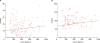 | Fig. 1Correlation between serum FGF21 concentrations and SYNTAX and GRACE scores. (A) Determination of the correlation between serum FGF21 levels and angiographic lesion severity, defined by the SYNTAX score. A Pearson correlation coefficient of 0.117 indicated no significant correlation (p=0.176). (B) Determination of the correlation between serum FGF21 levels and the risk of mortality, defined by the GRACE score. A Pearson correlation coefficient of 0.113 indicated no significant correlation (p=0.193).FGF; fibroblast growth factor, SYNTAX; SYNergy between percutaneous coronary intervention with (paclitaxel-eluting) TAXus stent and cardiac surgery, GRACE; Global Registry of Acute Coronary Events.
|
DISCUSSION
In this study, we found that mean serum FGF21 concentrations were significantly higher in 112 patients with ACS (with CAG-identified coronary artery lesions) than in 35 individuals without CAG-identified lesions, especially after adjustment for independent CVD risk factors including age, BMI, TC, LDL-C, and smoking. However, serum FGF21 concentrations did not significantly correlate with CAD severity (as assessed by SYNTAX scores) or risk of ACS mortality (as assessed by GRACE scores). Serum levels of FGF19, another endocrine subfamily member, were not associated with the presence or absence of CAG-identified lesions.
Results from several studies previously showed that FGF21 can be used as a biomarker of atherosclerotic processes.1718 In a study involving 670 patients, Chow et al.19 found that serum FGF21 levels were positively correlated with carotid intima-media thickness (IMT) in women (r=0.32; p<0.001) but not in men, and also demonstrated that the serum FGF21 level in women is an independent risk factor for increased carotid IMT (p=0.039). In a study involving 253 individuals, Shen et al.5 identified significantly higher levels of serum FGF21 in the group with CAD than in the non-CAD group (415.5 pg/mL [range, 258.0–693.7 pg/mL] vs. 321.5 pg/mL [239.1–497.6 pg/mL]; p<0.05). In a study of 87 patients with T2DM, Lenart-Lipińska et al.20 found significantly higher incidence of cardiovascular morbidity in those with serum FGF21 concentrations >240.7 pg/mL than in those with lower levels of FGF21 (cardiovascular morbidity hazard ratio=5.62; 95% confidence interval, 1.73–18.23; p=0.004). Our results now also demonstrate significantly higher serum FGF21 concentrations in patients with CAG-identified lesions than in those without such lesions.
Results from previous studies demonstrate correlation between serum FGF21 levels and CAD severity. Kim et al.10 showed that serum FGF21 levels measured in 120 patients are significantly correlated with both Gensini scores, defined by total narrowing of coronary artery lumens (r=0.271; p=0.014), and Extent scores, representing the percentage of coronary artery surface area affected by atheroma (r=0.217; p=0.041). However, FGF21 was not significantly associated with either Gensini (r=0.332; p=0.055) or Extent (r=0.296; p=0.084) scores in patients with diabetes in the final model. Similarly, our results indicate that serum FGF21 concentrations are not associated with angiographic severity (SYNTAX scores) or risk of mortality (GRACE scores). However, the number of patients involved in studies of the correlation between serum FGF21 and coronary artery severity is so far quite small, and further investigations will be required to clarify whether such a correlation exists, and if it does, to determine its precise nature.
There are some limitations in this study. As we mentioned above, relatively small subjects were enrolled in the current study. No statistical significant difference of serum FGF21 concentrations according diabetes status might be due to small sample size. Second, we collected serum samples prior to CAG procedures of each subjects. Because serum FGF21 displays a circadian rhythm with peak levels at night and decreases to nadir levels at morning,21 our data cannot reflect the influence of circadian rhythm of FGF21.
In conclusion, we found that patients with ACS had higher serum FGF21 levels than those without coronary artery lesions, regardless of established CVD risk factors including age, BMI, TC, and LDL-C. Our results suggest that FGF21 is an independent marker of ACS. However, serum FGF21 concentrations are not associated with angiographic lesion severity of ACS, nor with the risk of mortality. Studies with larger sample sizes will be required to confirm this observation.
 XML Download