

 PDF
PDF ePub
ePub Citation
 Print
Print


A 40-year-old male presented with stable angina of 5 months duration with normal resting electrocardiography and echocardiography. Antianginal treatment was started but he remained symptomatic despite optimal medical therapy. His clinical examination was unremarkable. Invasive coronary angiography revealed anomalous origin of left coronary artery arising from right sinus of Valsalva traversing anterior to right ventricle outflow tract (Figure 1, Movie 1) and coronary cameral fistula from diagonal branch draining into left ventricle. Right coronary artery was normal in origin and super-dominant with severe atherosclerotic narrowing in mid vessel (Figure 2).1) Aortic root angiogram revealed no coronary artery arising from left sinus of Valsalva (Figure 3). Coronary CT angiography revealed a single, common ostium of the right and left coronary artery arising from the right anterior sinus of Valsalva with anomalous course of the left coronary artery anterior to right ventricle outflow tract which is by definition considered a “single” coronary artery (Figure 3A and 3B) with fistulous communication between one of the distal branch of first diagonal to left ventricle2) Stress MIBI nuclear scan with adenosine was performed to rule out ischemia due to the anomalous vessel and the coronary cameral fistula. There was no stress induced ischemia in left anterior descending territory. Hence percutaneous coronary intervention of right coronary artery lesion was done. At 3 months follow up patient is asymptomatic. In summary this is a case with three in one coronary pathology including atherosclerotic stenosis, single coronary artery and coronary cameral fistula all in one. Multimodality imaging helped us to identify the culprit causing ischemia and to select the optimal treatment strategy.
Figures and Tables
Figure 1
Coronary angiogram. (A) Through right radial artery route left anterior oblique view showing anomalous origin of left anterior descending artery from right sinus of Valsalva traversing anterior to right ventricle outflow tract. There is a coronary cameral fistula (arrow) from distal branch of diagonal draining into left ventricle. (B) Through right radial artery route right anterior oblique view showing anomalous origin of left anterior descending artery from right sinus of Valsalva traversing anterior to right ventricle outflow tract (arrow).
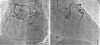
Figure 2
Coronary angiogram. (A) Right coronary artery was normal in origin and super-dominant, supplying up-to left circumflex artery territory having severe atherosclerotic stenosis (arrow) in mid right coronary artery. (B) Aortic root angiogram in left anterior oblique view showing no coronary artery origin from left aortic sinus.
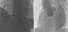
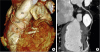
Figure 3
(A) Coronary CT angiography. Volume rendered image shows common ostium of the right and left coronary arteries arising from the right anterior sinus (solid arrow) with anomalous course of the left coronary artery anterior to RVOT (open arrow). Left anterior descending artery is giving early diagonal branch which has a fistulous communication between one of its distal branch to left ventricle (solid arrow). (B) The curved multi-planar reformatted image demonstrates that left anterior descending coronary artery is giving early diagonal branch (open arrow) which has a fistulous communication (long arrow) between one of its distal branch to left ventricle (solid arrow). PA: pulmonary artery, RVOT: right ventricle outflow tract.
SUPPLEMENTARY MATERIAL
Movie 1
Coronary angiogram through right radial artery route left anterior oblique view showing anomalous origin of left anterior descending artery from right aortic sinus traversing anterior to right ventricle outflow tract. There is a coronary cameral fistula from diagonal draining into left ventricle.

 XML Download