Journal List > Immune Netw > v.18(6) > 1109782

Jin and Yoon: From Bench to Clinic: the Potential of Therapeutic Targeting of the IL-22 Signaling Pathway in Atopic Dermatitis

Abstract

Atopic dermatitis (AD) is the most common pruritic inflammatory skin disease characterized by thickening of epidermis and dermis as well as by the infiltration of multiple pathogenic polarized T lymphocytes, including Th2, Th17, and Th22 cells. Significant progress has been made to develop targeted therapeutics for treating AD, e.g., Food and Drug Administration-approved dupilumab, an antibody for dual targeting of IL-4 and IL-13 signaling pathways. Additionally, a growing body of published evidence and a promising result from the early stage of the clinical trial with ILV-094, an anti-IL-22 antibody, strongly support the notion that IL-22 is a potential therapeutic target for treating AD. Moreover, we also experimentally proved that IL-22 contributes to the pathophysiology of AD by employing a murine model of AD induced by epicutaneous sensitization. Here, we review recent preclinical and clinical findings that have advanced our understanding of the roles of IL-22 and Th22 cells in skin inflammation. We conclude that blockade of IL-22 signaling may be a promising therapeutic approach for the treatment of AD.

Abbreviations

AD

atopic dermatitis

AHR

aryl-hydrocarbon receptor

CCR

C-C chemokine receptor

DC

dendritic cell

DLN

draining lymph node

H4R

histamine H4 receptor

IL-22R

IL-22 receptor

IL-23R

IL-23 receptor

LC

Langerhans cell

OVA

ovalbumin

INTRODUCTION

Atopic dermatitis (AD) is an inflammatory skin disease that affects up to 20% of children and 3% of adults (123). A hallmark of AD is dry and itchy skin with the disrupted skin barrier function. It is now considered that AD is a heterogeneous disease characterized by the activation of diverse cytokine signaling pathways, involving Th1, Th2, Th22, and Th17 cells, depending on the disease subtype (456). Notably, it has been reported that Asian patients with AD have much higher expression levels of Th22 and Th17 cell related cytokines in lesional skin, compared with those in American patients of the European origin (7). Additionally, new onset pediatric AD patients have robust mixed activation of Th2, Th22, and Th17 cells in AD skin lesions (89). Overall, these observations suggest that dysregulation of immune responses mediated by multi-polarized immune cells may participate in the diverse manifestations of AD.
IL-22 is a member of the IL-10 family of cytokines produced by Th17 and Th22 cells, innate lymphocytes that include γδ T cells and type 3 innate lymphoid cells (10111213141516). The IL-22 receptor (IL-22R) is expressed on epithelial cells, including keratinocytes, but not on immune cells, indicating an essential role for IL-22 signaling in mucosal barrier function (171819). IL-22 induces epithelial cell proliferation and expression of anti-apoptotic genes, and thus, promotes tissue repair activity and protects stem cells from injury (13202122). At the same time, it has been reported that the IL-22 signaling pathway participates in inflammatory skin diseases (23242526). For example, an intradermal injection of IL-22 in vivo causes keratinocyte proliferation and epidermal thickening (27). Administration of IL-22 in vitro also leads to keratinocyte proliferation and the thickening of human epidermis reconstituted in a three-dimensional matrix (2829), similar to the changes in the lesional skin of AD and psoriasis patients. Indeed, Il22 mRNA expression and T cells that produce IL-22 are significantly increased in skin lesions of patients with AD (303132). Additionally, serum IL-22 levels are also elevated in patients with AD (3334).
In line with these reports, we will discuss our recent findings on the contribution of IL-22/Th22 cells to allergic skin inflammation as well as Th22 cell polarization obtained in the murine model of AD induced by epicutaneous sensitization of allergen (35). We will present evidence that targeting of IL-22 signaling can be a plausible therapeutic option for AD. Finally, we will finish this review summarizing major clinical approaches currently being developed for treating AD.

THE SKIN AS A MAJOR ROUTE OF ALLERGEN SENSITIZATION FOR ALLERGIC DISEASES

The skin is considered as the largest immunological organ that acts as a barrier between the body and external environments, protecting against chemical and physical insults as well as against pathogenic microbes (3637). Defects in skin barrier function and abnormalities of skin immune systems give rise to skin inflammation and microbial infection (3839). AD generally tends to precede other allergic diseases such as asthma, allergic rhinitis and others, known as the atopic march (40414243), implying that the skin may be a crucial priming site in AD as well as in allergies of different parts of the body. In fact, most infants and children with AD history have a higher tendency to develop asthma, allergic rhinitis (4044) and other allergic conditions than infants without AD. Moreover, we and other groups experimentally proved that mechanical skin injury with tape stripping, a surrogate of scratching in AD, exacerbates the defect in skin barrier function, leads to the penetration of the allergen through the damaged skin, and induces the release of a range of cytokines and chemokines that drive immune responses to cutaneously immunized antigens (35454647). This AD-like skin inflammation subsequently stimulates allergic manifestations such as airway hyper-responsiveness (484950) and food allergy (5152). Overall, accumulated clinical and experimental findings unequivocally support the notion that the skin is a major sensitization site for allergic diseases that affect different parts of the body.

MOUSE MODEL OF AD TRIGGERED BY EPICUTANEOUS SENSITIZATION

Our understanding of human diseases has been enormously expanded by in-depth studies in animal models that are invaluable tools for unveiling pathogenic mechanisms and finding potential treatment targets. Therefore, to find potent target(s) and develop medicines for treating AD, it is essential to elucidate AD pathogenesis by establishing appropriate animal models that faithfully recapitulate the hallmark features of AD. To satisfy this demand, Spergel and colleagues (53) have developed a mouse model of AD induced by repeated epicutaneous sensitization of the injured skin inflicted by tape-stripping with ovalbumin (OVA). This model displays many features of human AD described below. Mechanical injury of the skin with tape stripping, a surrogate of scratching in patients with AD, enhanced the release of various pro-inflammatory cytokines and chemokines, which are regarded as important initiating factors for allergen-specific immune responses and allergic skin inflammation. Mice epicutaneously sensitized with allergen developed increased scratching behavior and skin lesions that exhibited many cardinal features of AD, including increased epidermal and dermal thickness, infiltration of a mixture of polarized CD4+ T cells and eosinophils as well as the expression of Th2 (53), Th17 (48), and Th22 (3550) cell derived cytokines with minimal or no change in IFN-γ level (53). Systemically, serum OVA-specific IgG1, IgE, and IgG2a were elevated in this model (53), as typically described in AD patients. Furthermore, OVA-sensitized mice developed higher airway hyper-responsiveness following a challenge with OVA (485053), frequently observed in asthmatic patients with AD history. Additionally, epicutaneous sensitization with allergen enhanced IgE-mediated mast cell degranulation and promoted mast cell-dependent anaphylaxis elicited by oral challenge (5152), which was similar to the food-induced anaphylaxis in patients with AD. Thus, this model has histological, immunological, and clinical features of human AD and can be utilized to gain better insights into the mechanisms of AD pathogenesis and allergic diseases associated with AD. Furthermore, this model can be utilized to find therapeutic targets and to develop medicines for the treatment of AD and allergic diseases associated with it.

POTENTIAL ROLE OF IL-22 AND TH22 CELLS IN AD

IL-22 is a member of the IL-10 family of cytokines, which was initially thought to be produced by Th1 cells (54). However, it was found that this cytokine is secreted in high quantities by Th17 cells (55). Interestingly, IL-22 and IL-17 are not always co-expressed and their expression are regulated differently in humans (5657). Furthermore, identification of Th22 cells producing IL-22 without the concomitant production of IFN-γ, IL-4, or IL-17 has attracted much attention due to their potential involvement in skin homeostasis and pathogenesis of AD (1516). Whereas the retinoid-related orphan receptor C is the master transcriptional factor for Th17 cell differentiation, the aryl-hydrocarbon receptor (AHR) is a critical factor for Th22 cell polarization in humans (16). Indeed, activation of the AHR induces a robust IL-22 expression, whereas it inhibits IL-17 production (16). Furthermore, it was reported that a subset of memory CD4+ T cells producing IL-22 express skin homing chemokine receptors C-C chemokine receptor (CCR) 10, CCR6, and CCR4 that promote their infiltration to skin (1516). Thus, it implies that Th22 cells may have a potential role in skin homeostasis and inflammation. Interestingly, the IL-22 receptor is expressed on epithelial cells, including keratinocytes in the skin, but not in immune cells (19), implying a potential role of IL-22 signaling in skin barrier functions contributed by keratinocytes. Th22 cells and IL-22-producing CD8+ cells are the main source of IL-22 in AD (31). Additionally, a high percentage of circulating Th22 cells and IL-22-producing CD8+ T cells from patients with AD co-express IL-13 (58). Moreover, the number of Th22 cells and IL-22+CD8+ T-cell positively correlates with AD disease severity (31), and IL-22, highly expressed in the affected skin of patients suffering from AD, is deeply involved in epidermal hyperplasia and barrier defects (5759). More importantly, therapeutic success at the early stage of the clinical trial of ILV-094, an anti-IL-22 antibody, demonstrated that IL-22 indeed contributes to the pathogenesis and symptoms of AD (6061). We also demonstrated that epicutaneous sensitization by the application of allergen to mechanically injured mouse skin resulted in elevated serum IL-22 levels and accumulation of CD3+CD4+IL-22+ T cells in allergen-sensitized skin, which is essential for epidermal thickening and keratinocyte proliferation, as both were absent in Il22−/− mice (35). In complementary experiments, we also showed that keratinocyte proliferation and epidermal thickening were similarly induced in OVA-challenged skin of naïve recipients of Th22 cells polarized in vitro (35). Furthermore, our recent findings demonstrated that epicutaneous immunization induced polarization of IL-22-producing T cells that subsequently contributed to airway hyper-responsiveness in allergen-challenged mice (50). These data support the view that targeting IL-22 in AD may be promising for the treatment of AD and other allergic diseases, including asthma.

PLAUSIBLE MECHANISMS OF TH22 CELL POLARIZATION IN THE SKIN

Since the discovery of the novel Th22 cell subset in human skin, the knowledge of IL-22/Th22 cell biology has increased considerably. A recent report demonstrated that human Langerhans cells (LCs) efficiently induce the expansion of memory Th22 cells and stimulate the polarization of Th22 cells from naive CD4+ T cells (62). Furthermore, recent findings showed that tissue-resident CD5-expressing dendritic cells (DCs), a subtype of the healthy human LCs and dermal skin cells can induce Th22 cells and cytotoxic T cells (63). In addition, it has been shown that the expression of CD1a, a lipid-presenting molecule, in a subtype of LCs is critical for the expansion of IL-22-producing T cells (64). However, the mechanism of differentiation of antigen-specific IL-22-producing T cells was not determined in that study. To examine in detail the mechanisms responsible for Th22 cell polarization, we utilized a mouse model of AD elicited by the application of antigen to mechanically injured skin. In this model, we have demonstrated a sequence of mechanisms leading to IL-22-dependent epidermal thickening and polarization of allergen-specific Th22 cells. Endogenous TLR4 ligands released by mechanical skin injury, a surrogate of scratching in AD, cause keratinocytes to release IL-23 that drives allergen-captured migratory skin DCs to produce endogenous IL-23 and polarize naïve CD4+ T cells to antigen specific CD4+ T cells. The latter cells, in turn, mount IL-22 response to cutaneously introduced antigen, causing keratinocyte proliferation and epidermal thickening, a hallmark feature of AD (35). We also observed that the IL-23 receptor (IL-23R) is expressed on a subpopulation of DCs in the skin and skin draining lymph nodes (DLNs) in mouse, but is not detectable on splenic DCs (35). We further proved that recombinant IL-23, as well as IL-23 released from explants of mechanically injured skin, directly polarizes DCs from skin DLNs in vitro to drive IL-22 production by naïve CD4+ T cells (35). More importantly, the relevance of this pathway to the pathophysiology of AD in humans is supported by the observation that human keratinocytes express biologically active IL-23 and our findings that a fraction of epidermal LCs and dermal DCs in normal human skin express IL-23R on their surface, as exogenous IL-23 polarizes LCs and drives IL-22 production by naïve CD4+ T cells (35). Overall, our data suggest that IL-23 released by keratinocytes following mechanical injury primes a subtype of IL-23R+ skin-derived migratory DCs to express endogenous IL-23, which polarizes them and initiates an IL-22 response to introduced antigen via damaged skin (35). The priming action of keratinocyte-derived IL-23 on skin DCs that induces an IL-22 immune response (35) parallels the priming action of keratinocyte-derived thymic stromal lymphopoietin (6566) and IL-33 (67) on DCs that stimulates a Th2 immune response. Thus, the release of cytokines by keratinocytes is a key early event in the development of adaptive immune response to cutaneous sensitization with allergen.

POTENTIAL THERAPEUTIC TARGETING OF IL-22/TH22 SIGNALING PATHWAYS IN AD

As mentioned above, IL-22 is highly expressed in the severely affected skin of individuals suffering from AD (60). Chronic AD is characterized by the conversion of the immune response from Th2 cell-dominated to mixed activation of Th1, Th22, and Th2 cells in the affected skin (3132). We also proved that IL-22 is a major player in epidermal acanthosis observed in the sensitized skin of mice (35) (Fig. 1). In line with these observations, a randomized, double-blind, placebo-controlled clinical study of an anti-IL-22 mAb (ILV-094) showed a substantial improvement in patients severely affected with AD (61). Thus, it is likely that a subset of patients with AD with more polarization toward Th22 cell cytokine pathways might be responsive to the direct blockade of IL-22 signaling pathway (6061). Other potential therapeutic approaches to the IL-22 mediated skin inflammation may include targeting of IL-23, a key cytokine in the process of Th22 cell polarization in the sensitized skin as described above (35) (Fig. 1). Furthermore, blockade of CC-chemokine ligand 20 and its cognate receptor CCR6 that attracts IL-22-producing cells to the sensitized skin can be another therapeutic option for AD (68) (Fig. 1). Finally, because the AHR is a key transcription factor for both Th22 polarization and IL-22 production (1516), its antagonists, including CH223191 (69), can be utilized to suppress Th22 cell polarization or to block IL-22 production for the treatment of AD that involves Th22 cells and IL-22 (Fig. 1).
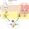
Figure 1

Potential therapeutic targeting of Th22/IL-22 signaling pathways in AD. Skin barrier defects caused by scratching or genetic mutations lead to penetration of external antigens and keratinocyte production of IL-23 via endogenous TLR4 ligand/TLR4 axis. A subset of IL-23R expressing DCs are activated and triggers AHR dependent Th22 immune response. Skin infiltrated CCR6+ Th22 cells induce epidermal hyperplasia and barrier dysfunction via IL-22/IL-22R signaling axis. Targeting TLR4/IL-23/Th22/IL-22 as well as CCL20/CCR6 pathways might be a promising strategy to overcome atopic skin inflammation.

in-18-e42-g001

ALTERNATIVE THERAPEUTIC APPROACHES FOR THE TREATMENT OF AD

Our increasing understanding of the pathogenic mechanisms of AD expanded the pipeline of new and more targeted therapies (70717273). Targeting therapies based on various mechanisms of action are being developed to suppress immune responses in AD (7071). Dupilumab, the first effective biologic targeting the IL-4/IL-13 receptor was approved by Food and Drug Administration for the treatment of moderate-to-severe AD (74). In a randomized, double-blind, placebo-controlled phase II study of nemolizumab targeting IL-31 signaling involved in itching, adult patients with moderate-to-severe AD showed the improvement of pruritus (7576). In addition, it was known that the JAK/STAT signaling axis is deeply involved in the dysregulation of immune responses in AD (7778). Based on this information, several therapeutic agents targeting TYK2, JAK1, JAK2, and JAK3 are being evaluated for treating moderate-to-severe AD (707273). The histamine/histamine H4 receptor (H4R) signaling has been linked to pruritus and inflammation in several preclinical AD murine models (798081). Thus, it can be a promising target for treating AD. In line with this assumption, oral administration of the H4R antagonist ZPL-389 significantly reduced pruritus in AD patients during clinical trials (82). Another type of H4R antagonist, JW1601, exhibited strong anti-pruritic and anti-inflammatory efficacies in several preclinical mouse studies (personal communication and unpublished data) and its phase I clinical trial will be shortly underway after the completion of good laboratory practice preclinical toxicity studies.

FUTURE PERSPECTIVES

In this review, we considered current available clinical and preclinical data supporting the notion that IL-22 may play a key role in skin inflammation by regulating epidermal thickness and skin barrier function. Thus, IL-22 blockade may prove beneficial to patients with AD. In particular, a recent clinical trial showed that treatment with fezakinumab, a monoclonal antibody against IL-22, consistently improved clinical disease scores in adults with moderate-to-severe AD (6061). Despite this benefit, it may be limited to the subtype of AD patients that are affected by dysregulated IL-22/Th22 cell immune response (60). Additionally, because individual patients experience AD symptoms of heterogeneous severity, exhibit different patterns of immune cell activation, and are influenced by distinct genetic risk factors, it is important to advance our knowledge of AD pathogenesis by using appropriate animal models and carefully designed clinical studies. Precise sub-phenotyping and determination of biomarkers in individual patients based on immunophenotypes and genomic patterns should be considered for further development and application of precision medicine in the management of AD.

Abbreviations

AD

atopic dermatitis

AHR

aryl-hydrocarbon receptor

CCR

C-C chemokine receptor

DC

dendritic cell

DLN

draining lymph node

H4R

histamine H4 receptor

IL-22R

IL-22 receptor

IL-23R

IL-23 receptor

LC

Langerhans cell

OVA

ovalbumin

ACKNOWLEDGEMENTS

Mirim Jin was supported by the Global Frontier Project Grants (NRF-2015M3A6A4065732) and the the Bio-Synergy Research Project (NRF-2018M3A9C4076473) of the Ministry of Science and ICT through the National Research Foundation of Korea.

Notes

Conflicts of Interest The authors declare no potential conflicts of interest.

Author Contributions

  • Writing - original draft: Jin M, Yoon J.

  • Writing - review & editing: Jin M, Yoon J.

References

1. Patient perspectives: atopic dermatitis (eczema). Pediatr Dermatol. 2016; 33:85–86.
2. Bieber T, Akdis C, Lauener R, Traidl-Hoffmann C, Schmid-Grendelmeier P, Schäppi G, Allam JP, Apfelbacher C, Augustin M, Beck L, et al. Global Allergy Forum and 3rd Davos Declaration 2015: atopic dermatitis/eczema: challenges and opportunities toward precision medicine. Allergy. 2016; 71:588–592.
crossref
3. Flohr C, Mann J. New insights into the epidemiology of childhood atopic dermatitis. Allergy. 2014; 69:3–16.
crossref
4. Leung DY, Guttman-Yassky E. Deciphering the complexities of atopic dermatitis: shifting paradigms in treatment approaches. J Allergy Clin Immunol. 2014; 134:769–779.
crossref
5. Brunner PM, Guttman-Yassky E, Leung DY. The immunology of atopic dermatitis and its reversibility with broad-spectrum and targeted therapies. J Allergy Clin Immunol. 2017; 139:S65–S76.
crossref
6. Mansouri Y, Guttman-Yassky E. Immune pathways in atopic dermatitis, and definition of biomarkers through broad and targeted therapeutics. J Clin Med. 2015; 4:858–873.
crossref
7. Noda S, Suárez-Fariñas M, Ungar B, Kim SJ, de Guzman Strong C, Xu H, Peng X, Estrada YD, Nakajima S, Honda T, et al. The Asian atopic dermatitis phenotype combines features of atopic dermatitis and psoriasis with increased TH17 polarization. J Allergy Clin Immunol. 2015; 136:1254–1264.
crossref
8. Brunner PM, Israel A, Zhang N, Leonard A, Wen HC, Huynh T, Tran G, Lyon S, Rodriguez G, Immaneni S, et al. Early-onset pediatric atopic dermatitis is characterized by TH2/TH17/TH22-centered inflammation and lipid alterations. J Allergy Clin Immunol. 2018; 141:2094–2106.
crossref
9. Esaki H, Brunner PM, Renert-Yuval Y, Czarnowicki T, Huynh T, Tran G, Lyon S, Rodriguez G, Immaneni S, Johnson DB, et al. Early-onset pediatric atopic dermatitis is TH2 but also TH17 polarized in skin. J Allergy Clin Immunol. 2016; 138:1639–1651.
crossref
10. Ouyang W, Rutz S, Crellin NK, Valdez PA, Hymowitz SG. Regulation and functions of the IL-10 family of cytokines in inflammation and disease. Annu Rev Immunol. 2011; 29:71–109.
crossref
11. Rutz S, Wang X, Ouyang W. The IL-20 subfamily of cytokines--from host defence to tissue homeostasis. Nat Rev Immunol. 2014; 14:783–795.
crossref
12. Cella M, Fuchs A, Vermi W, Facchetti F, Otero K, Lennerz JK, Doherty JM, Mills JC, Colonna M. A human natural killer cell subset provides an innate source of IL-22 for mucosal immunity. Nature. 2009; 457:722–725.
crossref
13. Sonnenberg GF, Fouser LA, Artis D. Border patrol: regulation of immunity, inflammation and tissue homeostasis at barrier surfaces by IL-22. Nat Immunol. 2011; 12:383–390.
crossref
14. Lee JS, Cella M, McDonald KG, Garlanda C, Kennedy GD, Nukaya M, Mantovani A, Kopan R, Bradfield CA, Newberry RD, et al. AHR drives the development of gut ILC22 cells and postnatal lymphoid tissues via pathways dependent on and independent of Notch. Nat Immunol. 2011; 13:144–151.
crossref
15. Duhen T, Geiger R, Jarrossay D, Lanzavecchia A, Sallusto F. Production of interleukin 22 but not interleukin 17 by a subset of human skin-homing memory T cells. Nat Immunol. 2009; 10:857–863.
crossref
16. Trifari S, Kaplan CD, Tran EH, Crellin NK, Spits H. Identification of a human helper T cell population that has abundant production of interleukin 22 and is distinct from TH-17, TH1 and TH2 cells. Nat Immunol. 2009; 10:864–871.
crossref
17. Kotenko SV, Krause CD, Izotova LS, Pollack BP, Wu W, Pestka S. Identification and functional characterization of a second chain of the interleukin-10 receptor complex. EMBO J. 1997; 16:5894–5903.
crossref
18. Kotenko SV, Izotova LS, Mirochnitchenko OV, Esterova E, Dickensheets H, Donnelly RP, Pestka S. Identification, cloning, and characterization of a novel soluble receptor that binds IL-22 and neutralizes its activity. J Immunol. 2001; 166:7096–7103.
crossref
19. Wolk K, Kunz S, Witte E, Friedrich M, Asadullah K, Sabat R. IL-22 increases the innate immunity of tissues. Immunity. 2004; 21:241–254.
crossref
20. Wolk K, Witte E, Witte K, Warszawska K, Sabat R. Biology of interleukin-22. Semin Immunopathol. 2010; 32:17–31.
crossref
21. Kronenberger B, Rudloff I, Bachmann M, Brunner F, Kapper L, Filmann N, Waidmann O, Herrmann E, Pfeilschifter J, Zeuzem S, et al. Interleukin-22 predicts severity and death in advanced liver cirrhosis: a prospective cohort study. BMC Med. 2012; 10:102.
crossref
22. Lindemans CA, Calafiore M, Mertelsmann AM, O'Connor MH, Dudakov JA, Jenq RR, Velardi E, Young LF, Smith OM, Lawrence G, et al. Interleukin-22 promotes intestinal-stem-cell-mediated epithelial regeneration. Nature. 2015; 528:560–564.
crossref
23. Kim Y, Lee J, Kim J, Choi CW, Hwang YI, Kang JS, Lee WJ. The pathogenic role of interleukin-22 and its receptor during UVB-induced skin inflammation. PLoS One. 2017; 12:e0178567.
crossref
24. Fukaya T, Fukui T, Uto T, Takagi H, Nasu J, Miyanaga N, Arimura K, Nakamura T, Koseki H, Choijookhuu N, et al. Pivotal role of IL-22 binding protein in the epithelial autoregulation of interleukin-22 signaling in the control of skin inflammation. Front Immunol. 2018; 9:1418.
crossref
25. Boniface K, Guignouard E, Pedretti N, Garcia M, Delwail A, Bernard FX, Nau F, Guillet G, Dagregorio G, Yssel H, et al. A role for T cell-derived interleukin 22 in psoriatic skin inflammation. Clin Exp Immunol. 2007; 150:407–415.
crossref
26. Fujita H. The role of IL-22 and Th22 cells in human skin diseases. J Dermatol Sci. 2013; 72:3–8.
crossref
27. Zhang W, Dang E, Shi X, Jin L, Feng Z, Hu L, Wu Y, Wang G. The pro-inflammatory cytokine IL-22 up-regulates keratin 17 expression in keratinocytes via STAT3 and ERK1/2. PLoS One. 2012; 7:e40797.
crossref
28. Boniface K, Bernard FX, Garcia M, Gurney AL, Lecron JC, Morel F. IL-22 inhibits epidermal differentiation and induces proinflammatory gene expression and migration of human keratinocytes. J Immunol. 2005; 174:3695–3702.
crossref
29. Sa SM, Valdez PA, Wu J, Jung K, Zhong F, Hall L, Kasman I, Winer J, Modrusan Z, Danilenko DM, et al. The effects of IL-20 subfamily cytokines on reconstituted human epidermis suggest potential roles in cutaneous innate defense and pathogenic adaptive immunity in psoriasis. J Immunol. 2007; 178:2229–2240.
crossref
30. Guttman-Yassky E, Lowes MA, Fuentes-Duculan J, Zaba LC, Cardinale I, Nograles KE, Khatcherian A, Novitskaya I, Carucci JA, Bergman R, et al. Low expression of the IL-23/Th17 pathway in atopic dermatitis compared to psoriasis. J Immunol. 2008; 181:7420–7427.
crossref
31. Nograles KE, Zaba LC, Shemer A, Fuentes-Duculan J, Cardinale I, Kikuchi T, Ramon M, Bergman R, Krueger JG, Guttman-Yassky E. IL-22-producing “T22” T cells account for upregulated IL-22 in atopic dermatitis despite reduced IL-17-producing TH17 T cells. J Allergy Clin Immunol. 2009; 123:1244–1252.e2.
crossref
32. Gittler JK, Shemer A, Suárez-Fariñas M, Fuentes-Duculan J, Gulewicz KJ, Wang CQ, Mitsui H, Cardinale I, de Guzman Strong C, Krueger JG, et al. Progressive activation of TH2/TH22 cytokines and selective epidermal proteins characterizes acute and chronic atopic dermatitis. J Allergy Clin Immunol. 2012; 130:1344–1354.
crossref
33. Hayashida S, Uchi H, Takeuchi S, Esaki H, Moroi Y, Furue M. Significant correlation of serum IL-22 levels with CCL17 levels in atopic dermatitis. J Dermatol Sci. 2011; 61:78–79.
crossref
34. Meephansan J, Ruchusatsawat K, Sindhupak W, Thorner PS, Wongpiyabovorn J. Effect of methotrexate on serum levels of IL-22 in patients with psoriasis. Eur J Dermatol. 2011; 21:501–504.
crossref
35. Yoon J, Leyva-Castillo JM, Wang G, Galand C, Oyoshi MK, Kumar L, Hoff S, He R, Chervonsky A, Oppenheim JJ, et al. IL-23 induced in keratinocytes by endogenous TLR4 ligands polarizes dendritic cells to drive IL-22 responses to skin immunization. J Exp Med. 2016; 213:2147–2166.
crossref
36. Di Meglio P, Perera GK, Nestle FO. The multitasking organ: recent insights into skin immune function. Immunity. 2011; 35:857–869.
crossref
37. Bos JD, Kapsenberg ML. The skin immune system its cellular constituents and their interactions. Immunol Today. 1986; 7:235–240.
crossref
38. Leung DY, Boguniewicz M, Howell MD, Nomura I, Hamid QA. New insights into atopic dermatitis. J Clin Invest. 2004; 113:651–657.
crossref
39. Boguniewicz M, Leung DY. Atopic dermatitis: a disease of altered skin barrier and immune dysregulation. Immunol Rev. 2011; 242:233–246.
crossref
40. Spergel JM, Paller AS. Atopic dermatitis and the atopic march. J Allergy Clin Immunol. 2003; 112:S118–S127.
crossref
41. von Kobyletzki LB, Bornehag CG, Hasselgren M, Larsson M, Lindström CB, Svensson Å. Eczema in early childhood is strongly associated with the development of asthma and rhinitis in a prospective cohort. BMC Dermatol. 2012; 12:11.
crossref
42. Bantz SK, Zhu Z, Zheng T. The atopic march: progression from atopic dermatitis to allergic rhinitis and asthma. J Clin Cell Immunol. 2014; 5:202.
crossref
43. Zheng T, Yu J, Oh MH, Zhu Z. The atopic march: progression from atopic dermatitis to allergic rhinitis and asthma. Allergy Asthma Immunol Res. 2011; 3:67–73.
crossref
44. Gustafsson D, Sjöberg O, Foucard T. Development of allergies and asthma in infants and young children with atopic dermatitis--a prospective follow-up to 7 years of age. Allergy. 2000; 55:240–245.
crossref
45. Oyoshi MK, He R, Li Y, Mondal S, Yoon J, Afshar R, Chen M, Lee DM, Luo HR, Luster AD, et al. Leukotriene B4-driven neutrophil recruitment to the skin is essential for allergic skin inflammation. Immunity. 2012; 37:747–758.
crossref
46. He R, Oyoshi MK, Garibyan L, Kumar L, Ziegler SF, Geha RS. TSLP acts on infiltrating effector T cells to drive allergic skin inflammation. Proc Natl Acad Sci U S A. 2008; 105:11875–11880.
crossref
47. Oyoshi MK, Larson RP, Ziegler SF, Geha RS. Mechanical injury polarizes skin dendritic cells to elicit a TH2 response by inducing cutaneous thymic stromal lymphopoietin expression. J Allergy Clin Immunol. 2010; 126:976–984. 984.e1–984.e5.
48. He R, Oyoshi MK, Jin H, Geha RS. Epicutaneous antigen exposure induces a Th17 response that drives airway inflammation after inhalation challenge. Proc Natl Acad Sci U S A. 2007; 104:15817–15822.
crossref
49. He R, Kim HY, Yoon J, Oyoshi MK, MacGinnitie A, Goya S, Freyschmidt EJ, Bryce P, McKenzie AN, Umetsu DT, et al. Exaggerated IL-17 response to epicutaneous sensitization mediates airway inflammation in the absence of IL-4 and IL-13. J Allergy Clin Immunol. 2009; 124:761–770.e1.
crossref
50. Leyva-Castillo JM, Yoon J, Geha RS. IL-22 promotes allergic airway inflammation in epicutaneously sensitized mice. J Allergy Clin Immunol. 2018; doi: 10.1016/j.jaci.2018.05.032.
crossref
51. Galand C, Leyva-Castillo JM, Yoon J, Han A, Lee MS, McKenzie AN, Stassen M, Oyoshi MK, Finkelman FD, Geha RS. IL-33 promotes food anaphylaxis in epicutaneously sensitized mice by targeting mast cells. J Allergy Clin Immunol. 2016; 138:1356–1366.
crossref
52. Bartnikas LM, Gurish MF, Burton OT, Leisten S, Janssen E, Oettgen HC, Beaupré J, Lewis CN, Austen KF, Schulte S, et al. Epicutaneous sensitization results in IgE-dependent intestinal mast cell expansion and food-induced anaphylaxis. J Allergy Clin Immunol. 2013; 131:451–460.e1-6.
crossref
53. Spergel JM, Mizoguchi E, Brewer JP, Martin TR, Bhan AK, Geha RS. Epicutaneous sensitization with protein antigen induces localized allergic dermatitis and hyperresponsiveness to methacholine after single exposure to aerosolized antigen in mice. J Clin Invest. 1998; 101:1614–1622.
crossref
54. Gurney AL. IL-22, a Th1 cytokine that targets the pancreas and select other peripheral tissues. Int Immunopharmacol. 2004; 4:669–677.
crossref
55. Liang SC, Tan XY, Luxenberg DP, Karim R, Dunussi-Joannopoulos K, Collins M, Fouser LA. Interleukin (IL)-22 and IL-17 are coexpressed by Th17 cells and cooperatively enhance expression of antimicrobial peptides. J Exp Med. 2006; 203:2271–2279.
crossref
56. Scriba TJ, Kalsdorf B, Abrahams DA, Isaacs F, Hofmeister J, Black G, Hassan HY, Wilkinson RJ, Walzl G, Gelderbloem SJ, et al. Distinct, specific IL-17- and IL-22-producing CD4+ T cell subsets contribute to the human anti-mycobacterial immune response. J Immunol. 2008; 180:1962–1970.
crossref
57. Eyerich S, Eyerich K, Pennino D, Carbone T, Nasorri F, Pallotta S, Cianfarani F, Odorisio T, Traidl-Hoffmann C, Behrendt H, et al. Th22 cells represent a distinct human T cell subset involved in epidermal immunity and remodeling. J Clin Invest. 2009; 119:3573–3585.
crossref
58. Teraki Y, Sakurai A, Izaki S. IL-13/IL-22-coproducing T cells, a novel subset, are increased in atopic dermatitis. J Allergy Clin Immunol. 2013; 132:971–974.
crossref
59. Akdis M, Palomares O, van de Veen W, van Splunter M, Akdis CA. TH17 and TH22 cells: a confusion of antimicrobial response with tissue inflammation versus protection. J Allergy Clin Immunol. 2012; 129:1438–1449.
crossref
60. Brunner PM, Pavel AB, Khattri S, Leonard A, Malik K, Rose S, Jim On S, Vekaria AS, Traidl-Hoffmann C, Singer GK, et al. Baseline IL-22 expression in patients with atopic dermatitis stratifies tissue responses to fezakinumab. J Allergy Clin Immunol. 2018; DOI: 10.1016/j.jaci.2018.07.028.
crossref
61. Guttman-Yassky E, Brunner PM, Neumann AU, Khattri S, Pavel AB, Malik K, Singer GK, Baum D, Gilleaudeau P, Sullivan-Whalen M, et al. Efficacy and safety of fezakinumab (an IL-22 monoclonal antibody) in adults with moderate-to-severe atopic dermatitis inadequately controlled by conventional treatments: a randomized, double-blind, phase 2a trial. J Am Acad Dermatol. 2018; 78:872–881.e6.
crossref
62. Fujita H, Nograles KE, Kikuchi T, Gonzalez J, Carucci JA, Krueger JG. Human Langerhans cells induce distinct IL-22-producing CD4+ T cells lacking IL-17 production. Proc Natl Acad Sci U S A. 2009; 106:21795–21800.
crossref
63. Korenfeld D, Gorvel L, Munk A, Man J, Schaffer A, Tung T, Mann C, Klechevsky E. A type of human skin dendritic cell marked by CD5 is associated with the development of inflammatory skin disease. JCI Insight. 2017; 2:e96101.
crossref
64. Kim JH, Hu Y, Yongqing T, Kim J, Hughes VA, Le Nours J, Marquez EA, Purcell AW, Wan Q, Sugita M, et al. CD1a on Langerhans cells controls inflammatory skin disease. Nat Immunol. 2016; 17:1159–1166.
crossref
65. Soumelis V, Reche PA, Kanzler H, Yuan W, Edward G, Homey B, Gilliet M, Ho S, Antonenko S, Lauerma A, et al. Human epithelial cells trigger dendritic cell mediated allergic inflammation by producing TSLP. Nat Immunol. 2002; 3:673–680.
crossref
66. Andersen CJ, Murphy KE, Fernandez ML. Impact of obesity and metabolic syndrome on immunity. Adv Nutr. 2016; 7:66–75.
crossref
67. Rank MA, Kobayashi T, Kozaki H, Bartemes KR, Squillace DL, Kita H. IL-33-activated dendritic cells induce an atypical TH2-type response. J Allergy Clin Immunol. 2009; 123:1047–1054.
68. Hedrick MN, Lonsdorf AS, Hwang ST, Farber JM. CCR6 as a possible therapeutic target in psoriasis. Expert Opin Ther Targets. 2010; 14:911–922.
crossref
69. Zhao B, Degroot DE, Hayashi A, He G, Denison MS. CH223191 is a ligand-selective antagonist of the Ah (Dioxin) receptor. Toxicol Sci. 2010; 117:393–403.
crossref
70. Paller AS, Kabashima K, Bieber T. Therapeutic pipeline for atopic dermatitis: End of the drought? J Allergy Clin Immunol. 2017; 140:633–643.
crossref
71. Weidinger S, Beck LA, Bieber T, Kabashima K, Irvine AD. Atopic dermatitis. Nat Rev Dis Primers. 2018; 4:1.
crossref
72. Bissonnette R, Papp KA, Poulin Y, Gooderham M, Raman M, Mallbris L, Wang C, Purohit V, Mamolo C, Papacharalambous J, et al. Topical tofacitinib for atopic dermatitis: a phase IIa randomized trial. Br J Dermatol. 2016; 175:902–911.
crossref
73. Guttman-Yassky E, Silverberg JI, Nemoto O, Forman SB, Wilke A, Prescilla R, de la Peña A, Nunes FP, Janes J, Gamalo M, et al. Baricitinib in adult patients with moderate-to-severe atopic dermatitis: a phase 2 parallel, double-blinded, randomized placebo-controlled multiple-dose study. J Am Acad Dermatol. 2018; DOI: 10.1016/j.jaad.2018.01.018.
crossref
74. Gooderham MJ, Hong HC, Eshtiaghi P, Papp KA. Dupilumab: A review of its use in the treatment of atopic dermatitis. J Am Acad Dermatol. 2018; 78:S28–S36.
crossref
75. Hajdarbegovic E, Balak DM. Anti-interleukin-31 receptor a antibody for atopic dermatitis. N Engl J Med. 2017; 376:2092–2093.
crossref
76. Kabashima K, Furue M, Hanifin JM, Pulka G, Wollenberg A, Galus R, Etoh T, Mihara R, Nakano M, Ruzicka T. Nemolizumab in patients with moderate-to-severe atopic dermatitis: Randomized, phase II, long-term extension study. J Allergy Clin Immunol. 2018; 142:1121–1130.e7.
crossref
77. Kahn J, Deverapalli SC, Rosmarin D. JAK-STAT signaling pathway inhibition: a role for treatment of various dermatologic diseases. Semin Cutan Med Surg. 2018; 37:198–208.
crossref
78. Damsky W, King BA. JAK inhibitors in dermatology: the promise of a new drug class. J Am Acad Dermatol. 2017; 76:736–744.
crossref
79. Ko K, Kim HJ, Ho PS, Lee SO, Lee JE, Min CR, Kim YC, Yoon JH, Park EJ, Kwon YJ, et al. Discovery of a novel highly selective histamine H4 receptor antagonist for the treatment of atopic dermatitis. J Med Chem. 2018; 61:2949–2961.
crossref
80. Rossbach K, Schaper K, Kloth C, Gutzmer R, Werfel T, Kietzmann M, Bäumer W. Histamine H4 receptor knockout mice display reduced inflammation in a chronic model of atopic dermatitis. Allergy. 2016; 71:189–197.
crossref
81. Ehling S, Roßbach K, Dunston SM, Stark H, Bäumer W. Allergic inflammation is augmented via histamine H4 receptor activation: the role of natural killer cells in vitro and in vivo. J Dermatol Sci. 2016; 83:106–115.
crossref
82. Vakharia PP, Silverberg JI. New therapies for atopic dermatitis: additional treatment classes. J Am Acad Dermatol. 2018; 78:S76–S83.
crossref
TOOLS
ORCID iDs

Mirim Jin
https://orcid.org/0000-0001-7268-3400

Juhan Yoon
https://orcid.org/0000-0002-2870-796X

Similar articles