

 PDF
PDF ePub
ePub Citation
 Print
Print


INTRODUCTION
Patients with congenital diaphragmatic hernia (CDH) have significant morbidity and mortality. The leading cause of CDH related morbidity and mortality is respiratory failure resulting from pulmonary hypoplasia. The severity of CDH varies widely depending on the degree of pulmonary hypertension and pulmonary hypoplasia. The outcomes of CDH have been shown to improve by introducing high-frequency oscillatory ventilation (HFOV), inhaled nitric oxide (iNO), extracorporeal membrane oxygenation (ECMO), and fetal interventions. [1]
According to recent studies, ventilator-induced lung injury which clinically presents as pneumothoraces may worsen both the short-term [23] and long-term outcomes [45] of CDH patients. In order to avoid the harmful effects of barotrauma, ventilatory management of neonates with CDH has universally shifted to gentle ventilation strategies permitting hypercapnia and hypoxia [678910]. Nevertheless, the occurrence of pneumothorax has remained relatively high. In recent studies, the frequency of pneumothorax occurrence was reported in the range of 17.5% to 36.1% [11121314]. As a fatal preoperative and/or postoperative complication, the incidence of pneumothorax occurrence should be kept low and management strategies for CDH patients need to be elaborated more.
We reviewed neonates with CDH managed at a single center in order to determine the risk factors associated with the outcomes of neonates with CDH.
METHODS
1. Patient selection
From February 1995 to July 2014, 64 neonates with CDH were managed at our institution. The medical records of these neonates were retrospectively reviewed. Patients who were diagnosed with CDH after the neonatal period were excluded.
2. Data collection
The primary outcome measure was the overall mortality at discharge of neonates with CDH. The overall mortality was classified into postoperative mortality and death without operation. The patient demographics, including gender, gestational age, birth weight, prenatal diagnosis with findings of liver herniation, and hernia side were reviewed. Whether or not the patient had undergone surgery was checked and the type of surgery was classified as either laparotomy, thoracoscopy, or thoracotomy. Whether or not the defect was fixed with primary closure or patch closure was also collected. While reviewing the clinical course of each patient, the use of ECMO, HFOV, and iNO was verified.
3. Treatment protocol
In the past, treatment strategies for CDH patients were not established well and were decided in clinical context. However, the framework of treatment strategies has been maintained.
When a neonate with CDH arrived or was born, nasogastric tube was inserted to prevent aspiration if needed and immediate intubation was performed if proper oxygenation was not achieved with supplementary oxygen and then the patient transferred to the neonatal intensive care unit (NICU). NICU care included the use of surfactants, pressure ventilation, HFOV, iNO for proper oxygenation and vasoactive agents for hemodynamic stability. ECMO was considered to rescue patients who did not respond to those treatments. Surgical repair of the CDH was postponed until respiratory/hemodynamic stability was achieved with NICU care. The approaching technique (open laparotomy, open thoracotomy, and minimally invasive thoracotomy) and the use of artificial patch was determined by the operating surgeon.
4. Statistical analysis
The associations of the selected variables with the outcomes were assessed with χ2 tests, Fisher's exact tests, logistic regression models, and proportional odds models. A multiple logistic regression analysis was conducted in order to verify the associations between overall mortality and sex, prematurity, low birth weight, right-sided lesion, prenatal diagnosis, liver herniation, ECMO, HFOV, nitric oxide (NO) ventilation, ipsilateral pneumothorax, and recent era. Statistical analysis was performed using SPSS 23.0 (IBM Corp., Armonk, NY, USA). For our analysis, a p value of <0.05 was used as a cutoff.
RESULTS
1. Patient demographics
Patient demographics are summarized in Table 1. According to the patient selection criteria, 64 neonates diagnosed with CDH were included with 44 (68.8%) of them being prenatally diagnosed. There were 40 males and 24 females, and the mean gestational age was 38.0 weeks while the mean birth weight was 2,865 g. The CDH lesion was on the left side in 44 cases (68.8%), on the right side in 18 cases (28.1%), and on both sides in 1 case (1.8%). There were 46 patients who underwent surgery, as 18 patients died prior to surgery.
Table 1
Patients demographics of neonates with CDH (n=64)
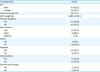
Values are expressed as number (%) or mean±standard deviation not otherwise specified.
ECMO, extracorporeal membrane oxygenation; HFOV, high-frequency oscillatory ventilation; iNO, inhaled nitric oxide.
![]()
In 6 cases, ECMO was adopted for treatment, while HFOV was used to treat 49 patients (76.6%), and NO was inhaled in 42 patients (65.6%).
2. Postoperative outcomes
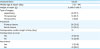
Postoperative outcomes are summarized in Table 2. Patients generally underwent surgery around the age of six days (6.2±12.4 days) and the average weight at repair was 2,996.0 g (2,996.0±562.6 g). Thirty-three patients (71.7%) were repaired by open laparotomy, 3 (6.6%) were repaired via open thoracotomy, and minimally invasive thoracoscopic repair was done in 10 cases (21.7%). Thirty-six patients (78.2%) underwent primary closure, while 10 patients (21.8%) required patch closure. The average postoperative length of stay was about 26 days (26.4±33.1 days). The postoperative survival rate was 67.4% (31/46). There were 5 cases of recurrence (10.9%, 5/46). Three of these were initially treated with the thoracoscopic approach while two were initially treated with laparotomy. The onset of recurrence varied from 9 to 424 days. There was only 1 patient who recurred within 1 month. Two patients recurred more than 1 year after the first surgery. For 2 patients, patch closure was needed to repair the defect.
Table 2
Postoperative outcomes (n=46)
![]()
3. Outcome CDH patients by era
All of the patients were divided into 3 groups by era. It can be seen that recent outcomes (2009–2014) have improved compared to earlier outcomes. From 1995 to 2001, the survival rate of CDH patients was 42% (8/19). From 2002 to 2008, the survival rate was 29% (6/21). For the most recent 6 years of data, 2009 to 2014, the survival rate was improved to 71% (17/24). According to logistic regression model, the recent era was a statistically significant risk factor (odds ratio [OR], 0.07; 95% confidence interval [CI], 0.01–0.52; p=0.009).
4. Risk factors for overall mortality
Table 3 represents risk factors for overall mortality. There were 2 statistically significant risk factors for overall mortality: On univariate analysis, right-sided lesion, the use of HFOV, the use of iNO, and ipsilateral pneumothorax were found to be significant risk factors. Of these risk factors, right-sided lesion (OR, 1.12–23.04) and ipsilateral pneumothorax (OR, 1.31–119.60) were both statistically significant risk factors according to multivariate analysis. The risk factors for postoperative mortality were also investigated. On univariate analysis, male sex, right-sided lesion, the use of HFOV or iNO, closure with patch, and pneumothorax were risk factors for postoperative mortality. However, there were no statistically significant risk factors on multivariate analysis.
Table 3
Risk factors for overall mortality
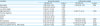
ECMO, extracorporeal membrane oxygenation; HFOV, high-frequency oscillatory ventilation; NO, nitric oxide.
![]()
DISCUSSION
Up until the mid-1990s, the main purpose of ventilator care for CDH patients was to induce respiratory alkalosis by hyperventilation. Because respiratory alkalosis helps lower pulmonary vascular resistance and pulmonary artery pressure, resulting in reverse ductal shunting [15]. Recent studies have demonstrated that ventilator associated lung injury due to hyperventilation could have harmful effects on survival and long term pulmonary function [45].
A permissive hypercapnia strategy has been advocated for the management of infants with PPHN for more than 25 years [16] and has been adopted for CDH patients since the late 1990s. Several case series have indicated that preductal oxygenation is the most important step to improve outcome with a permitting airway pressure limitation and hypercapnia [678910]. Through taking advantage of iNO, pulmonary vascular resistance and pulmonary arterial pressure can both be selectively lowered. Therefore, the hyperventilation strategy could be substituted with the gentle ventilation strategy, which is the recent mainstay of ventilator care in infants with CDH.
This study clarified the association between overall mortality and the occurrence of pneumothorax. We assume that the use of gentle ventilation strategies helped prevent the occurrence of pneumothorax and therefore may have contributed to improved survival. According to several case series of CDH, gentle ventilation strategies which limit airway pressure and permit hypercapnia with enhancing preductal saturation are the most important factors for improved survival [678910]. However, some studies have indicated that underlying disease status representing the degree of hypoplasia itself is the main cause of pneumothorax occurrence [17]. These results suggest that most of occurrences of pneumothorax are inevitable, regardless of respiratory management.
Other significant risk factor was right-sided lesion. Right-sided CDH has been reported as an important risk factor in several studies [1819] and the relationship also reproduced in this study. Some studies reported that right-sided CDH was difficult to diagnose prenatally than left-sided CDH [2021]. Also, right-sided CDH patients required more ECMO support than left CDH patients [1819]. This reflects more severe of pulmonary hypertension and pulmonary hypoplasia in right-sided CDH patients and could be a reasonable assumption for poorer outcomes. In this study, the number of right-sided CDH patients was only 20, which lacks statistical power to derive any significant results. In order to describe unique characteristics of right-sided CDH patients, larger studies should be conducted with unified treatment protocol.
In this study, the mortality rate of recent 6 years (2009–2014) was lower than that of the earlier 14 years (1995–2008). When analyzed according to era divided into three periods, the recent era showed better survival rate than that of the intermediate era (2002–2008; 71% vs. 29%, p=0.006) and demonstrated a tendency of lower mortality than that of the early era (1995–2008; 71% vs 42%, p=0.062). Compared to the recent 6 years, there were more patients who were not able to undergo surgery in the intermediate era (91.7% vs. 47.6%, p=0.002, analyzed by fisher's exact test). In the period of 1995 to 2001, more patients seemed not to be able to underwent surgery but there was only statistical insignificant tendency (91.7% vs. 73.7%, p=0.212, analyzed by fisher's exact test). The reason why a CDH patient cannot undergo surgery is preoperative instablity. We assume that disease severity itself contributed most to the patient's instability via pulmonary hypoplasia and/or pulmonary hypertension. Under discussions with pediatric surgeons and neonatal intensivists, gentle ventilation strategies were emphasized more and HFOV was adopted at an early stage to reduce the occurrence of pneumothorax. We suggest the possibility that these approaches could have positively affected outcomes of recent era.
As well-known risk factors, karyotype abnormality, cardiac anomaly and non-cardiac anomaly were also reviewed. However, karyotyping was only analyzed in 15 patients and there were only 2 cases of reported karyotype abnormality, which is insufficient number to derive statistical power. Forty-six patients were checked for cardiac anomaly and there were 11 patients with cardiac anomalies. Forty-seven patients were checked for non-cardiac anomaly and there were 19 patients with the non-cardiac anomaly. However, most of patients who were not examined were more prevalent within the mortality group, 13 of 18 patients for cardiac anomaly and 13 of 17 patients for non-cardiac anomaly, respectively. Therefore, statistical analysis was not performed for the relationship between the mortality and the cardiac/non-cardiac anomaly due to possible substantial bias.
The most important limitation of this study is that it is based on retrospective review. In addition, the number of cases was only 64, which is an insufficient number to induce statistical significance. As described above, the treatment strategies for CDH patients were not established well and were decided by clinical decisions. Because this study has been conducted for a long period, the rationale for clinical decision have been changed in some degree. These minor changes in clinical decision could have produced heterogeneity in this study over periods and may have affected statistical analysis.
In the future, a more elaborate prospective study with more patients is needed to reveal any causal relationship between the occurrence of pneumothorax and overall mortality and to examine the risk factors which lack statistical power in this study.
In conclusion, the survival rate of neonates with CDH at our center has improved over the last 2 decades. We assume that the main cause of this improvement is the adoption of the gentle ventilation strategy with permissive hypercapnia and permissive hypoxia. The risk factors for mortality include the occurrence of pneumothorax and right-sided lesion. Therefore, it is vital to prevent the occurrence of pneumothorax in treating CDH patients.
 XML Download