

 PDF
PDF ePub
ePub Citation
 Print
Print


Abstract
Calcinosis cutis is an uncommon disorder and defined as the deposit of insoluble calcium salts in the skin. It is classified as dystrophic, metastatic, idiopathic, and iatrogenic according to etiology. The dystrophic type is the most common and occurs in the previously damaged tissue, including connective tissue diseases, panniculitis, inherited disorders, benign and malignant tumors, a variety of scarring caused by burn, radiation, trauma, surgery, and keloid.
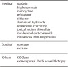
Treatment options are limited. Whereas medical therapy is usually not very effective, surgical intervention has shown to be beneficial and is indicated when painful masses, recurrent infection, ulcerations, functional impairment, and cosmetic concerns exist. Herein, we report two cases of dystrophic calcinosis cutis associated with longstanding, painful, ulcerated cutaneous lesions in the extremities successfully treated with surgical excision.
Figures and Tables
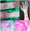 | Fig. 1Case 1(prior treatment). (A) An erythematous tender subcutaneous nodule in the forearm (B) A large plaque with focal ulcerations in the shin (C) Radio-opaque lesion(arrow) was observed on x-ray (D) Amorphous deposits of basophilic material in the dermis (arm, H&E, ×40) (E) Calcification and metaplastic ossification in severely degenerated tissue (shin, H&E, ×100)
|
References
1. Fairly JA. Cutaneous mineralization and ossification. In : Goldsmith LA, Katz SI, Gilchrest BA, Paller AS, Leffel DA, Wolff K, editors. Fitzpatrick's Dermatology in general medicine. 8th ed. NewYork: McGraw-Hill;2013. p. 1649.
2. Reiter N, El-Sabawi L, Leinweber B, Berghold A, Aberer E. Calcinosis cutis: part I. Diagnostic pathway. J Am Acad Dermatol. 2011; 65:1–12.
3. Reiter N, El-Sabawi L, Leinweber B, Berghold A, Aberer E. Calcinosis cutis: part II. Treatment options. J Am Acad Dermatol. 2011; 65:15–22.
4. Lee DJ, Rea TH, Modlin RL. Leprosy. In : Goldsmith LA, Katz SI, Gilchrest BA, Paller AS, Leffel DA, Wolff K, editors. Fitzpatrick's Dermatology in general medicine. 8th ed. NewYork: McGraw-Hill;2013. p. 2253.
5. Choi MW, Han TY, Lee JH, Son SJ. A case of calcinosis cutis showing a good response to topical sodium metabisulfite. Korean J Dermatol. 2018; 56:504–505.
6. Wu JJ, Metz BJ. Calcinosis cutis of juvenile dermatomyositis with incision and drainage. Dermatol Surg. 2008; 34:575–577.

7. Park YM, Lee SJ, Kang H, Cho SH. Large subcutaneous calcification in systemic lupus erythematosus : treatment with oral aluminum hydroxide administration followed by surgical excision. J Korean Med Sci. 1999; 14:589–592.
8. Valdatta L, Buoro M, Thioe A, Mortarino C, Tuinder S, Fidanza C, et al. Idiopathic circumscripta calcinosis cutis of the knee. Dermatol Surg. 2003; 29:1222–1224.
9. Lee SY, Kim DY, Cho BH, Lee SW. Dystrophic calcinosis cutis in a patient with squamous cell carcinoma secondary to postburn scar. Ann Plast Surg. 2001; 46:457–458.
10. Vitale A, Delcia G, Torre FL, Calcagno G, d'Alcontres FS. Massive gluteal calcinosis in a 10-year-old girl with juvenile dermatomyositis: successful surgical management. Plast Reconstr Surg. 2009; 124:456e–458e.
11. Bangert S, Hebert A. Surgical treatment of calcinosis cutis in juvenile dermatomyositis. J Am Acad Dermatol. 2007; 56:suppl 2. AB161.
12. Aksoy HM, Ozdemir R, Karaaslan O, Tiftikcioglu YO, Oruc M, Kocer U. Incidental idiopathic calcinosis cutis in a rhytidectomy patient. Dermatol Surg. 2004; 30:1145–1147.
 XML Download