

 PDF
PDF ePub
ePub Citation
 Print
Print


INTRODUCTION
Remote lung damage is of main significance after ischemiareperfusion (IR) period of intestinal and carry major importance [12]. Mediators and metabolites distributed from injured muscles (e.g., interleukin [IL]-1β, IL-6, IL-8, tumor necrosis factor [TNF]-α, and elastase-α1-antitrypsins) enhance the quantity of polymorphonuclear leukocytes in circulation [3]. These circulating cells in the lung tissue cause microvascular changes mediated by the neutrophils. Also, during reperfusion, skeletal muscle free radical formation leads to additional lung damage [123]. Raised plasma reactive oxygen species concentrations might induce oxidative damage in the capillary membranes of the lung by the peroxidation of lipids. Simultaneously, a defensive tissue response is provoked by inflammation, augmenting heat shock proteins expression. As a result, the IR leads to pulmonary edema by pulmonary hypertension, vasoconstriction, and increase in permeability of the alveolar membrane [45].
The advantageous results of hyperbaric oxygen (HBO) might be attributed to its capability to decrease activation of neutrophils and IR injury sequestration. In experimental studies on animals, HBO has been shown to reduce ischemic edema, increase aerobic metabolism to maintain energy-rich complexes, decrease vein neutrophil adhesion and restrain ongoing neighboring arteriolar ischemic muscle vasoconstriction [678]. HBO diminishes rat lung neutrophil sequestration with IR injury and suppress inflammatory processes [6789]. Iloprost (IL) is a prostacyclin analog and has been used in animal studies for its neuroprotective effect. IL mimics prostacyclin by inhibiting aggregation of platelets, cytoprotection, and vasodilatation [10]. IL pretreatment reduces injury of the lung following ischemia [56].
The present study aimed to evaluate the effects of combination therapy of HBO and IL on lung damage after tissue or organ IR, on serum TNF-α, myeloperoxidase (MPO), malondialdehyde (MDA) and soluble inter-cellular adhesion molecule-1 (sICAM-1) levels, and on IR induced neutrophil sequestration.
METHODS
Study design
Animal care complied with the Principles of Laboratory Animal Care as formulated by the National Society for Medical Research and the Guide for the Care and Use of Laboratory Animals (National Institutes for Health publication No. 5377-3, 1996). The experimental study was approved by the Animal Research Ethics Committee of Recep Tayyip Erdogan University Medical Faculty (Institution Review Board: 2015/45). All animals were given 5 days of adaptation to their environment prior to experiments. Room temperature was kept between 26℃ to 28℃.
Forty New Zealand white male rabbits, weighing 2.3–3.2 kg (mean, 2.8 kg), were randomly allocated into 1 of 5 study groups each containing eight animals. The HBO group received a single session of HBO treatment, the IL group received 25 ng/kg/min infusion of IL; the HBO + IL group received both HBO and IL; the control (IR) group received only 0.9% saline; and the fifth group was the sham group. HBO treatment and IL infusion were administrated during 60 minutes of ischemia and 60 minutes of reperfusion in the treatment groups.
Anesthesia and surgical technique
Initial anesthesia was achieved with intramuscular ketamine (50 mg/kg) and xylazine (5 mg/kg) without endotracheal intubation; then followed by 25 mg/kg fractionally in order to allow the animals to have spontaneous respiration. Body temperature was maintained close to 38℃ using a thermostatically controlled heated operation table. All animals received a similar volume of maintenance fluids (0.9% sodium chloride, 20 mL/hr) for the whole procedure. An arterial catheter (20 gauge) was placed in an ear artery to monitor blood pressure (Petas KMA 800, Ankara, Turkey) and arterial blood was sampled for gas analysis. Blood pressure was measured after the induction of anesthesia (preischemia), and 2 hours following the release of aortic cross clamping (postischemia) via the ear artery catheter. Arterial blood samplings measurements matched within the same time periods.
Animals were placed in the supine position. After sterile preparation, a 6- to 8-cm midline incision was made, and the superior mesenteric artery (SMA) and inferior mesenteric artery (IMA) was exposed through a transperitoneal approach with the abdominal contents reflected to the right. SMA and IMA were isolated from the left renal artery down to the aortic bifurcation. Each animal was given 150-U/kg heparin intravenously 5 minutes prior to SMA and IMA occlusion. Heparin was not reversed at the end of the procedure. In each rabbit, the SMA was isolated using atraumatic arterial bulldog clamps (Vascu-statt, Scanlan International, St. Paul, MN, USA). The ischemic phase was maintained with a complete occlusion of the SMA using atraumatic arterial bulldog clamps for 60 minutes, thereby interrupting the mesenteric blood flow. In all animals, except the sham controls (n = 8), the SMA was crossclamped just proximal to the aortic bifurcation using atraumatic arterial bulldog clamps (Vascu-statt, Scanlan International, St. Paul, MN, USA) for 60 minutes. The IMA was also clamped at its origin from the aorta, and loss of aortic pulse was confirmed by palpation.
The clamp was removed after 60 minutes and restoration of blood flow was verified visually. Six hours after the intervention, all animals were humanely killed by a lethal cardiac injection of pentobarbital (100 mg/kg), immediately after which a median sternotomy was used to extract lung tissues for histopathological examination by light microscopy and biochemical investigations by MDA and MPO assay. The right lungs were removed, fixed with 10% tamponated formalin and stored for 24 hours. A sagittal section was obtained at the level of the hilum. These tissues were fixed again with 10% formalin for 2 days.
HBO treatment procedure
Animals received HBO therapy in an animal monoplace chamber during periods of 60 minutes of ischemia and 60 minutes of reperfusion at 2.5 times atmospheric pressure. Before pressurization, 100% medical oxygen was flushed through the chamber for 10 minutes to displace ambient air. The oxygen pressure was then increased slowly and reached 2.5 ATA in 5 minutes.
Serum and tissue biochemical evaluation
Blood pH, pO2 (mmHg), pCO2 (mmHg), bicarbonate (HCO3) (mmol/L) and lactate dehydrogenase (LDH) (IU/L) values were determined 1 hour after the onset of ischemia and 1 hour after the onset of reperfusion. Blood pH, pO2, pCO2, HCO3, Na+ and K+ values were determined using a blood gas analyzer (Ciba Corning Blood Gas Analyzer Model 860, Ciba Corning Diagnostics Corp., Irvine, CA, USA). Plasma LDH levels were measured in lithium heparinized plasma using automated enzyme reactions (automated analysis for Hitachi System 717, Boehringer Mannheim Diagnostica, Mannheim, Germany).
TNF-α, sICAM-1, myeloperoxidase, and MDA assay
TNF-α (pg/mL) values were checked before ischemia, in the first hour of ischemia, and in the first and fourth hours of reperfusion; at the end of reperfusion period plasma and tissue MPA values, MDA values, and sICAM-1 levels were checked. TNF-α assays were performed according to the method described by Pizarro et al. [11] using the quantitative sandwich enzyme immunoassay technique (Quantikine M, R&D Systems, Minneapolis, MN, USA). sICAM-1 levels were measured by single stage photometric enzyme immunoassay method. Lipid peroxidation in the lung tissue was evaluated by measuring the level of MDA, which is the end product of lipid peroxidation. MDA was measured in plasma and the lung as an indicator of free radicals. Plasma and tissue (lung) MDA levels were determined with spectrophotometry using thiobarbituric acid-reactive substances [12]. MPO, an enzyme found primarily within neutrophils, is a sensitive marker for quantifying neutrophil content in tissue. MPO activity, a sensitive marker of PML infiltration in tissues, was calculated by hydrogen peroxide dependent oxidation of tetramethylbenzidine, a reaction catalyzed by MPO [13].
Histopathologic examination and lung neutrophil sequestration
After standard tissue preparation, 5-µm tissue sections were obtained. The specimens were stained with hematoxylin-eosin and examined with light microscopy (Eclipse E200; Nikon, Tokyo, Japan). All histopathological changes were detailed in each lung tissue, including inflammatory cell infiltration, alveolar edema, congestion, and preservation of the alveolar septum. At least 2 different sections were explored in each specimen. The same pathologist, who was blinded to the study, assigned a score of 0 to 4 on the basis of congestion, interstitial edema, polymorphonuclear leukocyte infiltration, and alveolar hemorrhage as follows: 0, normal histological appearance; 1, mild and focal changes; 2, moderate and multifocal changes; 3, marked and multi-focal changes; 4, marked and diffuse changes. Additionally, to determine the severity of inflammation that resulted from IR process, leukocyte infiltration was scored semi quantitatively according to the following scale: 0, no leukocyte infiltration; 1, <10 leukocytes; 2, 10–45 leukocytes; 3, >45 leukocytes (magnification <400) [14].
Statistical analyses
Data were analyzed using the SPSS ver. 10.0 (SPSS Inc., Chicago, IL, USA). All differences associated with a chance probability of 0.05 or less were considered statistically significant. All data are presented as mean ± standard deviation. Statistical analysis was performed using analysis of variance (ANOVA) test. One-way ANOVA test was followed by post hoc Dunnett test.
RESULTS
Physiological and hemodynamic parameters
There were not any statistically significant differences among groups for physiological parameters in the pre- and postischemic periods. Table 1 presents pH, pO2, pCO2, HCO3 levels. Blood pH, pO2, and HCO3 were significantly lower in control group compared to sham and treatment groups. Blood pO2 concentration was significantly elevated in both HBO and HBO+IL groups. There were not any significant differences in other parameters among HBO, IL, and HBO + IL groups.
TNF-α, sICAM-1, LDH, MPO, and MDA findings
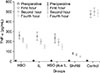
With regards to base (preoperative) TNF-α levels, no significant differences were detected among the 5 groups (P = 0.818). At the end of 1, 2, and 4 hours, all three treatment groups showed a significant reduction in TNF-α increase when compared to the control group (P = 0.001). However, when the 3 treatment groups were compared to each other, the changes in TNF-α levels at the end of 1, 2, and 4 hours was not statistically significant (P = 0.398). Increase in TNF-α levels in every single group under medication is evaluated via Friedman test. In all three groups, a meaningful reduction in TNF-α levels of test subjects was observed (P < 0.001 for all three groups) (Fig. 1). Plasma and lung MDA, MPO with plasma LDH, and sICAM-1 levels were significantly lower in the control group than those of HBO, IL, HBO + IL, and sham groups. Administration of IL and/or HBO inhibited MPO and MDA increase in plasma (P < 0.005) and in lung tissue (P < 0.005) (Table 2).
Light microscopic findings
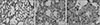
Histopathological results are shown in Table 3. Diffuse severe ischemic lung injury (P < 0.001) the histopathological score (P < 0.001) were significantly lower in the HBO, IL, HBO+IL, and sham groups when compared with the control group. Histopathological examination neutrophil sequestration was mostly detected in control group; while leukocyte infiltration in both sham and therapy groups was significantly lower than the control group (Fig. 2).
DISCUSSION
As the lungs and the liver have a considerable amount of macrophages, and are the primary organs in the recirculation pathway of translocated endotoxin, they are accepted as chief sources of TNF-α. TNF-α is believed to be a main mediator included in controlling the flow of the inflammatory procedures that conduct IR mediated injury of the lung [15161718]. Okusawa et al. [17] reported that intravenous injection of TNF-α triggers comparable lung damage to IR induced lung damage. Colletti et al. [18] emphasized that anti-TNF-α antibodies have a protective effect against IR induced lung damage. Additionally, it has been reported that an increase in TNF-α levels was observed in cases of IR damage, and a rapid upregulation of adhesion receptors increasing the adhesion capabilities of TNF-α activated neutrophil and endothelial cells and an increase in respiratory macrophage degranulation resulting in oxidative tissue damage [19]. Yang et al. [20] held high TNF-α levels from intestinal IR induced lung damage responsible. In the present study, serum inspections after IR with lung damage revealed an enhance in MPO, sICAM-1, and TNF-α levels. Our findings support our thesis that TNF-α might be in charge for lung damage after aortic IR.
The decrease of neutrophil activation might lessen the occurrence of IR induced lung damage. This theory has been favored by research that has shown that the depletion of neutrophils improves IR induced injury of the lung. The advantageous outcomes of HBO might be attributable to its capability to decrease neutrophil activation and sequestration in IR injury. HBO might accomplish its favorable effects by conserving energy-rich compounds, restraining hypoxicischemic-induced lipid peroxidation, decreasing leukocyte adherence, reducing sequestration in reperfused tissue, and adjusting the inflammatory process [721]. In an animal study, HBO was shown to decrease arterial neutrophil concentration, but did not change lung neutrophil sequestration [22]. Tjarnstrom et al. [9] revealed a significant reduction in lung neutrophil sequestration in an intestinal IR rat animal model. The enrollment of lung neutrophils began throughout the interval of ischemia and endured for the period of reperfusion for a delayed period of time [23].
Intestinal IR persuades a general activation of neutrophils [22]. This is revealed by reports showing that HBO decreases neutrophil activation and releases intercellular adhesion molecule-1 [2425]. Other mediators, i.e. decreased TNF-α, might also be blamable. Kamada [26] revealed that rheumatoid arthritis patients treated with HBO accomplished a significant healing, proposing that HBO has an immunosuppressive effect. This is strengthened by animal studies in which HBO inhibits the production of IL-1 [27]. One current study has revealed that HBO reduces the production of TNF-α, IL-6, and after immense hemorrhage [28]. Yang et al. [20] also reported that HBO hinders TNF-α production through IR damage.
IL acts as a membrane stabilizer and reduces neutrophils that are probable mediators of IR injury [16]. IL additionally reduces aggregation of white blood cells and vascular endothelium adhesion, superoxyde production from stimulated human neutrophils, and formation of free radicals in myocardium subjected to IR injury [16]. Since reduced microcirculation owing to the impairment of vascular endothelium of the lung documents a foremost pathologic characteristic, the prostanoids might be more efficient by altered properties such as leukocyte aggregation, platelet inhibition free radicals scavenging, and pulmonary vasodilatation [716]. In the present study, on test subjects treated with both HBO and IL not only a reduction in TNF-α levels but also a reduction in MPO and sICAM-1 levels is detected.
Rossman et al. [29] showed that mucosal and lung injury is reduced by perfusing the intestinal lumen with oxygenated perfluorocarbon during ischemia. Ueno et al. [30] revealed that HBO treatment following hepatectomy reduced activation of the neutrophils and enhanced the surgical outcomes. HBO should be given as quickly as possible throughout ischemia and reperfusion to gain a promising effect. Real time patient samples opting for therapeutic cardiovascular and cardiothoracic surgical modalities are also imperative to validate specific outcomes that will reveal critical insights as to a specific treatment regimen. However, results may not always hold good in difficult clinical scenarios where patients present with comorbidities such as smoking-induced lung collapse, chronic obstructive pulmonary disorders, type II diabetes mellitus, congenital abnormalities, and most importantly, non-small cell lung cancer. Therefore, it is important to design specific algorithms that will pave the way for the implication of effective and successful therapeutic strategies. In the present study, both HBO and IL were applied from the start of ischemia and positive effects were observed.
In conclusion, the present study has shown that both HBO and IL reduce the lung damage after aortic IR with regards to histopathological and biochemical aspects. This damage reduction effect of HBO and IL therapies was a result of reduced TNF-α production and reduced pulmonary neutrophil sequestration. Molecular cross-talks and interactions between different inflammatory cytokines will play a decisive role into disease pathogenesis and, therefore, it is extremely important to harness the effects of the same. Due to the experimental nature of the study, it is difficult to obtain conclusive long-term clinical implications. However, it is reasonable to speculate that through successful clinical trials in the future, it might be possible to ascertain therapeutic effects of HBO and IL for lung damage after aortic IR.


 XML Download