Journal List > World J Mens Health > v.37(1) > 1109671

Choi, Jeon, Kwon, Zhu, Lee, Choi, Jeong, Kim, Bae, Bae, Kim, Cho, Ha, Hong, Hwang, and Kim: Effect of Korean Herbal Formula (Modified Ojayeonjonghwan) on Androgen Receptor Expression in an Aging Rat Model of Late Onset Hypogonadism

Abstract

Purpose

Testosterone replacement therapy is an effective treatment for late-onset hypogonadism (LOH) despite a few contraindications and side-effects. The aim of this study was to determine whether modified Ojayeonjonghwan (KH-204, Korean herbal formula) improved LOH. KH-204 is a strong antioxidant herbal formula. We evaluated the effect of Korean herbal prescription on androgen receptor (AR) expression in an aged rat model of LOH.

Materials and Methods

Eighteen-month-old rats were used as aged LOH rat models. Eighteen Sprague-Dawley rats were randomly divided into three equal groups of six animals each and treated with one of the following: 1) normal control group (oral administration with distilled water, n=6), 2) KH-204 200 group (oral administration with 200 mg/kg of KH-204, n=6), and 3) KH-204 400 group (oral administration with 400 mg/kg of KH-204, n=6). After four weeks of treatment (once daily, distilled water or KH-204), serum testosterone levels, changes in testicular and epididymal weight, Western blotting analysis of AR expression and measurement of oxidative stress were examined.

Results

Treatment with the herbal formulation KH-204 200 mg/kg and 400 mg/kg (1) increased the weights of testis and epididymis; (2) increased the level of serum testosterone; (3) increased the level of superoxide dismutase and reduced the level of 8-hydroxy-20-deoxyguanosine; and (4) upregulated AR expression in testicular tissue.

Conclusions

KH-204 might be an effective alternative for LOH. It improves antioxidant mechanisms and increases testicular AR expression without side-effects.

INTRODUCTION

Late-onset hypogonadism (LOH) is a clinical and biochemical syndrome associated with aging and is characterized by decreased serum testosterone (T) [1]. In middle-aged or elderly males, low T may affect the function of multiple organ systems and result in physiological changes, such as cardiovascular risk, metabolic conditions, sexual dysfunction, and various other health issues [12345]. Sexual dysfunction including loss of libido, erectile dysfunction (ED) and reduced fertility, and diminished morning erection are the most sensitive and specific symptoms of LOH [15]. Other relevant symptoms include depression, irritability, insomnia, fatigue, sarcopenia, increased fat mass, low bone mass, and loss of vigor. To alleviate the symptoms of LOH and improve the quality of life, testosterone replacement therapy (TRT) is the gold standard when fertility and spermatogenesis are not required [1236]. Although TRT can be an effective treatment for LOH, it is known to have absolute or relative contraindications, such as breast cancer, prostate cancer, polycythemia, erythrocytosis, severe obstructive sleep apnea, or any life-threatening medical condition. In addition, the role of TRT in of the risk for prostate cancer, benign prostatic hypertrophy and cardiovascular disease is disputed [13]. The long-term effect of TRT is still uncertain.
To avoid these adverse effects of TRT and effectively treat LOH, traditional herbal medicines have been investigated as an alternative treatment [78]. Among the traditional herbal medicines, Ojayeonjonghwan (OJ) has been popularly used in a traditional Korean herbal prescription to treat patients with male sexual dysfunction including ED and infertility [910]. KH-204 is a modified OJ with Cornus officinalis Sieb. Et Zucc. (CO) replacing Plantago asiatica Linne [911]. CO has been used as a traditional medicine in various countries of Europe and Asia for treatment of a wide range of diseases, such as diabetes, urinary tract infection, sexual dysfunction, kidney and liver diseases, and so on [12]. Our previous studies demonstrated that KH-204 might ameliorate ED [1113] and infertility [1415]. In addition, we studied whether KH-204 had protective effects on the bladder of androgen-deprived rats [16]. On the basis of previous studies [1113141516], we hypothesized that KH-204 was a safe and effective alternative for treating LOH. However, the mechanisms underlying the effects of KH-204 on LOH are uncertain. We hypothesized that KH-204 may have positive effects on T production and androgen receptor (AR) expression in an aged rat model of LOH.

MATERIALS AND METHODS

1. Preparation of herbal formula (KH-204)

The main ingredients of KH-204 were obtained from the following five plants: 1) Cornus officinalis Sieb. et Zucc. (Cornaceae, CO; 32%), 2) Lycium chinense Miller (Solanaceae, LC; 32%), 3) Rubus coreanus Miquel (Rosaceae, RC; 16%), 4) Cuscuta chinensis Lam (Convolvulaceae, CC; 16%), and 5) Schisandra chinensis Baillon (Schisandraceae, SC; 4%). The product was developed and manufactured as a health supplement by the KEMIMEDI Co. Ltd (Seoul, Korea), a venture company developing oriental herbal medicines. Each herb (20 kg) was extracted in 200 L of distilled 30% ethanol and refluxed at 96℃ to 100℃ for 3 hours. The extract was filtered, and the water from the filtrate was removed using a rotary evaporator (R-210; BUCHI, Flawil, Switzerland) and a vacuum dryer (OV-11/12; Jeio Tech, Daejeon, Korea). All extracts were refrigerated. Marker compounds for each plant (CO, LC, RC, CC, and SC) include loganin, betaine, ellagic acid, hyperoside, and schisandrin, respectively. Marker compounds were confirmed by high performance liquid chromatography (HPLC). Cytotoxicity data of each herb and the HPLC chromatogram of each marker compound were described previously [1315].

2. Ethics statement

This study protocol was approved by the Institutional Animal Care and Use Committee in the School of Medicine, The Catholic University of Korea (approval number: CUMC-2015-0005-03).

3. Animals

Male Sprague-Dawley rats aged 18 months were obtained from Samtaco Bio Co. (Osan, Korea). The experiments were performed in accordance with the Standard Guidelines for the Care and Use of Laboratory Animals of the National Institutes of Health. The animals underwent acclimation for 1 week. They were raised in a controlled environment (temperature: 20.0℃±2.0℃, humidity: 65%±5%; 12 hours light 12 hours dark cycle) with ad libitum access to food and water. Eighteen-month-old aged rats were considered as aged LOH rat models [17]. Eighteen male Sprague-Dawley rats were randomly divided into three equal groups of six animals each and treated with one of the following: 1) Normal control group (oral administration with distilled water, n=6); 2) KH-204 200 group (treated with 200 mg/kg/d of KH-204, n=6), and 3) KH-204 400 group (treated with 400 mg/kg/d of KH-204, n=6). KH-204 was dissolved in distilled water and administered orally through an 8 F red Rob-Nel catheter once daily. After 28 days of once-daily oral administration of distilled water or KH-204, rats from all the groups were sacrificed under diethyl ether anesthesia and their testes and epididymides were excised and weighed.

4. Serum testosterone level analysis

Before the male rats were sacrificed, serum was collected from the inferior vena cava. Serum T levels were measured using an enzyme-linked immunosorbent assay (ELISA; Molecular Devices, San Jose, CA, USA) T detection kit (BioVendor-Laboratory Medicine Inc., Brno, Czech Republic) in accordance with the kit instructions [18].

5. Measurement of oxidative stress

Oxidative stress in testicular tissues was analyzed quantitatively by measuring the level of 8-hydroxy-2-deoxyguanosine (8-OHdG) and superoxide dismutase (SOD) activity. Using the DNeasy Blood and Tissue kit (Qiagen, Valencia, CA, USA), the total DNA was extracted from the testes according to the manufacturer's protocol. The 8-OHdG content in each sample was determined with a DNA oxidation kit (Highly Sensitive 8-OHdG Check ELISA kit; Japan Institute for the Control of Aging, Fukuroi, Japan) according to the manufacturer's instructions. After the final color was developed with the addition of 3,3′,5,5′-tetramethylbenzidine, absorbance was measured at 450 nm. Tissue sample concentration was measured using a standard curve and corrected for DNA concentration. The SOD activity (CuZnSOD and Mn SOD) in tissues was determined using a SOD Assay Kit-WST (Dojindo Laboratories, Kumamoto, Japan) and the decrease in the rate of superoxide-mediated reduction of nitroblue tetrazolium was monitored at 450 nm using a spectrophotometer [14].

6. Western blot analysis

Western blot analyses were used to determine the levels of AR protein in testis and prostate tissues. Tissues frozen in liquid nitrogen were homogenized in lysis buffer (50 mM Tris-HCl, 120 mM NaCl, 2 mM EDTA, 1 mM EGTA, and 1% Triton X-100). The protein concentration was determined using the bicinchoninic acid protein assay (Pierce Chemical Co., Dallas, TX, USA). Forty micrograms of total cellular protein was separated by 12% sodium dodecyl sulfate-polyacrylamide gel electrophoresis and electrophoretically transferred to nitrocellulose membranes. After blocking for 1 hour at room temperature with 5% skim milk in 0.1% tris-buffered saline with Tween 20, the membrane was incubated with β-actin antibody (1:1,000 dilution; Assay Designs, Ann Arbor, MI, USA) and anti-AR antibody (AR 1:1,000 dilution; Santa Cruz Biotechnology, Santa Cruz, CA, USA) overnight at 4℃. The β-actin was used as an internal control. Detection of bound antibody on each blot was evaluated with horseradish peroxidase-conjugated secondary antibody (goat anti-rabbit immunoglobulin G) visualized by hybond enhanced chemiluminescence (Amersham, Arlington Height, IL, USA). The densitometric analysis of band intensity was based on the Luminescent Image Analysis System (LAS-3000; Fujifilm, Tokyo, Japan) and analyzed using Multi Gauge 3.0 software (Fujifilm).

7. Statistical analysis

Statistical analyses were performed using IBM SPSS statistics for Windows ver. 20 (IBM Co., Armonk, NY, USA). The data are expressed as mean±standard deviation. One-way analysis of variance test with post hoc Dunnett test was used to analyze the statistical difference among the groups. A p-value < 0.05 was considered to indicate statistical significance.

RESULTS

1. Weights of testis, epididymis and prostate

No statistically significant difference in the mean weights of testis, epididymis and prostate was observed among the three groups after 4 weeks (Table 1). A trend toward higher testicular and epididymal weights was observed in KH-204 200 and 400 groups compared with those in the normal control group. Prostate weights were lower in the KH-204 treatment groups than in the control group (Table 1).

2. Serum testosterone levels

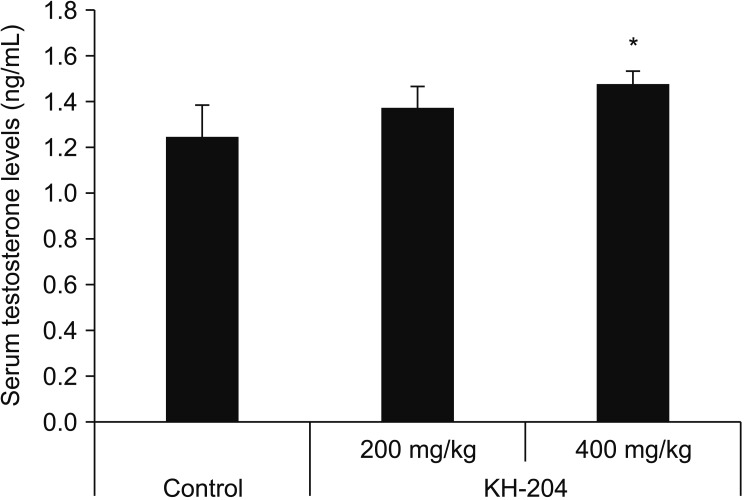
After treatment with KH-204, the serum T levels in the control and KH-204 200 and 400 groups were 1.25±0.14 ng/mL, 1.37±0.10 ng/mL, and 1.48±0.05 ng/mL, respectively, exhibiting a dose-dependent increase. The serum T level in the KH-204 400 group was significantly increased in comparison with the control group (p<0.05, Fig. 1).

3. Oxidative stress

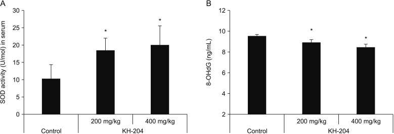
As shown in Fig. 2A, the mean levels of SOD expression were 10.31±4.00 U/mol in the control, 18.55±3.59 U/mol in the KH-204 200 group, and 20.07±5.57 U/mol in the KH-240 400 group. Testicular SOD activities were significantly increased in the two treated groups compared with the controls (p<0.005 each). The mean 8-OHdG level was 9.51±0.18 ng/mL in the control group, 8.92±0.25 ng/mL in the KH-204 200 group, and 8.47±0.26 ng/mL in the KH-204 400 group. The mean 8-OHdG expression was significantly lower in KH-204-treated rats compared with normal control rats (p<0.001, in each group, Fig. 2B). KH-204 treatment resulted in a dose-dependent reduction of oxidative stress markers in the testicular tissue.

4. Western blot analysis for tissue expression of androgen receptor

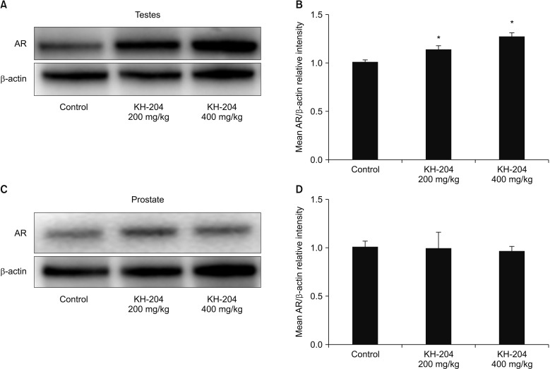
Expression of AR was measured in tissues of testis and prostate. A significant increase (p<0.005 and p<0.001) in normalized AR expression (mean AR/β-actin relative intensity: 1.13±0.04 and 1.26±0.04) was detected in the testes of rats exposed to KH-204 200 mg/kg/d and 400 mg/kg/d compared with the control rats (mean AR/β-actin relative intensity: 1.01±0.01) (Fig. 3A and 3B). Treatment with KH-204 showed a dose-dependent increase in AR expression of the testicular tissue. In the prostate, the mean AR/β-actin relative intensity was 1.01±0.06 in the control group, 0.99±0.16 in the KH-204 200 mg/kg/d group and 0.97±0.04 in the KH-204 400 mg/kg/d group. The prostates of the KH-204-treated rats were not significantly different from control rats (Fig. 3C and 3D).

DISCUSSION

LOH has been considered one of the most common endocrine disorders resulting in sexual dysfunction or infertility and is the most common form of male hypogonadism [19]. LOH in middle-aged and elderly men is a result of age-related changes in hypothalamic, pituitary and testicular factors and a slow decline in T production. Although the mechanisms by which serum T levels decline with advancing age are not well understood, previous studies [202122] and our data suggest altered redox balance in the Leydig cells (LCs) [23]. However, the pathophysiologic process of LOH development is complex and associated with many conditions, including metabolic disorders, chronic illness and aging. These conditions may be a cause of or result from LOH.
Aging is progressive and is accompanied by irreversible changes in physiological and metabolic processes ranging from the level of molecules to the organs and systems of life. Although the effects of aging on male reproductive system remain poorly defined, age-related alterations lead to progressive changes in hormonal production, spermatogenesis, and testes. One of the most well accepted aging theories is the free radical theory. Excessive oxygen free radical generation leading to lipid peroxidation, oxidative stress and damage to various intracellular molecules (lipids, DNA, and proteins) is considered as the major contributor to aging [24]. Imbalance in the production and elimination of reactive oxygen species (ROS) is characteristic of cellular aging, including LCs. Cao et al [21] reported that the antioxidant defense molecules such as SOD, and glutathione peroxidase were reduced in aged LCs. In addition, Chen et al [20] demonstrated that aging LCs produced significantly greater levels of ROS than LCs derived from young rats, and ROS may be involved in the reduced steroidogenic capacity of aging LCs. These studies suggested that alterations in the redox environment of aging LCs may reduce T production [2122], and administration of antioxidants can attenuate oxidative damage by scavenging free radicals in testes [20].
The present study showed that KH-204 treatment of aging rats increased the level of SOD and decreased the level of 8-OHdG. In addition, our study showed that KH-204 increased the serum T and AR expression in the testes of aging rats. Although the role of AR and T in the regulation of oxidative status in the aging gonad has yet to be established, the influence of aging on the expression of AR in the male reproductive tissues is interesting. ARs are regulated by the androgen levels in a tissue and cell-selective manner, and decline with age, although the regulatory mechanisms are not completely understood [25]. In reproductive tissues, testicular AR was expressed in the nucleus of Sertoli cells, LCs, and peritubular myoid cells, and prostatic AR was expressed in epithelial and stromal tissues [26]. While LCs exhibit the maximum AR expression at puberty, the number of ARs decrease, especially in genital tissues, with age [27]. Our results showed that KH-204-treated rats showed reduced prostatic weight, although the difference was not significant and was accompanied with limited changes in AR expression of the prostate tissue. Wu et al [28]. reported that their traditional medicine had anti-oxidative properties and attenuated overexpression of AR in a rat model of benign prostatic hyperplasia Yang et al [29] reported that herbal medications in the kidney yang-deficient model improved serum T levels in kidneys and testes. However, the underlying pharmacological mechanism was not established. These results from the testis are consistent with increased T levels and AR expression, generally, but the detailed relationships between aging, oxidative stress, and testicular functions remain to be to be elucidated.
Despite the efficacy of TRT in aging men with symptomatic LOH, the possibility of treatment-related adverse effects on cardiovascular disease, prostate cancer, and breast cancer remains uncertain. In addition, Tóthová et al [30] reported that exogenous T increases the oxidative stress and decreases anti-oxidative status in testes. In this respect, natural products or herbs (traditional herbal medicine) are considered a viable alternative treatment for LOH, without the adverse effects of TRT. Several studies have reported their possible mechanisms [7823]. However, the detailed mechanisms or pharmacological actions of these herbal medicines on LOH remain unclear.
Our previous studies suggested that KH-204 could be used as a safe alternative for ED and infertility. Sohn et al [11] reported KH-204 was effective in enhancing intracavernous pressure and nitric oxide-cyclic guanosine 3′,5′-monophosphate activity in penile tissues of spontaneously hypertensive male rats. Jang et al [13] demonstrated the antioxidant and hypolipidemic effects of KH-204 in effectively ameliorating ED by restoring endothelial function in a rat model of hypercholesterolaemia-induced ED. In addition, Kim et al [15] performed safety experiments with modified OJ, investigating the toxicity of five plant ingredients on the reproductive organs in male mice and suggested that oral administration of KH-204 was safe and positively influenced sperm parameters. In an experimental rat model of cryptorchidism, we previously reported that KH-204 increased the weight of the cryptochid testes, restored sperm quality and germinal cell layer thickness and decreased levels of 8-OHdG, heat shock protein 70 and apoptosis while increasing levels of SOD [14].
Based on these studies, we had established animal models of LOH to evaluate the efficacy of KH-204 on LOH. Our recent study showed that KH-204 treatment reduced oxidative stress and apoptosis in the androgen-deprived rat model of LOH. In that study, we suggested that KH-204 treatment degraded oxidative stress via nuclear factor erythroid 2-related factor 2/heme oxygenase-1 pathway and lowered the expression of transforming growth factor-beta 1/SMAD. This finding suggested that KH-204 may have anti-fibrotic functions in the testis. In addition, the present study showed that the KH-204 treatment in an aging rat model of LOH increased the levels of serum T and testicular AR expression and our data suggesting the antioxidant effect in testicular tissue are consistent with the results reported in the androgen-deprived LOH rat model [23].
Our study has a few limitations. First, while we attempted to identify the positive effects of herbal treatment on AR expression in the testicular tissue of aging male rats, we failed to evaluate the AR expression in different cell types of male reproductive tissues. Further experiments at the cellular and molecular level analyzing AR, estrogen receptor or glucocorticoid receptor expression and androgen actions are needed to elucidate the mechanisms of KH-204 in the LOH animal model. The expression of adrenocorticotropic hormone, luteinizing hormone, follicle stimulating hormone, corticosterone, and other androgens may explain the androgen activity axis. The interaction of activated glucocorticoid receptors triggering the activation of phosphatidylinositol-3 kinase/Akt pathway may explain the androgen activity of KH-204.
Second, we used only normal aged male rats as the control group to reproduce a more natural LOH animal model without considering other potential experimental groups, such as normal young male rats, androgen-deprived rats, or exogenous T treatment.
The small sample size did not allow us to perform various experiments for further investigation and analysis. Finally, our results may be different from other studies of LOH animal models showing genetic or species differences among different rat strains and humans [22]. Further long-term studies are required to verify the effects of KH-204 on the regulatory mechanisms of AR expression using experimental animal models of LOH.

CONCLUSIONS

In KH-204-treated aging rats, the testicular expression of AR was significantly increased. KH-204 treatment of aging rats may induce T synthesis via an antioxidant mechanism and increase the AR expression in testicular but not prostate tissue. KH-204 may represent a complementary and alternative non-hormonal health supplement to treat LOH without side-effects and without increasing the risks of prostatic disease.

ACKNOWLEDGEMENTS

This research was supported by the Bio & Medical Technology Development Program of the National Research Foundation (NRF) funded by the Ministry of Science & ICT (NRF-2018M3A9E8020861).

Notes

Disclosure: The authors have no potential conflicts of interest to disclose.

Author Contribution:

  • Research conception & design: Choi SW, Hwang SY, Kim SW.

  • Performing the experiments: Jeon SH, Kwon EB, Zhu GQ.

  • Data acquisition: Choi SW.

  • Data analysis and interpretation: Choi SW, Lee KW, Choi JB, Jeong HC, Kim KS.

  • Statistical analysis: Kwon EB, Choi SW, Bae SR, Bae WJ, Kim SJ, Cho HJ.

  • Drafting of the manuscript: Choi SW.

  • Critical revision of the manuscript: Ha US, Hong SH.

  • Receiving grant: Kim SW.

  • Approval of final manuscript: all authors.

References

1. Lunenfeld B, Mskhalaya G, Kalinchenko S, Tishova Y. Recommendations on the diagnosis, treatment and monitoring of late-onset hypogonadism in men: a suggested update. Aging Male. 2013; 16:143–150. PMID: 24188520.
2. Dean JD, McMahon CG, Guay AT, Morgentaler A, Althof SE, Becher EF, et al. The International Society for Sexual Medicine's process of care for the assessment and management of testosterone deficiency in adult men. J Sex Med. 2015; 12:1660–1686. PMID: 26081680.
crossref
3. Gray A, Feldman HA, McKinlay JB, Longcope C. Age, disease, and changing sex hormone levels in middle-aged men: results of the Massachusetts male aging study. J Clin Endocrinol Metab. 1991; 73:1016–1025. PMID: 1719016.
crossref
4. Feldman HA, Longcope C, Derby CA, Johannes CB, Araujo AB, Coviello AD, et al. Age trends in the level of serum testosterone and other hormones in middle-aged men: longitudinal results from the Massachusetts male aging study. J Clin Endocrinol Metab. 2002; 87:589–598. PMID: 11836290.
crossref
5. Wu FC, Tajar A, Beynon JM, Pye SR, Silman AJ, Finn JD, et al. Identification of late-onset hypogonadism in middle-aged and elderly men. N Engl J Med. 2010; 363:123–135. PMID: 20554979.
crossref
6. Corona G, Rastrelli G, Maggi M. Diagnosis and treatment of late-onset hypogonadism: systematic review and meta-analysis of TRT outcomes. Best Pract Res Clin Endocrinol Metab. 2013; 27:557–579. PMID: 24054931.
crossref
7. Amano T, Imao T, Takemae K. Clinical efficacy of Japanese traditional herbal medicine (Kampo) in patients with late-onset hypogonadism. Aging Male. 2010; 13:166–173. PMID: 20143961.
crossref
8. Zang ZJ, Ji SY, Dong W, Zhang YN, Zhang EH, Bin Z. A herbal medicine, saikokaryukotsuboreito, improves serum testosterone levels and affects sexual behavior in old male mice. Aging Male. 2015; 18:106–111. PMID: 25259618.
crossref
9. Park CS, Ryu SD, Hwang SY. Elevation of intracavernous pressure and NO-cGMP activity by a new herbal formula in penile tissues of aged and diabetic rats. J Ethnopharmacol. 2004; 94:85–92. PMID: 15261967.
crossref
10. Choi MJ, Choi BT, Shin HK, Shin BC, Han YK, Baek JU. Establishment of a comprehensive list of candidate antiaging medicinal herb used in Korean medicine by text mining of the classical Korean medical literature, “Dongeuibogam,” and preliminary evaluation of the antiaging effects of these herbs. Evid Based Complement Alternat Med. 2015; 2015:873185. PMID: 25861371.
crossref
11. Sohn DW, Kim HY, Kim SD, Lee EJ, Kim HS, Kim JK, et al. Elevation of intracavernous pressure and NO-cGMP activity by a new herbal formula in penile tissues of spontaneous hypertensive male rats. J Ethnopharmacol. 2008; 120:176–180. PMID: 18762238.
crossref
12. Ma W, Wang KJ, Cheng CS, Yan GQ, Lu WL, Ge JF, et al. Bioactive compounds from Cornus officinalis fruits and their effects on diabetic nephropathy. J Ethnopharmacol. 2014; 153:840–845. PMID: 24694395.
crossref
13. Jang H, Bae WJ, Kim SJ, Cho HJ, Yuk SM, Han DS, et al. The herbal formula KH-204 is protective against erectile dysfunction by minimizing oxidative stress and improving lipid profiles in a rat model of erectile dysfunction induced by hypercholesterolaemia. BMC Complement Altern Med. 2017; 17:129. PMID: 28235412.
crossref
14. Bae WJ, Ha US, Kim KS, Kim SJ, Cho HJ, Hong SH, et al. Effects of KH-204 on the expression of heat shock protein 70 and germ cell apoptosis in infertility rat models. BMC Complement Altern Med. 2014; 14:367. PMID: 25269420.
crossref
15. Kim SJ, Kim MR, Hwang SY, Bae WJ, Kim S, Hong SH, et al. Preliminary report on the safety of a new herbal formula and its effect on sperm quality. World J Mens Health. 2013; 31:254–261. PMID: 24459660.
crossref
16. Bae WJ, Ha US, Choi JB, Kim KS, Kim SJ, Cho HJ, et al. Protective effects of KH-204 in the bladder of androgen-deprived rats. World J Mens Health. 2015; 33:73–80. PMID: 26331123.
crossref
17. Jin HQ, Jiang F, Deng DM, Chen WX, Yang GZ, Zhuang TQ. [Regulatory effect of Bushenfang on the serum testosterone level of naturally aging rats and its mechanism]. Zhonghua Nan Ke Xue. 2011; 17:758–762. PMID: 21899001.
18. Miller VM, Zhu Y, Bucher C, McGinnis W, Ryan LK, Siegel A, et al. Gestational flu exposure induces changes in neurochemicals, affiliative hormones and brainstem inflammation, in addition to autism-like behaviors in mice. Brain Behav Immun. 2013; 33:153–163. PMID: 23880236.
crossref
19. Edelstein D, Sivanandy M, Shahani S, Basaria S. The latest options and future agents for treating male hypogonadism. Expert Opin Pharmacother. 2007; 8:2991–3008. PMID: 18001258.
crossref
20. Chen H, Cangello D, Benson S, Folmer J, Zhu H, Trush MA, et al. Age-related increase in mitochondrial superoxide generation in the testosterone-producing cells of brown Norway rat testes: relationship to reduced steroidogenic function? Exp Gerontol. 2001; 36:1361–1373. PMID: 11602210.
crossref
21. Cao L, Leers-Sucheta S, Azhar S. Aging alters the functional expression of enzymatic and non-enzymatic anti-oxidant defense systems in testicular rat Leydig cells. J Steroid Biochem Mol Biol. 2004; 88:61–67. PMID: 15026084.
crossref
22. Luo L, Chen H, Trush MA, Show MD, Anway MD, Zirkin BR. Aging and the brown Norway rat leydig cell antioxidant defense system. J Androl. 2006; 27:240–247. PMID: 16304208.
crossref
23. Bae WJ, Zhu GQ, Choi SW, Jeong HC, Bashraheel F, Kim KS, et al. Antioxidant and antifibrotic effect of a herbal formulation in vitro and in the experimental andropause via Nrf2/HO-1 signaling pathway. Oxid Med Cell Longev. 2017; 2017:6024839. PMID: 29075365.
24. Murugesan P, Muthusamy T, Balasubramanian K, Arunakaran J. Studies on the protective role of vitamin C and E against polychlorinated biphenyl (Aroclor 1254)--induced oxidative damage in Leydig cells. Free Radic Res. 2005; 39:1259–1272. PMID: 16298753.
crossref
25. Zhu LJ, Hardy MP, Inigo IV, Huhtaniemi I, Bardin CW, Moo-Young AJ. Effects of androgen on androgen receptor expression in rat testicular and epididymal cells: a quantitative immunohistochemical study. Biol Reprod. 2000; 63:368–376. PMID: 10906039.
26. Bhanmeechao C, Srisuwatanasagul S, Prapaiwan N, Ponglowhapan S. Reproductive aging in male dogs: the epididymal sperm defects and expression of androgen receptor in reproductive tissues. Theriogenology. 2018; 108:74–80. PMID: 29197295.
crossref
27. Carruthers M. The paradox dividing testosterone deficiency symptoms and androgen assays: a closer look at the cellular and molecular mechanisms of androgen action. J Sex Med. 2008; 5:998–1012. PMID: 18221290.
crossref
28. Wu X, Gu Y, Li L. The anti-hyperplasia, anti-oxidative and anti-inflammatory properties of Qing Ye Dan and swertiamarin in testosterone-induced benign prostatic hyperplasia in rats. Toxicol Lett. 2017; 265:9–16. PMID: 27866977.
crossref
29. Yang J, Wang Y, Bao Y, Guo J. The total flavones from Semen cuscutae reverse the reduction of testosterone level and the expression of androgen receptor gene in kidney-yang deficient mice. J Ethnopharmacol. 2008; 119:166–171. PMID: 18640256.
crossref
30. Tóthová L, Celec P, Ostatníková D, Okuliarová M, Zeman M, Hodosy J. Effect of exogenous testosterone on oxidative status of the testes in adult male rats. Andrologia. 2013; 45:417–423. PMID: 23121168.
crossref
Fig. 1

Effects of KH-204 on serum testosterone levels (n=6/group). All values represent mean±standard deviation, *p<0.05 compared with control group.

wjmh-37-105-g001
Fig. 2

Comparison of expression levels of oxidative stress markers as reflected by superoxide dismutase (SOD) (A) and 8-hydroxy-2-deoxyguanosine (8-OHdG) (B). All values are mean±standard deviation, *p<0.005 compared with control group.

wjmh-37-105-g002
Fig. 3

Western blots for expression of androgen receptor (AR) of KH-204 and control groups in male Sprague-Dawley rat testis and prostate tissue. In this figure, panel A and B show testis, whereas panel C and D present prostate data. Data are expressed as means. *Statistical significance in comparison with control group.

wjmh-37-105-g003
Table 1

Comparisons of testicular, epididymal, and prostatic weights in Sprague-Dawley male rats following KH-204 treatment

wjmh-37-105-i001
Variable Control KH-204 200 mg/kg KH-204 400 mg/kg
Testes weight (g) 2.196±0.109 2.265±0.129 2.235±0.120*
Epididymides weight (g) 0.865±0.067 0.925±0.109 0.924±0.203*
Prostate weight (g) 0.959±0.173 0.933±0.207 0.834±0.202*

Values are presented as mean±standard deviation. *Significant statistical difference (p<0.05) compared with the control group.

TOOLS
Similar articles