

 PDF
PDF ePub
ePub Citation
 Print
Print


Introduction
In paediatric dentistry, panoramic radiography is considered an important diagnostic tool for monitoring the development of dentition and detecting caries, trauma, and oral anomalies.1 Panoramic imaging does not require placement of an intra-oral film; hence, better cooperation and tolerance is expected from children.2
Modern advances in dental imaging have led to the development of various digital panoramic imaging techniques for minimizing the radiation dose and improving the quality of radiographs.3 Compared to conventional radiographic techniques, digital panoramic images have a number of advantages, including reduced radiation exposure, faster acquisition and processing time, the need for less storage space, and minimal environmental contamination.4 At the same time, the literature has suggested contradictory evidence related to image quality in conventional versus digitized radiographs.5
In digital panoramic radiography, the reduced radiation dose compromises the quality of the image, thereby affecting its diagnostic accuracy.6 For that reason, various techniques have been developed for enhancing the image quality without exposing the patient to additional radiation.7 A digital radiograph consists of anatomical structures with varying textures and intensity; as a result, these processing methods tend to enhance a single specific anatomical feature, while obscuring the visibility of other structures.8
Only a few recent studies have investigated whether image enhancement and processing techniques improved the overall image quality of complete panoramic radiographs, instead of specific anatomical structures.91011 The aim of the present study was to determine whether the application of a dedicated image processing technique to digital panoramic radiographs improved the subjective accuracy of anatomical structures in children with mixed dentition.
Materials and Methods
The study was carried out in compliance with the World Medical Association Declaration of Helsinki on medical research. Following approval from the local ethical committee (Approval no.: S57587), 50 panoramic radiographs were retrospectively collected from the Dentomaxillofacial Imaging Centre. The inclusion criteria were paediatric patients (6–12 years) with mixed dentition, for whom panoramic radiographs with good image quality were available and showed no craniofacial abnormalities. Patientspecific data were kept anonymous.
Image acquisition
Radiographic images were acquired using the VistaPano (Dürr Dental AG, Bietigheim-Bissingen, Germany) panoramic radiography device, operating at 70 kVp, 8–12 mA, and an exposure time of 13.5 s. All pre-processed images were saved for further analysis without applying any technique for image quality enhancement.
Image processing
Pre-processed panoramic radiographs were processed using the Smart Panoramic (S-Pan) technology, which accompanied the radiographic device. This technique enabled capturing multiple parallel layers of the radiograph from a single exposure. Each layer was then split into fragments automatically by the algorithm of the S-Pan technology, allowing it to reconstruct the image by selecting the most focussed and sharpest fragments. Thereafter, all the fragments that showed clear anatomical features were auto-recompiled into a single panoramic image.
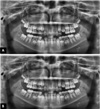
All pre- and post-processed data were saved as TIFF files for subsequent evaluation and comparison (Fig. 1).
Anatomical region selection
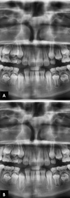
Similar anatomical structures were selected in both the pre- and post-processed images for assessment, including the dentinoenamel junction (DEJ), periodontal ligament (PDL) space, lamina dura, shape of the crown and roots of deciduous and permanent teeth, alveolar crest, and trabecular pattern. All selected structures were identified for evaluation within the maxillary and mandibular anterior and maxillary premolar regions (Fig. 2).
Image quality assessment
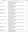
A modified version of the clinical image quality evaluation chart proposed by Choi et al. was used to evaluate the subjective radiographic image quality (Table 1).12 All the anatomical structures were scored from 1 to 5 (where 1=not visible and 5=clearly visible in all regions). All pre and post-processed panoramic radiographs were assessed on a Dell computer monitor (Dell P2312H, Dell Inc., Texas, USA) in a room with dimmed light at a viewing distance of 60 cm.
Reliability assessment
All images were assessed by 2 maxillofacial radiologists separately and blindly. Each image was evaluated twice by both observers (i.e., before and after processing) to determine the inter-observer reliability. For intra-observer reliability, each pre-processed and post-processed image was assessed by both observers at an interval of 2 weeks.
Statistical methods
The data were analysed using SPSS version 22.0 (IBM Corp., Armonk, NY, USA). The paired t-test was used to analyze differences in the observations of the anatomical structures between the pre- and post-processed radiographs, as well as the inter-and intra-observer variance. A P value of less than .05 was considered to indicate statistical significance. The intra and inter-observer agreement for anatomical structures was estimated using the kappa test, with the results classified as follows: poor, kappa <0.20; fair, kappa=0:21–0:40; moderate, kappa=0.41–0.60; good, kappa=0.61–0.80; and excellent, kappa=0.81–1.00.13
Results
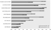
Table 2 and Figure 3 illustrate the subjective image quality assessment scores of the pre- and post-processed images in the maxillary and mandibular anterior region. The anterior region showed statistically significant differences for all anatomical structures between the pre- and post-processed images (P<.05). The score obtained from the processed images showed improved image quality compared to the original unprocessed data. The highest mean score difference was observed for the PDL space and lamina dura in permanent teeth (−1.86±1.01) and the lowest for the DEJ of the primary teeth (−0.26±0.60). The permanent tooth crown showed the highest post-processed score (4.56±0.76), whereas the DEJ of the primary teeth had the lowest score (1.20±0.86) based on the modified evaluation chart.
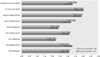
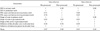
As shown in Table 3 and Figure 4, no significant differences were observed between the pre- and post-processed anatomical structures in the maxillary premolar region (P>.05), except for the root shape of permanent premolars (P=.01) and the trabecular pattern in alveolar bone (P=.001). The root shapes of permanent teeth showed the highest mean score difference (0.30±0.79), whereas the least difference was observed for the alveolar crest (0.02±0.92). The DEJ of the permanent teeth had the lowest score (3.10±1.39), whereas the alveolar crest showed the highest score (4.02±1.24) after being processed.
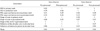
Table 4 and Table 5 present the Cohen kappa index for inter-observer and intra-observer reliability related to the pre- and post-processed images. The maxillary and mandibular anterior region showed excellent inter-observer (kappa≥0.82) and intra-observer≥0.86) agreement, whereas the premolar region showed good inter-observer (kappa≥0.63) and intra-observer agreement (kappa≥0.64) for both the pre- and post-processed images.
The post-processed images showed improved reliability when compared to the pre-processed data, but no significant differences were observed for inter-observer (anterior region, P=.93; premolar region, P=.97) or intra-observer reliability (anterior region, P=.86; premolar region, P=.06).
Discussion
In modern dentistry, regular conventional panoramic devices are being replaced by digital technology in order to obtain high-quality images, while minimizing patients' radiation exposure. At the same time, dose reduction directly affects image quality, which in turn can lead to the inaccurate identification of anatomical structures.14 Other reasons for diminished quality are related to noise and patient positioning during image acquisition.15 Image quality in paediatric patients with mixed dentition is of vital importance for diagnosis and treatment planning.14 To the best of our knowledge, no evidence is available in the literature on the effects of processing techniques on image quality in paediatric patients. Therefore, this study was conducted to analyse the impact of a state-of-the-art image processing technique on image quality in paediatric patients with mixed dentition.
In our study, post-processed digital panoramic radiographs allowed significantly better visualization of anatomical structures than standard non-processed digital images. This finding is in accordance with other studies that used processing techniques to improve image quality.161718 In contrast, according to Sabarudin and Tiau,4 no significant mean quality and scoring difference was observed between pre- and post-processed images related to the visualization of anatomical structures.
In comparison to conventional image processing techniques, which rely on filters, contrast, and brightness adjustments,19 our study involved a dedicated processing method. The most focussed segments were automatically identified and processed in this technique to generate a sharp panoramic image. This technique allowed better visualization of the maxillary and mandibular anterior region without any blurring by ghost image artefacts from the spinal column, as shown by the significantly higher evaluation scores obtained using modified clinical image quality evaluation charts for the processed images in the anterior region. At the same time, the technique provided overall good image quality, allowing proper visualization in the premolar region, in contrast to the pre-processed images. Improvements in inter-observer and intra-observer reliability were also observed after image processing.
In this study, only the anatomical structures in the maxillary and mandibular anterior and maxillary premolar region in patients with mixed dentition were selected for evaluation. This choice was made because structures in those regions are more prone to overlap and blurriness, which can compromise the diagnosis and treatment planning.202122
A blurred, shortened and narrowed appearance of the front teeth is a common error that occurs when the patient's head is positioned in front of the focus.23 As a result, studies performed in the mandibular premolar and posterior region showed minimal to non-significant distortion.2425
To compensate for these limitations, clinicians sometimes take additional intra-oral radiographs, which leads to more radiation exposure and is time-consuming. Therefore, we recommend the application of an image processing technique to improve image quality, instead of relying on supplementary radiographic investigations, especially in paediatric patients who are more vulnerable to radiation exposure.
In conclusion, the presented image processing technique can be regarded as a useful tool for improving the image quality of panoramic radiographs in paediatric patients with mixed dentition. Further investigations are required to study the application of the proposed technique for image quality in patients with dentofacial abnormalities.


 XML Download