

 PDF
PDF ePub
ePub Citation
 Print
Print


Introduction
Pulpal calcifications are diffuse or discrete calcified masses present in any portion of the pulp tissue (pulp chamber or root canal).1 They can be found in healthy, diseased, and even unerupted or impacted teeth in both the primary and permanent dentition.123 The formation of pulpal calcifications is not very well understood, and several aetiological factors have been proposed, including aging, pulp degeneration, inductive interactions between the epithelium and pulp tissue, genetic predisposition, long-standing irritants (caries, deep fillings, chronic inflammation, and abrasion), orthodontic tooth movement, trauma, periodontal disease, drugs, anaemia, arteriosclerosis, acromegaly, and Marfan syndrome. Additionally, a high prevalence of pulp stones has been reported in individuals with cardiovascular disease and kidney, gall, and salivary gland stones.3 The prevalence of pulpal calcifications varies widely, from 8% to 95% depending on the population, study design, and diagnostic method employed.23 Histologic studies have reported higher values than radiographic studies because calcified masses smaller than 200 µm cannot be seen on radiographs.456 The most commonly affected tooth is the first molar in both jaws, followed by the second molar. The least commonly affected teeth are the incisors and canines.24 The presence of pulpal calcifications does not affect the threshold of electric pulp testing7 and should not be considered to be a disorder requiring endodontic treatment.8 In the literature, panoramic, periapical, and bite-wing radiographs,9 as well as cone-beam computed tomography,1 have been used to identify pulpal calcifications.
The uncontrolled or subconscious movement of the jaw and its associated muscles during slumber (such as to grit or grind the teeth) is known as sleep bruxism (SB).10 The possible consequences of SB include masticatory muscle pain, headache, muscle fatigue, tooth wear, tooth hypersensitivity, teeth movement, scalloped tongue, and linea alba related to cheek biting.1011 Common means by which SB may be diagnosed include electromyography, surveys, dental examinations, and polysomnography. Among these methods, electromyography and polysomnography pairs are accepted as the highest quality and are the gold standard, although they suffer from prohibitive cost and availability issues for analysing a sample of any significant size.12 Females frequently experience SB,13 particularly those in the 20–45 age bracket,10 although its prevalence is inconsistent and ranges from 5% to 30%, depending on the diagnostic method and categorization used.14
Excessive occlusal force has been shown to cause changes in the pulp, such as hyperaemia, pulpitis, pulp necrosis, pulpal calcifications, and breaking of the periapical vessel.15 The objective of this study was to characterize the possible relationship between pulpal calcifications and SB, which causes long-standing irritation to the dentition.6 To our knowledge, no previous study on this topic has been published.
Materials and Methods
Participants
A total of 100 female patients between 20 and 31 years old were sampled for this analysis, which was performed with the approval of the Necmettin Erbakan University Faculty of Dentistry Research Ethics Committee. Furthermore, the analysis was performed according to the stipulations laid out by the Declaration of Helsinki (decision no.: 2017/12). The sampled individuals were taken from the Department of Oral and Maxillofacial Radiology of the Faculty of Dentistry at Necmettin Erbakan University, where they had been referred for a dental check-up. Informed consent was obtained from all participants included in the study.
The inclusion criteria were as follows: (a) women between 20 and 31 years old (a narrow range), (b) individuals without any missing teeth (except third molars). The exclusion criteria were as follows: (a) patients who had crowns, bridges, or endodontically treated teeth, (b) painful temporomandibular joint disorders, (c) mental disorders, (d) use of psychotropic substances, (e) abuse of substances such as alcohol or drugs, (f) the presence of gross malocclusion with concomitant orthodontic processes.
Fifty-nine SB patients comprised the study group, and 41 individuals without SB who satisfied the above criteria were selected independently and included in the control group.
Clinical assessment
The clinical assessment was performed in 2 phases: 1) an interview and 2) a physical examination. In phase 1 (the interview), participants were asked about regular or frequent tooth grinding sounds occurring during sleep, muscle fatigue, temporal headache, transient morning jaw muscle pain, and jaw locking upon awakening. In phase 2 (the physical examination), a visual examination was conducted for the presence of abnormal tooth wear.
SB was diagnosed via the following classification, as stipulated by the American Academy of Sleep Medicine.1617 In this system, criteria A and B must be met. Criterion A is the presence of regular or frequent tooth-grinding sounds occurring during sleep, and criterion B is 1 or more of the following: 1) tooth wear consistent with tooth grinding during sleep, 2) transient morning jaw muscle pain or fatigue, and/or a temporal headache, and/or jaw locking upon awakening consistent with tooth grinding during sleep.
Image evaluation
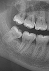
The diagnosis of pulpal calcifications (both pulp stone in the pulp chamber and pulpal sclerosis in the root canal) was made on digital panoramic radiographs. All digital panoramic radiographs were obtained using a 2D Veraviewpocs (J Morita MFG Corp., Kyoto, Japan) machine. Only high-quality images without any superposition were included. The dental status of each tooth was scored as intact, carious, restored, or restored and carious. Definite radio-opaque bodies inside the pulp cavity and/or root canals were recorded as pulpal calcifications (Fig. 1).
The examination for pulpal calcifications was repeated by the same oral radiologist in the same manner (presence or absence) at a 1-week interval.
Data analysis
All statistical analyses were performed using SPSS version 21.0 (IBM Corp., Armonk, NY, USA). Binary logistic regression analysis was used to determine the risk factors for pulpal calcifications. The primary outcome (dependent) variable was the presence of pulpal calcifications (yes/no), and the predictor (independent) variable was SB. The Spearman correlation coefficient was applied, and the Pearson chi-square test was used for categorical variables. To test intra-examiner reproducibility, Cohen kappa analysis was conducted. All samples were re-examined for the presence of pulpal calcifications at a 1-week interval and the same results were obtained following the second examination (κ=1). P values <.05 were considered to indicate statistical significance.
Results
A total of 2800 teeth from 100 participants were assessed, of which 1652 teeth were from the 59 SB patients and 1148 were from the 41 non-SB patients. The mean age of the participants was 24.17±3.2 years. Sixty-one percent of the patients had at least 1 dental pulpal calcification. In total, 213 (7.6%) of the 2800 teeth had pulpal calcifications. There was no statistically significant relationship between SB and the presence of pulpal calcifications (P>.05). In individuals diagnosed with SB, the total number of pulpal calcification was 129, while in non-SB patients, it was 84 (Table 1). The number of teeth with a pulpal calcification per person was 2.18 in the SB patients (129/59) and 2.04 (84/41) in the non-SB patients. Binary logistic analysis was showed that SB was not a risk factor for the presence of pulpal calcifications (odds ratio, 1.19; 95% CI: 0.52–2.69, P>.05).
There was no correlation between age and number of the pulpal calcifications (P>.05). However, a significant relationship was found for location, as 153 teeth with pulpal calcifications were in the maxilla, compared to 60 in the mandible (P<.001). Pulpal calcifications were found to be the most prevalent in the first molars and scarcest in the first premolars, both in the mandible and the maxilla. No pulpal calcifications were observed in the incisors or canine teeth. The distribution of pulpal calcifications according to tooth type is shown in Table 1.
Restorations and caries
A total of 116 teeth were restored: 65 in the mandible and 51 in the maxilla. A statistically significant relationship was found between the presence of pulpal calcifications and restorations (P<.01). with 2.5 times more pulpal calcifications observed in restored teeth than in non-restored teeth. There was no significant association between the presence of pulpal calcifications and tooth caries (P>.05) in the 60 teeth with caries, of which 38 were in the maxilla and 22 in the mandible. No tooth in the sample had both caries and a restoration. The distribution of caries and restorations are shown in Table 2.
Discussion
Pulpal calcifications are commonly encountered during endodontic treatment procedures, and especially large calcifications can block access to root canals and change the internal anatomy of the tooth.1 The aetiology of pulpal calcifications remains controversial, and the present study was performed to characterize the relationship between the presence of pulpal calcifications and SB in females. The sample of this study consisted of only females with a narrow age range (20 and 31 years), because age and sex have been discussed as predisposing factors in the literature, with a higher prevalence of pulpal calcifications observed in females than in males.2618 As age increases, the frequency of pulpal calcifications has been reported to increase.19 However, the only reliable way to determine the effect of aging is to through longitudinal follow-up.2 In a recent study,20 it was found that pulp samples from older individuals showed higher concentrations of alkaline phosphatase, which could trigger calcification. Controlling for sex is also important in an analysis of SB because it has been reported to vary between males and females, with females more frequently experiencing SB.13
The reported prevalence of pulpal calcification is quite variable (8%–95%).236 Some studies presented the prevalence based on individuals, and others based on the number of teeth.2 The present study showed that 61% of patients had at least 1 dental pulpal calcification, while 213 (7.6%) of the 2800 teeth had pulpal calcifications. The maxillary first molars presented the highest frequency of pulpal calcifications (44%), followed by the mandibular first molars (15%). This result can be explained by the fact that the first molars are the first permanent teeth to erupt, and are exposed to more stress.2
Molar teeth have the largest pulp volume and blood supply of all teeth,19 which leads to more vascular wall injuries when they are irritated by restorations or deeper caries.1 They have the largest surface area bearing occlusal forces, which may cause them to experience degenerative changes.6 Furthermore, 2.5 times more pulpal calcifications were seen in restored teeth in this study. This finding is consistent with the study of da Silva et al.,1 who observed 2.1 times more pulpal calcifications in restored teeth. They also found that deeper restorations presented a higher frequency of pulpal calcifications.
Pulpal calcifications were found more frequently in the maxillary arch (P<.05) This may be explained by the higher number of restorations in the maxillary arch.1 Long-standing irritants such as caries, restorations, and trauma could trigger pulpal calcifications.5 Sener et al.2 found that the prevalence of pulpal calcifications was higher in women than in men. The researchers attributed this result to the more frequent occurrence of bruxism in women, and they hypothesised that bruxism can be a longstanding source of irritation to the dentition. Moreover, it has been reported that vascular damage is followed by mineralization, which may act as a nidus for further mineralization and pulpal calcification formation.21 In this study, the hypothesis that SB caused vascular injury through occlusal trauma was not investigated. It was shown that SB was not a risk factor for the presence of pulpal calcifications (P>.05). Although no relationship was found between SB and pulpal calcifications, pulpal calcifications were slightly more common in individuals with SB. This was a cross-sectional study and there were no data about the duration of SB in the participants, which would be useful to consider as a variable. Since no previous study has done so, prospective analyses including patients' follow-up radiographs and information about the duration of SB are needed to arrive at a more precise judgment.
Most previous studies on the prevalence of pulpal calcifications were conducted with bitewing and periapical radiographs. A limitation of this study was that pulpal calcifications were detected by panoramic radiographs, which cause distortion.22 However, panoramic radiography has the benefit of enabling the simultaneous examination of all teeth with a single exposure; furthermore, it uses minimal ionizing radiation, which makes it an excellent imaging method for screening for pulpal calcifications.23 This study reflected the results obtained from patients referred to our dental school for routine dental examinations, and additional imaging was not applied in order to protect patients.
In conclusion, pulpal calcifications were detected in 61% of the participants and in 7.6% of the teeth. The first molars were the most frequently affected teeth in both arches. The presence of a restoration significantly increased the likelihood of these calcifications. Within the limitations of this study, it was shown that SB was not related to pulpal calcifications. In the future, prospectively-designed studies with larger samples, including follow-up radiographs and information about the duration of SB, are required.


 XML Download