

 PDF
PDF ePub
ePub Citation
 Print
Print


Introduction
Dental implants are considered to be an ideal option for the rehabilitation of edentulous jaws due to their stable outcomes and high success rates.1 Despite the high predictability of dental implants, complications are still encountered. These complications may be associated with various factors, such as treatment planning, the anatomy of the site, and the surgical technique and experience of the practitioner.2 A thorough clinical and radiological examination plays an important role in avoiding complications and enhancing the success of the treatment.
Intraoral radiography, panoramic radiography, computed tomography (CT), and cone-beam CT (CBCT) are frequently-used imaging modalities in treatment planning that improve long-term treatment success.3 Of these modalities, panoramic radiography has been widely used, since it has the advantages of being cost-effective, readily available, and providing high-resolution images.4 Panoramic radiography is a unique tool in terms of offering a large amount of information about the dentition and the jaws with a small radiation dose.5 However, distortions in the horizontal plane and magnification in the vertical plane are inevitable consequences of the working principle of panoramic radiography devices. In addition, incorrect patient positioning and technical or processing errors have substantial effects on the accuracy of images.6 Furthermore, 2-dimensional (2D) radiographic examinations are incapable of displaying the available bone width and the exact relationships with neighboring anatomical structures.7 Additionally, structures outside the focal trough are blurred and appear as shadows and artifacts on panoramic radiography.
The emergence of dentomaxillofacial software applications for presurgical planning was a major milestone in oral and maxillofacial surgery.8 Since the introduction of CBCT in the late 1990s, 3-dimensional (3D) technology with a lower radiation dose has become widely used in oral and maxillofacial surgery practice.9 CBCT scans have several advantages over 2D images and CT, including real-size data, the potential for generating a comprehensive set of 2D images, the ability to perform vertical scanning with the patient in a natural seated position, isotropic voxel size, less disturbance from metal artifacts, and Digital Imaging and Communications in Medicine compatibility. Furthermore, CBCT obtains a large amount of data in a relatively short time period of exposure to X-ray radiation and provides high-resolution images in several orthogonal planes for accurate measurements.9 Hence, CBCT offers a smaller dose of radiation, lower costs, and better energy efficiency than CT. Moreover, it is suitable not only for preoperative diagnoses, but also for real-time intraoperative assessments.10
A precise radiographic assessment of the jaws is crucial for presurgical planning and implant placement. Panoramic radiography and CBCT are the most frequently used imaging modalities in dental implantology. Although a considerable amount of research has been published on the use of panoramic radiography and CBCT in dental implantology, debate continues about the best imaging modality in presurgical implant planning. Some of these studies concluded that panoramic radiography was a reliable and safe technique for determining bone height, while others claimed that presurgical planning using panoramic radiography might lead to an inaccurate designation of the length of the implants, which could eventually result in complications such as nerve injury and sinus perforations. 9111213
Therefore, the aim of this study was to evaluate the correlations between measurements made using panoramic radiography and CBCT based on certain anatomical landmarks of the jaws, with the goal of preventing complications due to inaccurate measurements in the pre-surgical planning phase of dental implant placement.
Go to : 
Materials and Methods
This study was approved (reference number: 631/01112 017) by the Clinical Research Ethics Committee of the University of Akdeniz, Antalya, Turkey, and written informed consent was obtained from each patient. A retrospective study was conducted of 56 patients (30 male, 26 female; 32–79 years old, mean age of 58 years) who underwent preoperative panoramic radiography and CBCT evaluation for dental implant surgery between August 2016 and September 2017 at the Department of Oral and Maxillofacial Surgery.
The inclusion criteria were the availability of clear digital panoramic radiography and CBCT images showing at least 1 edentulous region in the neighborhood of the maxillary sinus, nasal floor, mental foramen, and/or mandibular canal in any quadrant. Images that demonstrated positioning errors, artifacts, unequal magnification or geometric distortion, and unclear anatomical structures were excluded from the study data.
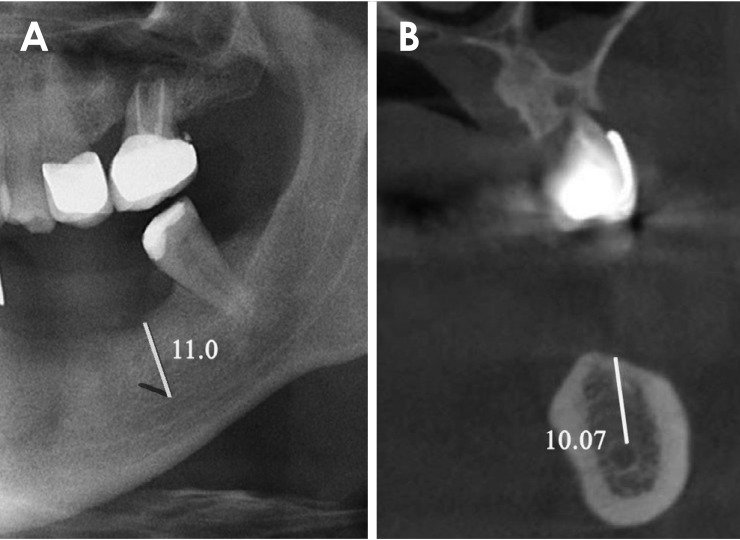
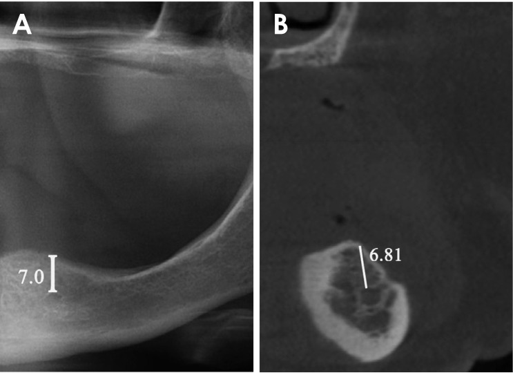
The digital panoramic radiographs were obtained using the same device (Planmeca ProMax® 2D; Planmeca, Helsinki, Finland) with a 1.2 magnification ratio, a voltage of 60 kV, a current of 5 mA, and a minimum exposure time of 17 s. The CBCT images were taken using the Planmeca Promax® 3D (Planmeca, Helsinki, Finland) at a voltage of 80 kV, a current of 12 mA, a scanning field of 16×5 cm, and a scanning time of 12 s. All measurements were performed using the Planmeca Romexis® Viewer v.3.8.1 (Planmeca, Helsinki, Finland) on panoramic images at 1 : 1 magnification and on CBCT scans in the coronal and sagittal planes. The shortest vertical distances between the alveolar crest and neighboring anatomical structures were measured, as follows: 1. The shortest distance between the bottom of the left and/or right maxillary sinus and the alveolar crest (Fig. 1). 2. The shortest distance between the bottom of the left and/or right nasal floor and the alveolar crest (Fig. 2). 3. The shortest distance between the top of the mandibular canal and the alveolar crest in the area of the left and/or right mandibular second molar (Fig. 3). 4. The shortest distance between the top of the left and/or right foramen mentale and the alveolar crest (Fig. 4).
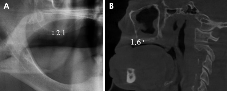 | Fig. 1Measurement of the shortest vertical distance between the alveolar crest and the bottom of the maxillary sinus on a panoramic image (A) and a cone-beam computed tomography image (B).
|
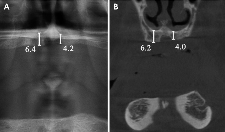 | Fig. 2Measurement of the shortest vertical distance between the alveolar crest and the bottom of the nasal floor on a panoramic image (A) and a cone-beam computed tomography image (B).
|
Measurements were independently performed by 2 oral and maxillofacial surgeons, and the mean values were used for the analysis. The intraclass correlation coefficient (ICC) was used to determine interobserver and intraobserver reliability.
Data were analyzed using SPSS version 22 (IBM Corp., Armonk, NY, USA). The normality of the data was assessed by a visual inspection of histograms, QQ-plots, box plots, and the Shapiro-Wilk test. The paired t-test was utilized to compare the measured values on panoramic radiography and CBCT images. The level of statistical significance was set at P<.05. Pearson correlation analysis was used to analyze the relationships between the data obtained using the 2 methods. The correlation coefficient (r) between the paired samples was calculated, and the measures were considered to be closely correlated if r was between 0.5 and 1.
Go to :
Results
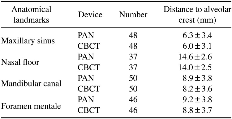
After the eligibility criteria were assessed, the final sample was composed of 30 men (53.6%) and 26 women (46.4%), who ranged in age from 32 to 79 years (mean, 58 years). Statistically significant differences were not observed for sex or age (P>.05). The number of measurements of each anatomical region and the mean values of the distances between the anatomical structures and the alveolar crest on panoramic radiography and CBCT images are shown in Table 1.
The ICC values for intraobserver reliability were 0.96 and 0.98, respectively, and the ICC value for interobserver reliability was 0.89. The ICC values indicated good to excellent reliability.14
Statistically significant differences were found between the measurements on panoramic and CBCT images for all anatomical structures (P<.05) (Table 2). The correlation coefficients (r) between the paired samples obtained from panoramic radiography and CBCT were closely correlated (P<.05), with r values varying between 0.921 and 0.979 for the different anatomical structures (Table 3).
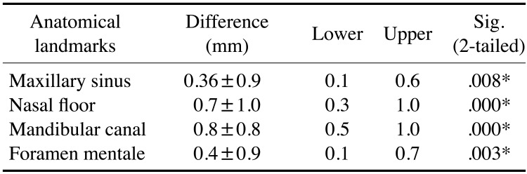
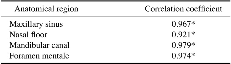
In the maxillary sinus, a statistically significant difference was found between the measured values on panoramic radiography and CBCT images (t47=2.758, P<.05). On average, these measurements on panoramic images were 0.36 mm higher than those made on CBCT images (95% CI, 0.097–0.620 mm). The measured values on panoramic and CBCT images for the distance between the floor of the maxillary sinus and the alveolar crest were very closely and positively correlated (r=0.967, P<.05).
Similarly, panoramic radiography and CBCT showed statistically significant differences in measurements at the region of the nasal floor (t36=4.054, P<.05). On average, these measurements on panoramic images were 0.67 mm higher than those on CBCT images (95% CI, 0.337–1.012 mm). The measured values on panoramic and CBCT images for the distance between the nasal floor and the alveolar crest were very closely and positively correlated (r=0.921, P<.05).
In the region of the mandibular canal, there was also a statistically significant difference between the measurements made on panoramic radiography and CBCT images (t49=6.723, P<.05). On average, these measurements on panoramic images were 0.76 mm higher than those on CBCT images (95% CI, 0.533–0.988). The measured values on panoramic and CBCT images for the distance between the mandibular canal and the alveolar crest were very closely and positively correlated (r=0.979, P<.05).
Finally, the difference between the measured values on panoramic radiography and CBCT images was statistically significant for the foramen mentale (t45=3.197, P<.05). On average, these measurements on panoramic images were 0.4 mm higher than those on CBCT images (95% CI, 0.149–0.655). The measured values on panoramic and CBCT images for the distance between the foramen mentale and the alveolar crest were very closely and positively correlated (r=0.974, P<.05).
Go to :
Discussion
Various imaging modalities are currently available in oral and maxillofacial radiology, of which intraoral radiographs, panoramic radiography, CT, and CBCT are the most commonly preferred techniques in dental implantology. Intraoral radiographs have been widely used, and have the advantages of low cost and ready availability. However, the inherent 2D nature of intraoral radiographs may lead to anatomical superimposition and geometric distortion.15 Isidor16 reported that due to superimposition, it was not possible to detect an unfavorable marginal bone level or the absence of osseointegration on 2D images. Furthermore, several studies have shown that the restricted preoperative diagnostic ability of 2D images in dental implant surgery may cause implant failure.17 Osseous destruction as a result of periodontal disease or peri-implantitis is an increasing problem in the long-term success of implant treatment.18 Three-dimensional images also show interproximal alveolar bone levels in the oro-vestibular direction, which is an essential criterion for follow-up of the peri-implant bone.19
Panoramic radiography provides a comprehensive 2D radiological examination of the jaws. The main advantages of panoramic radiography include a low radiation dose, relatively short exposure time, and comfort and simplicity of examination.20 However, the lower image quality compared to intraoral radiographs, geometric distortions such as unequal magnification and elongation, superimposition of the cervical spine, and the presence of ghost images are among the disadvantages of panoramic radiography.21 Laster et al.22 reported that horizontal measurements on panoramic radiography may be unreliable due to distortion and overlapping.
The outcomes of implant treatment have become highly predictable in recent years.23 Nonetheless, the relationship of implants with important vital structures can significantly affect the success of the surgical procedure. Thus, restricting the preoperative diagnostic examination to 2D imaging methods may potentially cause implant failure.17 Tang et al.21 recommended 3D imaging in cases where implant surgery posed a risk of damaging vital structures. Jacobs and Quirynen7 and Hassan and Jacobs24 also suggested using CT to evaluate extensive structures, such as the maxillary sinus. Similarly, Dreiseidler et al.25 reported that the image quality of CT and CBCT was superior to that of panoramic radiography, although CT and CBCT had the disadvantages of not being available in many local hospitals due to technical requirements and high cost.
In recent years, maxillofacial CBCT has been widely used in dental implantology.26 CBCT is advantageous due to its high spatial resolution, short scanning time, and rapid image acquisition.27 Monsour and Dudhia6 reported that patients undergoing CT examinations were exposed to a higher radiation dose than those who underwent examinations using panoramic radiography and CBCT.
To date, the use of different imaging techniques for pre-implant evaluation has been analyzed in several studies. Kopecka et al.28 compared the use of panoramic radiography and CBCT in assessing the interantral bone height available for dental implant placement and reported that panoramic radiography was sufficient in the incisor region, but not in the canine region. In a cadaveric study, Hu et al.11 compared the measurement errors on panoramic radiography and CBCT images and found that the mean presurgical measurement error was significantly smaller for CBCT than for panoramic radiography in the maxillary region, whereas it did not differ significantly in the mandibular region.
In contrast, Renton et al.29 reported that a majority of idiopathic trigeminal neuropathies (90%) were found in patients who had undergone pre-surgical 2D radiographic evaluation, while only 10% of cases presented such neuropathies after pre-surgical CBCT had been taken. In accordance with Renton et al.,29 Angelopoulos et al.30 also reported that CBCT images enabled a more precise evaluation of the mandibular canal.
In another study, Tang et al.21 compared the magnification rate of panoramic radiography in measuring different maxillofacial loci to that of CBCT and reported that the distances measured by panoramic radiography were closely correlated with those measured by CBCT. Similarly, the authors found strong positive correlations between panoramic radiography and CBCT in measurements of alveolar bone height in the regions of the maxillary sinus, nasal fossa, mandibular canal, and foramen mentale.
Vazquez et al.31 investigated the incidence of altered mental nerve sensation after implant placement in the posterior segment of the mandible using only panoramic radiographs for the preoperative evaluation. They reported that when a safety margin of at least 2 mm above the mandibular canal was respected, panoramic radiography was sufficient to evaluate the available bone height prior to the insertion of posterior mandibular implants. Regarding the mandibular canal region, Gerlach et al.32 also recommended a vertical safety margin of at least 1.7 mm when planning surgery using CBCT-based data. In accordance with the recommendations of Vazquez et al.31 and Gerlach et al.,32 the present study revealed that the average difference between panoramic radiography and CBCT ranged from 0.36 mm to 0.76 mm. However, in cases where the available bone is not sufficient, it may be necessary to place an implant deeper than the safety measurement, meaning that it is not always possible to respect a safety margin of 2 mm.33 In such cases, although the current study revealed that the average difference between panoramic radiography and CBCT was less than 1 mm, the use of CBCT is recommended for more accurate planning.
In conclusion, the results of this study support the idea that panoramic radiography might provide sufficient information on bone height for preoperative implant planning in routine cases or when CBCT is not available. However, an additional CBCT evaluation might be helpful in cases where a safety margin cannot be respected due to insufficient bone height.
Go to :
 XML Download