

 PDF
PDF ePub
ePub Citation
 Print
Print


Introduction
Oral carcinoma cuniculatum (CC) is a rare, unacquainted variant of oral squamous cell carcinoma (OSCC).1 It is similar to cutaneous CC, which was first described by Aird et al. in 1954, in its clinicopathological findings and biological behavior.2
Apart from the 3 cases of cutaneous CC described by Aird et al.2 on the plantar aspect of the foot, this type of tumor has been reported at other sites, including the face, esophagus, abdomen, palm, leg, penis, and cervix.34567
In 1977, Flieger and Owiński8 were the first to report CC in the oral cavity, but they failed to establish the relationship of this tumor to ordinary OSCC because of its unique clinical features, including locally aggressive behavior with both endophytic and exophytic growth patterns. In addition, it has a unique histological architecture that comprises well-differentiated epithelial cells with no or rare cytological atypia, unlike OSCC.9
Oral CC is a locally aggressive tumor formed of multiple, branching, keratin-filled crypts (rabbit burrows or cuniculi) lined by well-differentiated hyperplastic stratified squamous epithelium with minimal cellular atypia, but evident local bone invasion and rare metastasis.10
The World Health Organization (WHO) 2005 classification of tumors included oral CC as a new variant of OSCC characterized by the presence of keratin-filled branching crypts and keratin cores, resulting in the tumor having a cuniculatum architecture, similar to rabbit burrows.11
Although it is a variant of OSCC, the diagnosis of oral CC has remained very difficult and challenging because of clinicians' lack of awareness and familiarity with the tumor, which in turn has resulted in under-reporting of oral CC and its suggested low incidence.12
Correlation of histological findings with clinical and radiographic features is essential for the diagnosis of oral CC and to avoid confusing it with other tumors, mainly verrucous carcinoma (VC), well-differentiated SCC, papillary squamous cell carcinoma (PSCC), and other tumors included in the histological differential diagnosis of oral CC.910111213
The aim of the current study was to conduct a systematic review of the studies describing oral CC by pooling patients from multiple studies, with the goal of illustrating and further distinguishing the clinical features, etiologic agents, predisposing factors, imaging features, histopathological findings, treatment, and follow-up and survival rates of this distinctive rare tumor.
Materials and Methods
A systematic review of the published cases of oral CC in the literature was performed according to the Preferred Reporting Items for Systematic Reviews and Meta-Analyses (PRISMA) statement, an evidence-based set of items for presenting these types of studies.14
Search strategy
A broad literature searches in Medline (full text), the Dentistry and Oral Sciences Source via EbscoHost research databases, and PubMed was performed. Relevant studies published from 1954, the first mention of CC in the literature, to January 2018 were identified. These electronic databases were searched using both MeSH terms and free text words. A search strategy was developed that combined the terms (carcinoma cuniculatum) AND (palatal OR palate OR gingiva OR tongue OR oral OR maxilla OR maxillofacial OR mandible OR mandibular OR ramus OR alveolar OR jaw). In the advanced search, all fields were included in the PubMed search and all texts in Dentistry and Oral Sciences Source databases and in Medline (for full texts). Three independent researchers, including 1 oral pathologist and 2 oral radiologists, examined the retrieved hits and discarded duplicates. The references of relevant articles were screened for papers missed in the initial search.
Selection criteria
The inclusion criteria were: articles published in the English language, with human subjects, that described oral CC in the oral or maxillofacial region. All titles and abstracts were analyzed. Full-text articles were obtained only for abstracts found to satisfy the inclusion criteria. Articles published in languages other than English, those regarding non-human subjects, and studies on any variant of OSCC other than oral CC were excluded from the review.
Data extraction
Individual and aggregated data describing the clinical features, etiologic agents, predisposing factors, radiographic features, histopathological and immunohistochemical features, treatment, and follow-up and survival rates of patients with oral CC were collected. Other variables were also gathered, including author names, publication year, and number of cases. The quality and internal validity of the studies were independently assessed by the authors according to the CARE (CAseREport) Statement and Checklist Tool (Fig. 1).15 Any disagreement among the authors was resolved by discussion.
Results
Search results
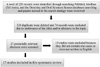
The search strategy retrieved 229 hits; 27 were relevant for our study, of which 10 studies were excluded because they were not written in the English language81617181920 or they did not contain any oral cases.21222324 This resulted in a total of 17 articles that were included in the systematic review,1391013252627282930313233343536 which presented 43 cases (Fig. 2).
Clinical features
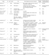
The clinical features of the included cases are presented in Table 1. The number of included cases was 43 (19 males and 24 females) with a mean age of 62.7 years (range, 7–81 years). The most common sites were the mandibular gingiva in 19 cases (44.2%), followed by the tongue in 10 cases (23.2%), the maxillary gingiva in 6 cases (13.9%), the mandible in 4 cases (9.3%), the vestibule in 2 cases (4.6%), the maxillary region in 1 case (2.3%), and the buccal mucosa in 1 case (2.3%).
The clinical presentations of the tumors varied, and patients presented with various symptoms as single entities or co-existing with each other. The most common symptom was pain in 21 cases (48.8%), followed by ulceration in 16 cases (37.2%), swelling in 13 cases (30.2%), induration in 9 cases (20.9%), a red and white lesion in 8 cases (18.6%), white patches/areas in 7 cases (16.3%), exudation in 7 cases (16.3%), leukoplakia in 5 cases (11.6%), bleeding in 4 cases (9.3%), erythroleukoplakia in 2 cases (4.6%), lichen planus in 1 case (2.3%), leukoplakia and lichen planus in 1 case (2.3%), soreness in 1 case (2.3%), bone exposure in 1 case (2.3%), teeth mobility in 1 case (2.3%), restriction of tongue mobility with dysphagia and difficulty with articulation in 1 case (2.3%), and gingival enlargement in 1 case (2.3%). Roughly one-fourth of the cases showed a nodular, pebbly, cobblestone, verrucous, or cauliflower-like surface.
The lesion duration was recorded in most cases, with a mean of 19.5 months. However, the duration of the lesion was not recorded in 13 cases.
Thirteen cases (30.2%) were reported to be in tobacco smokers, alcoholics, or both, while 1 case (2.3%) reported to be in a smokeless tobacco user and in 26 cases (60.5%), the patient did not consume any alcohol or smoked. The corresponding information was missing in the remaining cases.
In addition, the size of the lesion was reported in only 7 of the 43 cases; it ranged from 1 to 6 cm.
Imaging findings
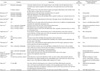
The imaging findings of the tumors are listed in Table 2. In 25 of the 43 cases, no imaging modality was used. Computed tomography (CT) was the modality of choice in 5 cases involving the bone, where it showed osteolytic lesions. The tumors were occasionally well-defined, but in 14 cases the lesions were almost ill-defined and caused bone destruction (32%). Erosion of the underlying bone occurred in tumors located within soft tissue. Magnetic resonance imaging (MRI) was the modality of choice in 3 cases involving the tongue and the gingiva to detect the actual extent of the lesion. MRI showed tumorous lesions of heterogeneous intensity affecting most of the tongue, but sparing most of the extrinsic muscles, and also showed inflammation in the gingiva. Both CT and MRI were used together in 2 other cases. Panoramic radiographs were used in 8 cases as screening tools through which the abnormality was detected. Panoramic findings included an ill-defined irregular osteolytic radiolucent lesion in 6 cases (13.9%), erosion of the superficial cortical bone in 1 case (2.3%), and a ‘moth-eaten’ osteolytic lesion in 1 case (2.3%).
Preoperative diagnosis
The preoperative diagnoses are listed in Table 2. They ranged from inflammatory conditions such as granuloma, abscess, or osteomyelitis in 5 cases to reactive epithelium with hyperkeratosis or pseudoepithelomatous hyperplasia in 1 case and malignancy, including VC and OSCC, in 5 cases. The diagnosis changed from an inflammatory condition to malignancy in 3 cases. In 3 cases, the preoperative diagnosis was a keratocyst, and it was a mandibular cyst in 1 case. No preoperative diagnoses were reported in 28 cases.
Histopathological features
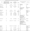
The histopathological features of the tumors are listed in Table 3. Histopathological features consistent with oral CC were reported in all 43 cases (100%). These comprised well-differentiated squamous epithelium that extended deep into the connective tissue with multiple, branching, keratin-filled crypts (rabbit burrows or cuniculi), with absent or mild cytological atypia that was usually limited to the basal and parabasal layers. In 39 cases (90.7%), an inflammatory stromal reaction mainly consisting of neutrophils was observed, along with discharging abscesses reported in 28 cases (65.1%).
The margins were free from tumor in 35 cases (81.4%) and showed infiltration in 2 cases (4.6%); however, no data were reported from the remaining 6 cases (13.9%). Cases that were reported to involve infiltration, invasion, destruction, and penetration of the surrounding tissue and cases with deep projections or down-growths with an invasive front were considered to be locally invasive. All cases showed local invasion, which did not extend any further in 39 cases (90.7%), while the remaining 4 cases showed metastasis: 3 to the regional lymph nodes and 1 to the lungs.
Immunohistochemical findings or special staining
The immunohistochemical findings are listed in Table 3. Neither special nor immunohistochemical staining was used in 37 cases (86%). Positive Gomori methenamine silver (GMS) and periodic Acid-Schiff (PAS) staining indicated the presence of fungal organisms resembling Candida both within the crypts and the epithelium in 1 case. In 1 case, there was a high rate of positive p53 immunoexpression (indicative of mutations in the TP53 gene, where tumors with more than 10% positively stained p53 cells are highly suggestive of malignant features), which was found in more than 90% of basal and suprabasal cells, whereas it was minimally expressed with no mutations in another case and showed negative results for expression in 2 other cases. High positive Ki-67 expression (a protein that increases with cell division preparation; the more positive cells for Ki-67, the more quickly a tumor is dividing, indicating the uncontrolled cell proliferation associated with malignant cells) in the basal and suprabasal layers was reported in 1 case, while low levels of expression were reported in 4 cases. In addition, findings for p16 expression (a protein that acts as a tumor suppressor by decelerating the cell progression from the G1 phase to the S phase) were negative in the single tested case. Furthermore, human papillomavirus (HPV) was tested in 3 cases, with negative results.
Growth pattern
Both exophytic and endophytic growth patterns were histologically reported in 15 cases (34.9%), while an endophytic pattern was reported in 7 cases (16.3%), an exophytic pattern was reported in 2 cases (4.6%), and records were missing in the remaining cases.
Treatment and follow-up
Results are shown in Table 4. Complete surgical resection with a safety margin was the treatment of choice in 42 cases (97.7%), with a subtotal maxillectomy or mandibulectomy performed in cases with bone invasion, in addition to neck dissection in cases with enlarged lymph nodes. Only one case was treated by chemotherapy and radiotherapy.
Following the surgical removal of the tumor, 37 patients (86%) continued to attend follow-up appointments. The follow-up duration ranged from 4 months to 14 years, with a mean of 31.1 months. Of the 37 patients, 29 (67.4%) were reported to be disease-free and did not show recurrence or mortality. However, 4 cases (9.3%) showed local recurrence. In the cases of recurrence, the treatment was second surgical removal in 1 case, second surgical removal with concurrent chemotherapy in 1 case, concurrent chemoradiotherapy in 1 case, and chemotherapy in 1 case.
Differential diagnosis
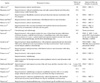
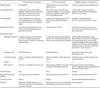
The criteria for the differential diagnosis between oral CC and other similar lesions were collected from the included articles (Table 5). A reliable knowledge of these criteria could provide the basis for a proper differential diagnosis between oral CC and other similar lesions.
Discussion
Oral CC is a rare variant of OSCC that was added to the WHO classification in 2005;11 however, most oral clinicians are unfamiliar with this entity and often misdiagnose it, leading to fatal outcomes due to faulty treatment of the affected patients.
The current systematic review is the first to systematically analyze the existing oral CC cases described in the English-language literature in an effort to illustrate and further characterize the etiologic agents, predisposing factors, clinical and imaging features, histopathological findings, treatment, and follow-up and survival rates. The goal of this review was to enable oral clinicians to diagnose this rare entity promptly and to differentiate it from other tumors with similar features, facilitating its adequate curative treatment.
A total of 55 reported oral CC cases were found in the literature between 1954 and January 2018, of which only 43 cases were included for analysis in the current systematic review, as the remaining 12 cases81617181920 were described in studies not written in the English language.
Although Hutton et al.29 reported the presence of oral CC in a 7-year-old patient, the present search showed a predominance in the sixth and seventh decades of life, with a mean age of 62.7 years. This finding is consistent with those of Sun et al.,9 Pons et al.,10 and Padilla and Murrah.13
In the current review, oral CC was found to occur with a slight female predominance: 24 females (55.8%) and 19 males (44.2%). This matches the research of Sun et al.9 on 15 cases, Padilla and Murrah13 on 10 cases, and Fonseca et al.33 on 2 cases. However, a slight male predominance of oral CC was reported by Pons et al.,10 who studied 3 cases, all of whom were males. Since oral CC is a variant of OSCC, and OSCC has a male predominance, this female predominance is surprising.11
The current analysis revealed that the mandibular gingiva was the most common site for oral CC, as it was reported in more than half of the cases. This finding is in agreement with those of Pons et al.,10 Padilla and Murrah,13 and Fonseca et al.33 However, Sun et al.9 reported the tongue to be the most common site.
The most common clinical presentation was pain,3910262730313335 followed by ulceration,13913262829303233 swelling,913262930323336 and induration.91336 This differs from the clinical presentation of OSCC, which may vary according to the affected intraoral subsite and for which pain is not the most common feature. This tendency is also distinct from VC, which usually presents as a well-demarcated, thin white keratotic plaque which quickly thickens and develops papillary (blunted tips) or verruciform (pointed tips) surface projections and from PSCC, for which hoarseness and airway obstruction are the most common presenting symptoms, as the larynx and the hypopharynx are among the most common sites of involvement.11
The size of oral CC ranged from 1 to 6 cm; however, this range was derived from only 7 cases,3132526283436 which is insufficient for a conclusive estimation of the average size of these tumors.
The duration of the lesion was recorded in 90.7% of the cases,13910132528313233343536 with a mean of 19.5 months. This relatively long duration most likely indicates the slowly growing clinical course of oral CC,9103336 which matches VC; however, this finding is in contrast to the behavior of OSCC, which shows a rapidly growing course.11
In the current analysis, 13 patients (30.2%) were tobacco smokers, alcoholics, or both,391325263136 and 1 patient (2.3%) was a smokeless tobacco user.13 Smoking and alcohol consumption are known to be predisposing factors for oral malignancy;11 however, a clear etiology for oral CC has yet to be established.
Premalignant lesions/conditions as leukoplakia, erythroplakia, and lichen planus had been found in 10 cases (23.2%), suggesting that they underwent a malignant transformation to oral CC, which matches with what has been reported by several authors.19132526293133
Preoperative imaging was reported for 18 cases (41.7%); some studies used panoramic radiographs,1101325262733 while others used CT,102529303136 MRI,383132 or both.35 Most of the lesions were osteolytic and almost ill-defined, with cortical destruction and erosion of the underlying bone in tumors located in soft tissue. Ill-defined borders of a lesion indicate an inflammatory condition or malignancy; therefore, a correlation with the clinical features of the lesion is mandatory. Moreover, erosion of the underlying bone indicates the infiltrative nature of the lesion, as found in malignant conditions.
It is worth mentioning that the preoperative diagnoses were incorrect in all cases. To a large extent, this occurred due to clinicians' lack of awareness about this entity. This also may have occurred due to the improper biopsy depth as biopsies were taken only from the superficial part of the lesion in some cases.
Regarding the histopathological features, all the included cases exhibited the same features specified by the WHO classification11 and the diagnostic system purposed by Chen et al.,39 wherein the presence of a defined set of histological features is required for the accurate diagnosis of oral CC and its differentiation from other similar lesions, such as VC and PSCC. In all cases, oral CC had a distinctive histopathologic appearance that revealed multiple branching keratin-filled crypts resembling rabbit burrows or cuniculi (thus the name cuniculatum), lined by well differentiated squamous epithelium with absent or minimal cytological atypia and normal to little mitosis. Moreover, micro-abscesses with discharge of yellowish secretion through the crypts and a prominent stromal reaction, mainly consisting of neutrophils, were reported in most cases.39101325262730313233343536
Oral CC has a locally aggressive invasive nature due to its deep epithelial projections or down-growths that extend into the connective tissue. In the current analysis, all the cases showed local invasion, with evidence of bone erosion and destruction noted radiographically, and 4 cases showed metastasis: 3 to the regional lymph nodes and 1 to the lungs.9 In our opinion, the occurrence of regional lymph node and/or distant metastasis should be taken into consideration, as the early diagnosis and knowledge of appropriate treatments will prevent further spread of the tumor.
The presence of mutated forms of p53 was found to be associated with malignant features;4041 yet, studies on its association with oral CC are lacking, since only 4 studies reported findings on p53 expression. These included case reports on 4 patients, with discordant results, as 2 studies showed positive results for p53 expression,332 while the other 2 showed negative results.2528 Similar conflicting reports on Ki-67 expression, which is associated with cell proliferation in malignancy, were found: 1 study reported its increased expression,32 while other studies reported its consistently low expression.252833 Regarding p16, which is a tumor suppressor, it was tested in only 1 study,3 which reported negative results for its expression. Extensive further research on the immunohistochemistry of oral CC is required.
Although a correlation between HPV and cutaneous CC has been observed,3738 such a correlation was not established in oral cases, where no record of HPV presence was reported, except in 3 of the included cases,252832 all of which showed negative results. As a result, the role of HPV in oral CC pathogenesis remains unclear.
The growth pattern of the lesion is reflected in its clinical presentation; both exophytic and endophytic growth patterns were recorded in 15 cases (34.9%),10132834 which most commonly presented as ulcerations, red and white lesions, and pain of a relatively long duration, in contrast to the prominent exophytic growth of VC.11
Regarding the treatment of oral CC, complete surgical resection with a safety margin was the treatment of choice. Subtotal maxillectomy or mandibulectomy19252628293135 and/or neck dissection1026273136 was performed in patients with extensive tumors because of the tendency of oral CC for local invasion. This treatment yielded effective results and an excellent (disease-free) prognosis in 29 of the 37 patients who continued to attend follow-up appointments for a duration ranging from 4 months to 14 years following surgery.
However, 4 patients (9.3%) experienced local recurrence.931 Three cases of tumor recurrence were reported by Sun et al.;9 1 patient underwent repeated surgical resection following the initial recurrence, but a second recurrence occurred in the following year, together with partial malignant transformation to OSCC (hybrid tumor of oral CC and conventional OSCC), and the patient died of uncontrolled cancer with cachexia 6 months later. The second patient received concurrent chemoradiotherapy after recurrence, but the tumor was not controlled and metastasis developed to the deep cervical lymph nodes and the lungs, ultimately leading to death. The third received chemotherapy after recurrence, but the tumor had increased in size and treatment was not effective. Suzuki et al.31 reported a case in which multiple recurrences occurred after multiple surgical excisions, and finally the patient was treated unsuccessfully with chemotherapy before his death from aspiration pneumonia due to the involvement of the parapharyngeal space. The aforementioned findings encompass 3 cases of mortality: 1 case died of lung metastasis,9 while the other 2 died due to the increased tumor size.931 Therefore, mortality from oral CC is thought to primarily result from locally destructive growth, rather than metastasis.
Furthermore, such findings underscore the necessity of the cautious use of chemotherapy and/or radiotherapy, especially in the treatment of recurrent cases that did not respond to these types of treatment. This is supported by the assumption that radiotherapy may provoke anaplastic transformation.931 It is worth noting that recurrent oral CC demands careful attention due to its more resistant and aggressive behavior than its primary form.
In conclusion, oral CC is a rare, well-differentiated variant of OSCC with unique clinical and histopathological features. It has a good prognosis following appropriate surgical treatment with a relatively low recurrence rate. However, it shows resistant behavior in cases of recurrence. Proper knowledge of the diagnostic criteria of this lesion, together with a thorough correlation of the histological findings with the clinical and imaging features, is essential for its diagnosis and differentiation from other similar lesions. Due to its locally aggressive and invasive nature, it is advisable to perform multiple deep biopsies of the suspected lesions in addition to a thorough sampling of these biopsies to avoid underdiagnosis.

 XML Download