

 PDF
PDF Citation
 Print
Print


INTRODUCTION
Endometrial cancer (EC) is the most common gynecological malignancy with increased incidence rate in developed countries [1]. Almost 80% ECs are of endometrioid endometrial adenocarcinoma (EEA) histology and diagnosed early with good prognosis [2]. However, there is still a subset of patients failed to reach 5-year overall survival even for those with low-grade or early-stage EEA [3456]. After the initial diagnosis 15% of women in grade I and 25% in grade II EEA died from the disease in 5 years [35]. Also, among patients with grade I–II endometrioid ECs, approximately 3% in stage I and 20% in stage II did not survive over 5 years [4]. The current risk assessment and therapeutic decision-making majorly rely on the International Federation of Gynecology and Obstetrics (FIGO) grade, stage and histology [7]. Thus, this assessment is still suboptimal that some patients with poor prognosis might be left undertreated. More improved means are urgently needed, to precisely identify high-risk patients with grade I–II EEA.
Estrogen receptor (ER) and progesterone receptor (PR) are most validated prognostic biomarkers for endometrial cancer [891011]. Gene expression of ESR1 and PGR were found significantly correlated to the protein expression of ERα and PR by immunohistochemistry (IHC), respectively [12131415]. A prospective study evaluating ER and PR on curettage specimens from 832 endometrial carcinoma patients reported that ER and PR double loss independently predicted lymph node (LN) metastasis and shorter disease-specific survival (DSS) [16]. Other studies also associated the negativity of ER and PR with poor survival in endometrial carcinoma even in low-grade subtypes [891011171819], However, no clinical risk-stratified analysis has been done for grade I–II ECs patients with ER/PR loss. It is still unclear whether patients of this subgroup may need more intervention after surgery. Currently ER and PR status have not been implemented into any clinical guideline [202122]. It has not been investigated, whether integrating ER and PR into clinical risk stratification can assist to select high-risk patients with grade I–II EEA.
Integrating genomic classification into clinicopathological risk assessment has shown improved prognostic value [8232425]. The Cancer Genome Atlas (TCGA) research network had significantly improved risk stratification by discriminating ECs into four molecular categories: POLE ultra-mutated tumors, microsatellite instability tumors, copy-number low tumors and copy-number high tumors [26]. Nevertheless, whether TCGA classification has prognostic value in grade I–II EEA cases, and whether it correlates to expression of ER and PR are unclear.
Our study was to investigate whether ER and PR double negativity (ER/PR loss) could improve the risk stratification for patients with grade I–II EEA. We aimed at 1) the clinical characteristics for patients with ER/PR loss in grade I–II EEA; 2) the prognostic value of ER/PR loss in grade I–II EEA and in currently clinical risk groups; and 3) the prognostic value of TCGA genomic subgroups in grade I–II EEA and its association between ER/PR loss and TCGA classification.
MATERIALS AND METHODS
1. Patient cohort-Shanghai Ob&Gyn Hospital (Shanghai cohort)
A total of 1,124 patients with endometrial cancer underwent surgery from January 2008 to June 2014, in Shanghai Obstetrics and Gynecology Hospital of Fudan University (Shanghai Ob&Gyn Hospital). Among those patients, we retrospectively enrolled 903 cases into our study based on the inclusion criteria: diagnosed as primary EEA of grade I–II; no previous (neoadjuvant) chemotherapy; underwent trans-abdominal or laparoscopic hysterectomy; available postoperative formalin-fixed, paraffin-embedded tumor tissue; and available clinicopathological data and follow-up data. All of the 903 patients were re-staged according to the FIGO 2012 staging guidelines by XZL and XJC. Grading was performed on hysterectomy specimen according to World Health Organization (WHO) pathological classification (2014), based on percentage of solid growth and nuclear atypia. Clinical and histopathological data as well as follow-up data were collected by experienced clinicians. The study protocol was approved by the Ethics Committees of Shanghai Obstetrics and Gynecology Hospital of Fudan University (approval number: 2017-94). All patients underwent informed written consent for the use of their biospecimens and clinicopathological data for research purpose.
Progression-free survival (PFS) was defined as the time from the diagnosis (the operation date) to first recurrence of disease. Overall survival (OS) was defined as the time interval between the diagnosis and EEA-correlated death or loss to follow-up. Overall observation time was the time interval between the diagnosis and the last contact (March 17th, 2017), defined as death or last follow-up.
2. Immunohistochemical staining and evaluation
ER, PR and p53 expression were assessed by IHC on formalin-fixed paraffin-embedded (FFPE) tumor samples for 903 patients with grade I–II EEA. We assessed p53 expression for excluding potential unidentified serous ECs which significantly led to poor prognosis [27]. FFPE samples were collected according to standard operation procedures established in the hospital. IHC were carried out with the IHC Protocol F Program of Leica BOND-MAXTM Detection System (Leica Biosystems, Wetzlar, Germany) according to the manufacturer's instruction. The primary mouse monoclonal antibodies for ER (clone EP1, 1:150), PR (clone PgR636, 1:500) and p53 (clone DO-7, 1:200) were purchased from DakoCytomation (Glostrup, Denmark). IHC procedures were performed as we previously described [28]. Staining in normal endometrial tissues surrounding the tumor served as internal positive control to avoid false negative results.
The expression of ER, PR and p53 were evaluated by two experienced pathologists (QZ and YQQ) who were blinded to the patient characteristics and outcome. In case of disagreement, results were discussed in a multi-headed microscope till a consensus was reached. Staining of ER and PR was considered as positive if the tumor cell nuclei were stained in ≥1% of the cells [293031], corresponding to the definition of positivity currently in clinical use for breast carcinoma. p53 expression was graded according to the proportion of positive stained tumor nuclei as previously described [242532]: “p53 loss”, <1% of positive stained tumor cell; “p53 normal”, 1%–50% stained tumor cells; and “p53 over-expression”, >50% discretely-distributed and strong-stained tumor cells.
3. Patient cohort-TCGA (TCGA cohort)
Publicly available gene expression data of endometrial carcinoma were obtained from the project of TCGA research network (https://cancergenome.nih.gov). Within the TCGA cohort of fully evaluable cases (n=548), totally 218 patients were diagnosed as grade I–II EEA, and 140 of these 218 patients had information of four TCGA genomic sub-phenotypes: POLE ultra-mutated phenotype (POLE; n=9, 6.4%), microsatellite instability (MSI; n=40, 28.6%), copy-number low (CN low; n=84, 60.0%), and copy number high (CN high; n=7, 5.0%). All of the 218 patients with grade I–II EEA had mRNA expression data of ESR1 and PGR. The low quartile was considered as a cut-off for high and low ESR1 and PGR, as described previously [14]. Patients with low expression of both ESR1 and PGR were grouped as “ESR1/PGR low” (n=36), whereas those with high ESR1 and/or high PGR were grouped as “ESR1/PGR high” (n=182).
4. Statistical analysis
Statistical analysis was performed by SPSS 22.0 (SPSS, Inc., Chicago, IL, USA) The Shapiro-Wilk test was used to ascertain whether continuous variables had a normal distribution. Continuous variables were summarized by means and standard deviations, and categorical variables were presented as frequency with percentage. The intra-group differences of continuous variables were investigated by Student's t-test when normally distributed, or Mann-Whitney U test when non-normally distributed. χ2 test or Fisher's exact was used to analyze the difference between categorical variables. Estimates of PFS and OS rates and 95% confidence intervals (CIs) were calculated using the Kaplan-Meier method. Log Rank test were used for univariate statistical comparisons. Adjusted hazard ratios (HRs) and 95% CI were estimated with the Cox proportional hazards model. A 2-tailed p value of less than 0.05 was considered as statistically significant.
RESULTS
The clinical and pathological characteristics of 903 patients were summarized in Table 1. After a median follow-up of 59.0 months (0-109.0) of follow-up, eighty-two (9.1%) patients were lost contact with us. Among 821 patients with complete follow-up, in total 41 (5.0%) patients experienced disease recurrence, of whom 17 (41.5%) died eventually due to EEA.
Table 1
Comparison of clinical characteristics between “ER or PR positive” and “ER/PR loss” groups
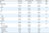
Data shown are median (range) or number (%).
ER, estrogen receptor; PR, progesterone receptor; BMI, body mass index; FIGO, International Federation of Gynecology and Obstetrics; MI, myometrial invasion; LN, lymph node; TH, total hysterectomy with or without bilateral salpingooophorectomy; TH + pLN dissection, TH plus pelvic lymph node dissection; ETH + pLN dissection, extensive total hysterectomy with bilateral salpingooophorectomy plus pelvic lymphnode dissection with or without para-aortic lymph node dissection; Chemotherapy, paclitaxel + cisplatin; Radiotherapy, external beam radiotherapy.
*Significant difference p<0.05; †p value: difference between “ER or PR positive” group and “ER/PR loss” group; ‡All variables were analyzed among 903 patients except for BMI and tumor size. Missing data for 132 cases for BMI, and 161 for tumor size.
![]()
1. ER/PR loss correlates with a more aggressive clinical subtype in grade I–II EEA
The ER and PR expression were detected in the nuclei of the tumor cells, as showed in Fig. 1A-D. Thirty-five cases with both ER and PR negative expression were grouped as “ER/PR loss”, whereas the other 868 with either ER or PR positive expression were grouped as “ER or PR positive”. As presented in Table 1, patients with ER/PR loss were more likely to have deeper myometrial infiltration (≥50%, p=0.012), severer FIGO stage (II–IV, p=0.004), pelvic LN metastasis (p=0.016), vascular invasion (p=0.015), and relapse (p=0.001), compared with “ER or PR positive” group.
 | Fig. 1Representative photomicrographs of immunohistochemical staining for estimation of ER-α, PR, and p53: (A) ER loss, (B) ER positive, (C) PR loss, (D) PR positive, (E) p53 loss, (F) p53 normal, and (G) p53 over-expression (A-D: original magnification ×40; E,F: original magnification ×20).ER, estrogen receptor; PR, progesterone receptor.
|
2. ER/PR loss independently predicts poor survival in early and late stages of grade I–II EEA
With the median follow-up of 59 months (0–109), the median PFS and OS were 48 months (36–59) and 48 months (36–59) for ER/PR loss patients, and 63 months (60–65) and 64 months (61–66) for patients with ER or PR positive expression, respectively. The ER/PR loss was significantly correlated with poor PFS in univariate analysis (HR=5.253; 95% CI=2.21–12.52; p<0.001; Fig. 2A). Other factors with prognostic influence on PFS were age, FIGO stage, grade, myometrial invasion (MI), LN metastasis, vascular invasion, and ovary metastasis (Table 2). In multivariate analysis, ER/PR loss independently predicted poor PFS (HR=3.77; 95% CI=1.48–9.56; p=0.005; Table 2). Further, we found patients with ER/PR loss had significantly higher risk of death in both univariate (HR=7.593, 95% CI=2.55–22.60; p<0.001; Fig. 2B) and multivariate analysis (HR=5.56; 95% CI=1.37–22.55; p=0.016; Table 3).
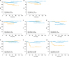 | Fig. 2Estimated PFS and OS for grade I–II EEA patients (A, B) according to ER/PR status and within risk subgroups based on FIGO stage: stage IA (C, D), stage IB (E, F) and stage II–IV (G, H) (Kaplan-Meier estimation).PFS, progression-free survival; OS, overall survival; ER, estrogen receptor; PR, progesterone receptor; FIGO, International Federation of Gynecology and Obstetrics.
|
Table 2
Uni- and multivariate survival analysis of PFS for 821patients† with grade I–II EEA according to Cox proportional hazards regression model
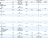
Notes: In total of 806 patients were included into multivariate analysis.
PFS, progression-free survival; EEA, endometrioid endometrial adenocarcinoma; HR, hazard ratio; CI, confidence interval; BMI, body mass index; ER, estrogen receptor; PR, progesterone receptor; MI, myometrial invasion; FIGO, International Federation of Gynecology and Obstetrics.
*Significant difference p<0.05; †In total of 821 patients with available follow-up data.
![]()
Table 3
Uni- and multivariate survival analysis of OS for 821patients† with grade I–II EEA according to Cox proportional hazards regression model
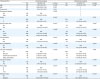
Notes: In total of 806 patients were included into multivariate analysis.
OS, overall survival; EEA, endometrioid endometrial adenocarcinoma; HR, hazard ratio; CI, confidence interval; BMI, body mass index; ER, estrogen receptor; PR, progesterone receptor; MI, myometrial invasion; FIGO, International Federation of Gynecology and Obstetrics.
*Significant difference p<0.05; †In total of 821 patients with available follow-up data.
![]()
As both FIGO stage and grade have been considered essentially for classic model of risk stratification, we subsequently investigated the prognostic value of ER/PR loss in different FIGO stages. We observed ER/PR loss was still associated with poor PFS in patients with stage IA (n=695, p=0.041; Fig. 2C) and stage II–IV (n=129, p<0.001; Fig. 2G), even after adjusting by age, MI, LN metastasis, and vascular invasion (HR=5.54; 95% CI=1.28–23.89; p=0.022 and HR=5.77; 95% CI=1.57–21.27; p=0.008, respectively). There was no association between ER/PR status and PFS in patients with stage IB (Fig. 2E), which might due to the small amount of sample (n=79). The prognostic value of ER/PR status on OS was only observed among patients with stage II–IV (p<0.001, Fig. 2H), rather in stage IA and IB (Fig. 2D and F). In multivariate analysis, the risk of death was dramatically increased in patients with both ER/PR loss and FIGO stage II–IV (HR=9.875; 95% CI=1.88–52.02; p=0.007), adjusted for MI, LN metastasis, vascular invasion, and ovary metastasis. In addition, for a subgroup of 578 patients in grade I and stage IA without LN metastasis and vascular invasion, ER/PR loss was an independent and significant predictor for both PFS (HR=11.22; 95% CI=2.48–50.81; p=0.002) and OS (HR=16.77; 95% CI=1.83–153.63; p=0.013) adjusted for age.
3. No correlation between p53 over-expression and ER/PR status in grade I–II EEA
To investigate if the poor prognosis of ER/PR loss group might result from potential unidentified serous ECs, we performed immunohistochemistry of p53 on available tissue samples (n=845). Representative photomicrographs illustrating p53 expression were presented in Fig. 1E-G. In ER/PR loss group (n=34), we observed 0 (0%) patients with “p53 over-expression” compared with 11 (1.4%) patients in group of positive ER/PR (n=811), respectively. We found no significant correlation between p53 over-expression and ER/PR status (Fisher's exact test, p=0.272).
4. No correlation between ESR1/PGR and TCGA classification in grade I–II EEA from TCGA cohort
In TCGA cohort, we firstly performed analyses on 218 patients with grade I–II EEA. Between patients with high ESR1/PGR (n=182) and low ESR1/PGR (n=36), we observed no significant difference in FIGO stage and grade. Low ESR1/PGR significantly correlated with poor PFS rather than OS in both univariate (HR=2.33, 95% CI=1.06–5.13; p=0.035; Fig. 3B) and multivariate analyses (HR=2.40; 95% CI=1.08–5.32; p=0.031), adjusted by FIGO stage. Secondly, we evaluated prognostic value of TCGA genomic classification in grade I–II EEA. In TCGA cohort, totally 140 patients of grade I–II EEA had information of four TCGA genomic subtypes. We found no prognostic value of TCGA genomic classification on both PFS (p=0.704; Fig. 3C) and OS (p=0.772) for grade I–II EEA patients. Lastly, we investigated if ESR1/PGR expression was association with TCGA classification in grade I–II EEA. Among the 140 patients with TCGA genomic information, 21 had low ESR1/PGR and 119 had high ESR1/PGR. The distribution of TCGA genomic subgroups in patients with low ESR1/PGR and high ESR1/PGR was showed in Fig. 3D. We found no correlation between ESR1/PGR status and TCGA classification (χ2 test, p=0.303).
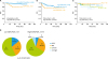 | Fig. 3Prognostic values of ER/PR status, ESR1/PGR status and TCGA genomic subgroups in grade I–II EEA patients (Kaplan-Meier estimation) and association between TCGA classification and ESR1/PGR expression(χ2 test): (A) estimated PFS according to ER/PR status by immunohistochemistry in Shanghai cohort, (B) estimated PFS according to mRNA expression of ESR1/PGR status in TCGA cohort, (C) estimated PFS according to TCGA genomic subgroups in TCGA cohort, and (D) distribution of TCGA genomic subgroups in low- and high-ESR1/PGR groups and the association between TCGA classification and ESR1/PGR status.ER, estrogen receptor; PR, progesterone receptor; TGCA, The Cancer Genome Atlas; EEA, endometrioid endometrial adenocarcinoma; PFS, progression-free survival.
|
DISCUSSION
In grade I–II EEA, we found the ER/PR loss was a strong and independent risk factor for recurrence and death. These prognostic values were further observed in early and late stages and might be independent of TCGA classification. These findings are clinically important, because ER/PR loss may help identify high-risk patients from presumed low-risk group for in-time and tailored treatment. Also, immunohistochemistry of ER and PR are low-cost and easily applied for most medical centers. To our knowledge, this is the first large-scale study on the prognostic impact of ER/PR loss in grade I–II EEA integrated with currently clinical risk stratification.
The strengths of our study are including a large-scale homogenous population of grade I–II EEA and integrating molecular biomarkers with standard risk stratification. The prognostic values of ER and PR in ECs were mostly evaluated across all histological subtypes [1617193334]. Due to the tumor heterogeneity and the histological-subtype specific of ER and PR, possible prognostic effects should be analyzed in a subtype-specific manner [19]. A few studies with low-grade subgroup analyses among patients with endometrioid histology presented independently prognostic value of ER and PR, or their combination as a panel [16171835]. However, these subgroup-analyses were based on relatively small amounts of patients or curettage specimens, and no stratification analyses were followed. It is still unclear whether ER/PR can help identify patients that currently left undertreated in grade I–II EEA. In our study, we observed ER/PR loss had independently prognostic value in both presumed low-risk and high-risk subgroups. Grade I–II EEA patients in stage IA, IB, and II–IV were commonly considered with low-, intermediate- and high-risk, and provided with different therapeutic regimens after surgery. According to our findings, women with ER/PR loss should be shifted into a higher risk group or at least be paid on more clinical attention: those in stage IA may need adjuvant therapies after surgery rather than simple observation; and those in stage II–IV may need more frequent follow-up or more effective therapeutic protocols. Besides, we observed ER/PR loss predicted poor PFS and OS even in grade I and stage IA EEA patients, suggesting fertility preserving therapy requested by young patients with ER/PR loss should be considered carefully and re-assessed by clinical specialists. Thus, we suggest the cost-effective immunohistochemistry of ER/PR should be considered to apply routinely as prognostic biomarker panel to meet clinical needs, particularly for patients with grade I–II EEA.
The optimal cut-off for prognostic biomarkers would ideally be clinically validated against patient outcome in prospective clinical trials, or in systematically collected archived specimens from randomized clinical trials [36]. According to this, the optimal cut-offs for ER and PR staining have not been defined in ECs, hindering the evaluation on their prognostic values between studies [837]. Historically, many investigators had defined 10% or greater nuclear staining as the threshold for positive ER and PR [1824383940]. However, extended studies using ≥1% as such cut-off showed significantly better survival for patients with ≥1% ER-positive cells compared with patients with complete negative cells [29303141]. Also 67% of the exactly 10% ER-positive cancers and 24% of the 1%–9% ER-positive cancers showed ESR1 mRNA levels and gene signatures that were consistent with >10% ER-positive cancers, suggesting IHC might have underestimate ER positivity in 1%–9% ER-positive cancers [37]. Hence, we use ≥1% as the definition of positive ER and PR to provide more precise and robust evidence for clinical use.
In our study, ER/PR loss was found associated with poorer survivals in grade I–II EEA. Herein, we suspected if this subtype might include unidentified serous ECs that usually led to unfavorable prognosis. Some ambiguous endometrial carcinomas have overlapping histological features of serous and endometrioid carcinoma [274243]. On these challenging cases, lack of consensus on diagnosing histotype and grade between expert pathologists leads to vast difference in assessing prognosis and treatment for the same patient. These cases usually involve uterine serous versus FIGO grade II endometrioid carcinoma [27]. Using the FIGO grading system for endometrial endometrioid carcinomas, a tumor composed predominantly of glands or papillae (architecture grade I), even in the presence of high nuclear grade (nuclear grade III), would be classified as FIGO grade II endometrioid carcinoma [27]. p53 immunohistochemistry was showed with differential value in these ambiguous ECs that “favor serous” ECs have more p53 over-expression than “favor endometrioid” ECs [2732]. Ambiguous cases with p53 over-expression had a more aggressive clinical course than cases without p53 over-expression, which had similar 3-year PFS (94%) and DSS (100%) to that of FIGO grade II endometrioid carcinomas (85%) [27]. Nevertheless, no difference in p53 expression was observed between “ER/PR loss” and “ER or PR positive” groups in grade I–II EEA, suggesting the poor prognosis in ER/PR loss group may not result from unidentified serous ECs.
Improved risk assessment in ECs by integrating TCGA classification with clinicopathological factors has been addressed [232425]. In TCGA cohort, we found no prognostic value for TCGA classification in grade I–II EEA. This might due to the relatively small (n=140) series of grade I–II EEA had assigned TCGA genomic subgroups. In addition, to analyze the potential association between TCGA classification and ER/PR loss, we defined the low ESR1/PGR group in TCGA cohort. Studies showed mRNA expression of ESR1 and PGR were significantly related to ER and PR protein expression by IHC [121315]. We defined the lower quartile of mRNA expression as the cut-off for low ESR1/PGR [14], which achieved a closest HR (2.40; 95% CI=1.08–5.32) as that of ER/PR loss group in our cohort (HR=3.77; 95% CI=1.48–9.56), with sufficient samples (n=36) for analyses. Other cut-off values used previously were the median [1444], the first tertile [45] or the log2-converted value 10.18 [37]. However, none of them achieved statistical appropriateness in our study. We subsequently found no association between TCGA subgroups and ESR1/PGR status, suggesting ER/PR loss may predict poor prognosis independent of TCGA classification in grade I–II EEA. Nevertheless, expression of ESR1/PGR mRNA and ER/PR protein were not identified in same cohort. Whether the low ESR1/PGR group in TCGA cohort could represent for ER/PR loss in our cohort needs further validation.
Studies showed ER loss correlated with alternated phosphoinositide 3 kinase/mammalian target of Rapamycin (PI3K/mTOR) pathway 19. ER or PR loss might enable epithelia cells to become more like mesenchymal cells with increased ability to invade and resist apoptosis [1446]. Given PI3K/mTOR inhibitors today are in phase I/II trial in advanced ECs (www.clinialtrials.gov, November 2017), PI3K/mTOR inhibitors might also have potential as the target therapy for grade I–II EEA with ER/PR loss.
Despite the strength of the large and homogenous population, a robust cut-off of ER/PR and improved risk stratification, this study has some limitations. Firstly, our study is a retrospective single-center study. Moreover, the mRNA expression of ESR1/ PGR and the IHC of ER/PR were not performed in the same cohort that might generate bias. The cut-off for low ESR1/PGR expression corresponding to ER/PR loss also needs further validation. Currently we have no tissue for all the patients to run RNA-sequencing or polymerase chain reaction (PCR), although no study yet reports “real world” TCGA classification in grade I–II EEA, either. Given that RNA-sequencing is expressive and large-scale tissue collection is time-consuming, thus, our study still has merits for using public TCGA data to explore potential correlation. Since we found ER/PR status may not correlate with TCGA subgroups in grade I–II EEA patients, further studies may shift their priorities to other biomarkers when funding was limited, instead of beginning with high-cost investigations in coordinate with TCGA classification.
In conclusion, based on a large-scale and homogenous population, we found ER/PR loss strongly and independently predicted poor survival for patients with grade I–II EEA, in both early and late stages. This prognostic value might be independent of TCGA molecular classification. Grade I–II EEA patients with ER/PR loss may need more post-surgery intervention to improve their outcome. Fertility preserving therapy for these women may also need re-consideration. Herein, integrating cost-effective immunohistochemistry of ER/PR into future randomized clinical trials and clinical algorithms may improve risk stratification for tailored therapeutic management.
 XML Download