

 PDF
PDF ePub
ePub Citation
 Print
Print


INTRODUCTION
Maxillofacial prostheses replace missing facial structures resulted from congenital malformations, trauma, or cancer surgery. Maxillofacial prostheses can provide patients the opportunity to restore self-esteem and social reintegration.12 For an esthetically pleasing maxillofacial prosthesis, the essential requirements are good edge adaptation, harmony with facial expressions, natural form and surface texture, and good color match with the surrounding skin.3 Among these factors that impact the final esthetic result of a prosthesis, an accurate matching of both color and translucency of the prosthetic material to those of patient's skin is extremely important. Although the shape of maxillofacial prosthesis is not perfectly harmonious with facial tissues, accurate color and translucency match between maxillofacial prosthesis and skin under different lighting conditions provides natural appearance.4
Coloring maxillofacial prosthetic material to reproduce skin shade and translucency has been conventionally achieved by subjectively assessing the patient's skin color and adding pigments manually into silicone elastomer prosthetic material until an acceptable color is shown. In this technique, the assessment of the color match is performed by holding a piece of colored silicone elastomer next to the face.5 This chairside, visual, trial and error method is generally time consuming, is extremely difficult even for an experienced and skilled prosthodontist, and can be affected by the illumination used for color matching.56 Furthermore, multiple trials may be required to achieve proper color and increased pigment loading may compromise translucency of the mixture.
The ultimate goal of work in this area is the creation of an objective, precise, repeatable color measuring and mixing for silicone elastomers that closely match to the human skin color independent of light source. Quantifying the skin color and pigmentation by using colorimeter and spectrophotometer and then establishing a pigment formulation using this data is the procedure of this work.78 This approach was first reported more than 25 years ago.7 In the recent literature, a few studies described the use of spectrophotometric data of skin to establish the pigment formulation that matches the skin.789 These attempts included creating a database using commercially available pigments for maxillofacial silicones and preparing a recipe with a color formulation software for a measured skin area. Although these studies bring valuable information, some shortcomings should be in consideration such as color measuring problems of the translucent skin with spectrophotometer, the use of a color formulation software developed primarily for industrial field, and difficulty in achieving a good match between skin and silicone in terms of translucency.910 Since the structure of human skin contains complex characteristics such as translucency and heterogeneity, a computerized color measuring and matching system specific to human skin color was necessary. Recently, a color matching system namely “e-skin system” has been specially developed for in maxillofacial prosthodontics. The system include a special spectrocolorimeter which gives a code for the measured skin area. This code is entered to an online calculator (http://www.spectromatch.com/sign-in/) and a color formulation called “recipe” is provided. The use of the system has been mentioned in a case report in the literature11; however, clinical outcomes of the system has not been objectively investigated.
The purpose of the present study was to evaluate accuracy in color and translucency matching of pigment formulae determined by computerized color formulation software across different skin color samples. The null hypothesis was that a close color match between target and replicate colors would be obtained for tested skin colored silicones.
MATERIALS AND METHODS
A platinum-catalized, addition curing silicone (M511, Technovent Ltd., Newport, UK) was colored to simulate 28 different colors of human skin. Twenty of these 28 skin colors were obtained by mixing the pigments by mixing the each of prefabricated skin shade pigments into the silicone and 8 were prepared by mixing different pigments. These 28 colors were utilized in this study to serve as “target skin colors” (TC). Twenty target skin colors were fabricated using all the human skin tones supplied by the manufacturer. To fabricate the other eight target skin colors, frequently used eight skin colors were selected and modified with fiber flocking pigments (Fig. 1). The pigments used for each of 28 skin shade are shown in Table 1. Two components of the silicone (Part A and Part B) were combined at a 10:1 ratio by weight as recommended by the manufacturer. Once combined silicone components were thoroughly mixed, one of 20 intrinsic skin shades (Color 1–20 in Table 1) was added 0.2% by weight.12 Remaining 8 skin colors (Color 21–28 in Table 1) including intrinsic skin shades and flocking pigments in concentration of totally 0.2% by weight prepared by the authors to simulate heterogeneous skin color arising from veins, vessels, and pigmentation. The colored silicone was poured into disk shaped stone molds with dimensions of 4 mm thickness and 18 mm in diameter. The molds were placed into an oven, held at 100℃ for 1 hour for polymerization, and then allowed to cool to room temperature. Polymerized silicone disks were removed from the molds, evaluated under magnification (Loupe opt-on, Orange Dental, Biberach, Germany) for porosity. Excess material at the edges of disk shaped specimens were trimmed using scissors and cleaned in an ultrasonic cleaner (Erosonic Energy, Euronda, Vincenza, Italy) in distilled water for 10 minutes to remove dental stone residue. For each TC group, five specimens were prepared (n = 5).
Using a spectrocolorometer (e-skin, Spectromatch Ltd., Bath, UK), each specimen was measured and the color code displayed on the screen was noted down. For each specimen, 3 consecutive measurements were made and the repetitive color code among 3 measurements was assigned to be used for “replicate skin color” (RC) fabrication. The spectrocolorometer was calibrated with its own inherent calibration tile in accordance with standard procedure suggested by the manufacturer before these repeated measurements. The measurement area of 4 mm was used for the spectrocolorometer.
To prepare RC for each specimen, the color code of the specimen was entered into E-skin system's on-line calculator (http://www.spectromatch.com/sign-in/) The recipe was given by the calculator. To serve as an example, Figure 2 shows the steps to obtain the recipe for a specimen (Specimen C25-1). The silicone, catalyst, and paste pigments (QuickWeigh LSR, Spectromatch Ltd., Bath, UK) were measured on a balance with a weight tolerance to 00.000 g (FZ120i, A&D Company, Ltd., Tokyo, Japan) according to the recipe obtained from on-line calculator. The mixture was blended to achieve a homogenous color. The colored silicone was processed as described for TC specimen preparation.
To evaluate color and translucency agreement between each TC and corresponding RC (TC-RC pair), the color of each specimen was measured with a spectrophotometer (Konica Minolta Cm2300d, Minolta Konica, Tokyo, Japan). Measuring characteristics of the spectrophotometer were standard illuminant D65, illumination geometry d/8 degree, 10° colorimetric standard observer, and measurement area of 8 mm in diameter. Measurements of each specimen were performed on standard white (L: 97.17, a: −0.11, b: 0.16) and black (L: 11.78, a: 3.3, b: 14.24) backgrounds using the CIE L*a*b* color space. L*, a*, and b* values of the specimens where L = darkness (0 to 100, 100 is lightest), a = green/red (+ is red, − is green), and b = blue/yellow (+ is yellow, − is blue) were recorded. The spectrophotometer was calibrated with its own white calibration tile in accordance with standard procedure suggested by the manufacturer prior to each specimen.
The color difference (CIELAB Delta-E) between each TC-RC pair was calculated from following equation13:
CIELAB Delta-E values derived from the TC-RC pairs were compared with one-way ANOVA to determine whether mean CIELAB Delta-E values of each color group was significantly different from those of the other groups. Significant differences among the group means were determined by the Tukey multiple-comparison test after ANOVA.
For assessing the agreement between L*, a*, and b* values of TC and RC silicone specimens, a two-way mixed average measures intraclass correlation coefficient (ICC) for consistency were calculated (confidence intervals set at 95%). For the L*, a*, and b*, the measured ICC was greater than 0.98, indicating excellent reliability between TC and RC silicone specimens (P < .001). In addition, ICC was also calculated for the TP values. Interpretation of ICC scores was based on Cicchetti's recommendations in which ICC of < 0.40 is poor, 0.40 – 0.59 is fair, 0.60 – 0.74 is good, and 0.75 – 1.00 is excellent.14
TP of TC and RC silicone specimens were compared with Paired t-test. The significance level was defined at 0.05. All statistical analyses were conducted with SPSS v24 (IBM SPSS Statistics for Windows, Version 24, IBM Corp., Armonk, NY, USA).
RESULTS
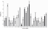
Descriptive statistics and statistical comparison of the CIELAB Delta-E values were shown in Table 2. CIELAB Delta-E values calculated for each target color-replicate color pairs were ranged between 1.64 and 7.88 (Fig. 3). An ICC of > 0.9 was obtained for interrater and intrarater agreements in L*, a*, and b*. For the TP, the value was 0.663, indicating good reliability (P < .001) (Table 3). Descriptive statistics and statistical comparison of the TP are shown in Table 4. The mean TP value of replicate colors (8.876) were significantly higher than that of target colors (7.653).
DISCUSSION
To evaluate the color matching performance of a color reproduction system, the differences between original L*, a*, b* values and reproduced L*, a*, b* values were calculated. The color difference unit, namely Delta-E, was used as a guideline to determine degree of color match (smaller Delta-E means closer match between two colors). In practice, interpretation of Delta-E values are made according to the “perceptibility” and “acceptability” thresholds. The perceptibility is the Delta-E value at which observers can distinguish the color difference. Acceptability threshold is the highest Delta-E value that color difference is defined as acceptable by observers. The findings of this study demonstrated that a mean CIELAB Delta-E value of 3.83 was calculated between a total of 140 target and their corresponding replicate silicone specimens. This Delta-E value falls into the range of acceptability threshold values reported for maxillofacial silicones in the study by Paravina et al.,15 which is the one study reporting color difference thresholds in this field. They reported that Delta-E value of 3.0 and 4.4 is considered as acceptability threshold for light and dark maxillofacial silicone samples, respectively. Another study conducted by Leow et al.16 evaluated color difference thresholds for silicone light and dark hand prostheses and reported 1.8 and 2.6 Delta-E for light and dark silicone prostheses, respectively. However, these studies can provide very limited information to establish a standard evaluation method and thresholds for color differences of silicone prostheses. Further research is needed to determine the clinically acceptable Delta-E for maxillofacial silicones by performing several analyses for tooth colored dental materials.1718192021222324
In the present study, the accuracy of a computerized color matching system in terms of color and translucency matching was evaluated for 28 different target skin colors. Twenty of these 28 skin colors were premixed skin colors and 8 were premixed skin colors modified with flocking colorant agents to simulate vascular appearance of the skin. A wide range of skin colors were used in the study because a possible relationship between skin tone and performance of computerized color matching system has been observed.1516 Previous research on replication of human skin color were also conducted on a wide range of skin colors from extremely light to dark skin tones simulating multiracial skin tones.58916 Coward et al.7 reported that color matching is more challenging in darker skin tones as individuals with darker skin tones or lower L* values revealed higher Delta-E values. On the other hand, Xiao et al.9 reported reduced accuracy of color reproduction using a 3D color printing system for extremely dark and bright shin colors while majority of 14 tested skin colors were closely reproduced. In another study, Xiao et al.25 proposed 3D colour image reproduction system for the automated manufacturing of facial prosthetics using 3D additive manufacturing techniques. They reported that using this method to produce facial prostheses provides good colour reproduction as well as accurate shape and fine texture compared to standard methods. Paravina et al.15 also reported higher threshold for dark skin replications that were more chromatic. In the present study, contrary results were obtained; that is, dark color replications showed lower CIELAB Delta-E values compared with light replications, which may indicate some relationship between target color values and functionality of the system. Considering the increased melanin pigment density of dark skin colors, replication of its color by the tested color matching systems' brown or its related pigments may be more applicable.
Beside CIELAB Delta-E value, indicating threshold between TC and RC silicone specimens, agreement between L*, a* and b* was also analyzed. Each color components (L*, a*, b*) of the compared silicone pairs showed excellent reliability as the ICC was greater than 0.98. In color science, it has been reported that visual thresholds for lightness depend on the L* value of a sample.22 To this respect, an ICC value of 0.987 for L* showed high agreement or low threshold between TC and RC silicone specimens.
The translucency parameters of target and replicate color specimens were significantly different as replicate colors were found more translucent. This difference may be resulted from different compositions of coloring agents used in fabrication of two groups. Colorants used in target colors composed of pigments dissolved in liquid silicone and those in replicate colors are composed of pigments dispersed into a silicone base. Difference in filler content of colorant agents may influence translucency of colored silicone specimens. Hungerford et al.5 reported that filler content of the polymers has an impact on refractive index of a specimen as well as pigments and coloring agents incorporated into the material. The ICC value of 0.663 for TP values indicated the differences in translucency between TC and RC silicone specimens.
Human skin, which is a partially translucent and functional multilayered biomaterial, has complex optical properties.10 The deep penetration of the light through the skin makes it impossible to measure and calculate translucency of skin as translucent materials at a certain thickness. In human skin, light is not reflected within the detection area of measuring spot of color measuring device, and sub-surface scattering and absorption occur during measurement, which is so-called “edge loss”. For computerized color matching of maxillofacial silicones, concerns may arise on optical behaviors of silicone and skin. However, edge-loss error has also been reported for translucent pigmented elastomers.6 Furthermore, edge-loss on silicone and human skin could be similar during measurements.23 Ishikawa-Nagai et al.24 reported that a large illuminated area and a small measurement area is the most appropriate method for measuring translucent samples to reduce edge-loss. In our spectrophotometer, measurement area of 8 mm in diameter and 11 mm illuminating area may minimize the edge-loss error.
For a close color reproduction of a silicone maxillofacial prosthesis, several factors take part in color matching process, like mixing appropriate pigments in required amounts. The silicone mixing technique is one of the reported factors affecting color reproducibility as presence of air voids and pores within the silicone scatter the reflected light, affecting the total color of specimen.26 Hatamleh and Watts26 reported that manually mixed specimens contained greater number and volume of pores compared with mechanical mixing under vacuum. In our study, manual mixing of the silicone for both specimen groups might have led to color and translucency differences in addition to using coloring agents with different compositions.
In order to reproduce skin color precisely, the area of the target measurement is a critical issue. For an indistinguishable maxillofacial prosthesis from surrounding skin, color measurement and color mixing should be performed considering color differences of skin across the defect and face. Further research is needed for larger area of a target measurement.
The final decision on the maxillofacial prostheses is left to patients and clinicians; therefore, perceptional evaluation is also an important factor. In this study, CIELAB Delta-E values and TP values between target and replicate silicone specimens were evaluated objectively. Further, the clinicians' perception of a color match should be evaluated as a final verification. Another limitation of the study is the use of silicone specimens as target color instead of human skin. Different optical properties and spectral differences of human skin require further research conducted on human subjects.
CONCLUSION
The computerized color measuring and matching system specific to human skin color resulted in low color differences (CIELAB Delta-E) between target and replicate color silicone specimens. L*, a*, b*, and TP values of TC and RC silicone specimens were in excellent or good agreement. The system can provide many conveniences in the field of maxillofacial prosthodontics by eliminating the risk of errors resulting from subjective color assessment and coloring and by shortening time for color mixing. Further research is necessary to examine the system on human subjects as assessed both objectively by spectrophotometer and subjectively by individuals experienced in the fabrication of maxillofacial prostheses.


 XML Download