Abstract
Posttraumatic facial deformities (PTFDs) are very difficult to correct, and if they do occur, their impact can be devastating. It may sometimes be impossible for patients to return to normal life. The aim of surgical treatment is to restore the deformed bone structure and soft tissue to create symmetry between the affected side and the opposite side. In the process of managing PTFD, correcting enophthalmos is one of the most challenging aspects for surgeons because of difficulties in overcoming the scar tissue and danger of injuring to the optic nerve. In this article, surgical options for reconstruction of the medial wall, floor, lateral wall, and roof of the orbit are described. To optimize aesthetic improvement, additional cosmetic procedures such as facial contouring surgery, blepharoplasty and rhinoplasty can be used. Plastic surgeons should join emergency trauma teams to implement an overall treatment plan containing rational strategies to avoid or minimize PTFD.
Go to : 
REFERENCES
1. Wikipedia. Face [Internet]. [place unknown]: Wikipedia;2018. [cited 2018 Nov 23]. Available from:. https://en.wikipedia.org/wiki/Face.
2. Chua DY, Park SS. Posttraumatic nasal deformities: correcting the crooked and saddle nose. Facial Plast Surg. 2015; 31:259–269.

3. Kim TG, Chung KJ, Lee JH, Kim YH, Lee JH. Clinical outcomes between atrophic and nonatrophic mandibular fracture in elderly patients. J Craniofac Surg. 2018; 29:e815–e818.

4. Kim YH, Jung CY, Chung KJ, Lee JH, Kim TG. A systematized strategy in corrective rhinoplasty for the Asian deviated nose. Ann Plast Surg. 2017; 79:7–12.

6. Kim TH, Kang SJ, Jeon SP, Yun JY, Sun H. Usefulness of indirect open reduction via a transconjunctival approach for the treatment of nasal bone fracture associated with orbital blowout fracture. Arch Craniofac Surg. 2018; 19:102–107.

7. Kang CM, Han DG. Correlation between operation result and patient satisfaction of nasal bone fracture. Arch Craniofac Surg. 2017; 18:25–29.

8. Kang CM, Han DG. Objective outcomes of closed reduction according to the type of nasal bone fracture. Arch Craniofac Surg. 2017; 18:30–36.

9. Kawamoto HK Jr. Late posttraumatic enophthalmos: a correctable deformity? Plast Reconstr Surg. 1982; 69:423–432.
10. Hazani R, Yaremchuk MJ. Correction of posttraumatic enophthalmos. Arch Plast Surg. 2012; 39:11–17.

11. Kim YH, Ha JH, Kim TG, Lee JH. Posttraumatic enophthalmos: injuries and outcomes. J Craniofac Surg. 2012; 23:1005–1009.
12. Wolfe SA. The influence of Paul Tessier on our current treatment of facial trauma, both in primary care and in the management of late sequelae. Clin Plast Surg. 1997; 24:515–518.
13. Kang DH, Jung DW, Kim YH, Kim TG, Lee J, Chung KJ. Kirschner wire fixation for the treatment of comminuted zygomatic fractures. Arch Craniofac Surg. 2015; 16:119–124.

14. Choi SH, Kang DH, Gu JH. The correlation between the orbital volume ratio and enophthalmos in unoperated blowout fractures. Arch Plast Surg. 2016; 43:518–522.

15. Kim YH, Park Y, Chung KJ. Considerations for the management of medial orbital wall blowout fracture. Arch Plast Surg. 2016; 43:229–236.

17. Clauser L, Galie M, Pagliaro F, Tieghi R. Posttraumatic enophthalmos: etiology, principles of reconstruction, and correction. J Craniofac Surg. 2008; 19:351–359.
18. Chen CT, Huang F, Chen YR. Management of posttraumatic enophthalmos. Chang Gung Med J. 2006; 29:251–261.
19. Manson PN, Clifford CM, Su CT, Iliff NT, Morgan R. Mecha-nisms of global support and posttraumatic enophthalmos: I. The anatomy of the ligament sling and its relation to intramuscular cone orbital fat. Plast Reconstr Surg. 1986; 77:193–202.
20. Hur SW, Kim SE, Chung KJ, Lee JH, Kim TG, Kim YH. Combined orbital fractures: surgical strategy of sequential repair. Arch Plast Surg. 2015; 42:424–430.

21. Choi WK, Kim YJ, Nam SH, Choi YW. Ocular complications in assault-related blowout fracture. Arch Craniofac Surg. 2016; 17:128–134.

22. Park BC, Kim YH, Kim TG, Lee JH, Kim MM. Treatment of posttraumatic facial deformity patient with Brown's syndrome: case report. J Korean Cleft Palate Craniofac Assoc. 2010; 11:33–36.
23. Choi JW, Kim N. Clinical application of three-dimensional printing technology in craniofacial plastic surgery. Arch Plast Surg. 2015; 42:267–277.

24. Kim Y, Kim H, Kim YO. Virtual reality and augmented reality in plastic surgery: a review. Arch Plast Surg. 2017; 44:179–187.

25. Lee SC, Park SH, Han SK, Yoon ES, Dhong ES, Jung SH, You HJ, Kim DW. Prognostic factors of orbital fractures with muscle incarceration. Arch Plast Surg. 2017; 44:407–412.

26. Cha JH, Moon MH, Lee YH, Koh IC, Kim KN, Kim CG, Kim H. Correlation between the 2-dimensional extent of orbital defects and the 3-dimensional volume of herniated orbital content in patients with isolated orbital wall fractures. Arch Plast Surg. 2017; 44:26–33.

27. Choi SH, Kang DH, Gu JH. The correlation between the orbital volume ratio and enophthalmos in unoperated blowout fractures. Arch Plast Surg. 2016; 43:518–522.

28. Kellman RM, Bersani T. Delayed and secondary repair of posttraumatic enophthalmos and orbital deformities. Facial Plast Surg Clin North Am. 2002; 10:311–323.

29. Jain A, Rubin PA. Evaluation and management of posttraumatic enophthalmos. Oper Tech Plastic Reconstr Surg. 2002; 8:259–266.
30. Cheon JS, Seo BN, Yang JY, Son KM. Retrobulbar hematoma in blowout fracture after open reduction. Arch Plast Surg. 2013; 40:445–449.

31. Girotto JA, Gamble WB, Robertson B, Redett R, Muehlberger T, Mayer M, Zinreich J, Iliff N, Miller N, Manson PN. Blindness after reduction of facial fractures. Plast Reconstr Surg. 1998; 102:1821–1834.

32. Cheon JS, Seo BN, Yang JY, Son KM. Retrobulbar hematoma in blowout fracture after open reduction. Arch Plast Surg. 2013; 40:445–449.

33. Kim YH, Jung DW, Kim TG, Lee JH, Kim IK. Correction of orbital wall fracture close to the optic canal using computer-assisted navigation surgery. J Craniofac Surg. 2013; 24:1118–1122.

34. Jung DW, Chung KJ, Kim YH. The use of a transparent corneal protector permits early detection of mydriasis to prevent blindness during orbital wall fracture surgery. Arch Plast Surg. 2013; 40:791–792.

35. Senese O, Boutremans E, Gossiaux C, Loeb I, Dequanter D. Retrospective analysis of 79 patients with orbital floor fracture: outcomes and patient-reported satisfaction. Arch Craniofac Surg. 2018; 19:108–113.

36. Yoon SH, Lee JH. The reliability of the transconjunctival approach for orbital exposure: measurement of positional changes in the lower eyelid. Arch Craniofac Surg. 2017; 18:249–254.

37. Kim YH, Seul JH. An analysis of delayed correction of 25-cases of post traumatic ocular displacement. J Korean Soc Plast Reconstr Surg. 1997; 24:1016–1030.
38. Tessier P. Inferior orbitotomy: a new approach to the orbital floor. Clin Plast Surg. 1982; 9:569–575.
39. Kim YH, Kim SE, Kim TG, Lee J. Expansion orbitotomy: another approach to the orbital floor. J Craniofac Surg. 2013; 24:1397–1398.
40. Kim YH, Kim TG, Lee JH, Nam HJ, Lim JH. Inlay implanting technique for the correction of medial orbital wall fracture. Plast Reconstr Surg. 2011; 127:321–326.

41. Kim YH, Lee JH, Park Y, Kim SE, Chung KJ, Lee JH, Kim TG. Reconstruction of medial orbital wall fractures without subperiosteal dissection: the “push-out” technique. Arch Plast Surg. 2017; 44:496–501.

42. Rowe NL. The treatment of maxillary fractures when reduction and fixation have been delayed. Rowe LN, Killy HC, editors. editors.Fracture of the facial skeleton. 2nd ed.Edinburg: Livingstone;1968. 439.
43. Yoon SH, Jeong E, Chung JH. Malar relocation with reverse-L osteotomy and autogenous bone graft. Arch Craniofac Surg. 2017; 18:264–268.

44. Song SH, Kwon H, Oh SH, Kim SJ, Park J, Kim SI. Open reduction of zygoma fractures with the extended transconjunctival approach and T-bar screw reduction. Arch Plast Surg. 2018; 45:325–332.

45. Kim T, Yeo CH, Chung KJ, Lee JH, Kim YH. Repair of lower canalicular laceration using the mini-monoka stent: primary and revisional repairs. J Craniofac Surg. 2018; 29:949–952.
46. Kim TG, Chung KJ, Kim YH, Lim JH, Lee JH. Medial canthopexy using Y-V epicanthoplasty incision in the correction of telecanthus. Ann Plast Surg. 2014; 72:164–168.

47. Byun IH, Byun D, Baek WY. Facial flap repositioning in posttraumatic facial asymmetry. Arch Craniofac Surg. 2016; 17:240–243.

48. Shrotriya R, Puri V. Instrumentation in maxillofacial surgery: few practical tips. Arch Plast Surg. 2017; 44:573–574.

50. Kim J, Yang HJ, Kim JH, Kim SJ. Reduction of the isolated anterior wall of the maxillary sinus fracture with double urinary balloon catheters and fibrin glue. Arch Craniofac Surg. 2017; 18:238–242.

52. Wolfe SA. Autogenous bone grafts versus alloplastic material in maxillofacial surgery. Clin Plast Surg. 1982; 9:539–540.
53. Moon SJ, Suh HS, Park BY, Kang SR. Safety of silastic sheet for orbital wall reconstruction. Arch Plast Surg. 2014; 41:362–365.

54. Yang JH, Chang SC, Shin JY, Roh SG, Lee NH. Use of resorbable mesh and fibrin glue for restoration in comminuted fracture of anterior maxillary wall. Arch Craniofac Surg. 2018; 19:175–180.

55. Kontio R, Lindqvist C. Management of orbital fractures. Oral Maxillofac Surg Clin North Am. 2009; 21:209–220.

56. Choi WC, Choi HG, Kim JN, Lee MC, Shin DH, Kim SH, Kim CK, Jo DI. The efficacy of bioabsorbable mesh in craniofacial trauma surgery. Arch Craniofac Surg. 2016; 17:135–139.

57. Swanson A. Stunning photos show why S. Korea is the plastic surgery capital of the world [Internet]. Washington, DC: The Washington Post;2015. [cited 2018 Nov 23]. Available from:. https://www.washingtonpost.com/news/wonk/wp/2015/05/16/stunning-photos-show-why-south-korea-is-the-plastic-surgery-capital-of-the-world/?utm_term=.8ed5f938f5f5.
58. Hwang K, Park JL. Purpose of zygoma reduction: not just for a smaller cheek bone. J Craniofac Surg. 2018; 29:537–538.
59. Rohrich RJ, Coberly DM, Fagien S, Stuzin JM. Current concepts in aesthetic upper blepharoplasty. Plast Reconstr Surg. 2004; 113:32e–42e.

60. Park J, Yun S, Son D. Changes in eyebrow position and movement with aging. Arch Plast Surg. 2017; 44:65–71.

61. Innocenti A, Melita D, Ghezzi S, Ciancio F. Extended transconjunctival lower eyelid blepharoplasty with release of the tear trough ligament and fat redistribution. Plast Reconstr Surg. 2018; 142:235e–236e.

62. Elbarbary AS, Ali A. Medial canthopexy of old unrepaired naso-orbito-ethmoidal (noe) traumatic telecanthus. J Craniomaxillofac Surg. 2014; 42:106–112.

63. Kim YJ, Lee KH, Choi HL, Jeong EC. Cosmetic lateral canthoplasty: preserving the lateral canthal angle. Arch Plast Surg. 2016; 43:316–320.

64. Chae SW, Yun BM. Cosmetic lateral canthoplasty: lateral canthoplasty to lengthen the lateral canthal angle and correct the outer tail of the eye. Arch Plast Surg. 2016; 43:321–327.

65. Kim MS. Effective lateral canthal lengthening with triangular rotation flap. Arch Plast Surg. 2016; 43:311–315.

66. Chen CT, Hu TL, Lai JB, Chen YC, Chen YR. Reconstruction of traumatic nasal deformity in Orientals. J Plast Reconstr Aesthet Surg. 2010; 63:257–264.

67. Kim JY, Yang HJ, Jeong JW. A New Technique for conchal cartilage harvest. Arch Plast Surg. 2017; 44:166–169.

68. Lee CA, Kim JW. Reconstruction of the alar-facial groove using a nasolabial flap and medial directional force with a 'tissue-adding' effect. Arch Plast Surg. 2017; 44:469–470.

69. Kim YK, Shin S, Kang NH, Kim JH. Contracted nose after silicone implantation: a new classification system and treatment algorithm. Arch Plast Surg. 2017; 44:59–64.

70. Agrawal KS, Pabari M, Shrotriya R. A refined technique for management of nasal flaring: the quest for the holy grail of alar base modification. Arch Plast Surg. 2016; 43:604–607.

71. Jang YJ, Kim SM, Lew DH, Song SY. Simple correction of alar retraction by conchal cartilage extension grafts. Arch Plast Surg. 2016; 43:564–569.

72. Wright EJ, Khosla RK, Howell L, Lee GK. Rhinoplasty education using a standardized patient encounter. Arch Plast Surg. 2016; 43:451–456.

73. Chung KJ, Kim YH, Kim TG, Lee JH, Lim JH. Treatment of complex facial fractures: clinical experience of different timing and order. J Craniofac Surg. 2013; 24:216–220.
Go to : 
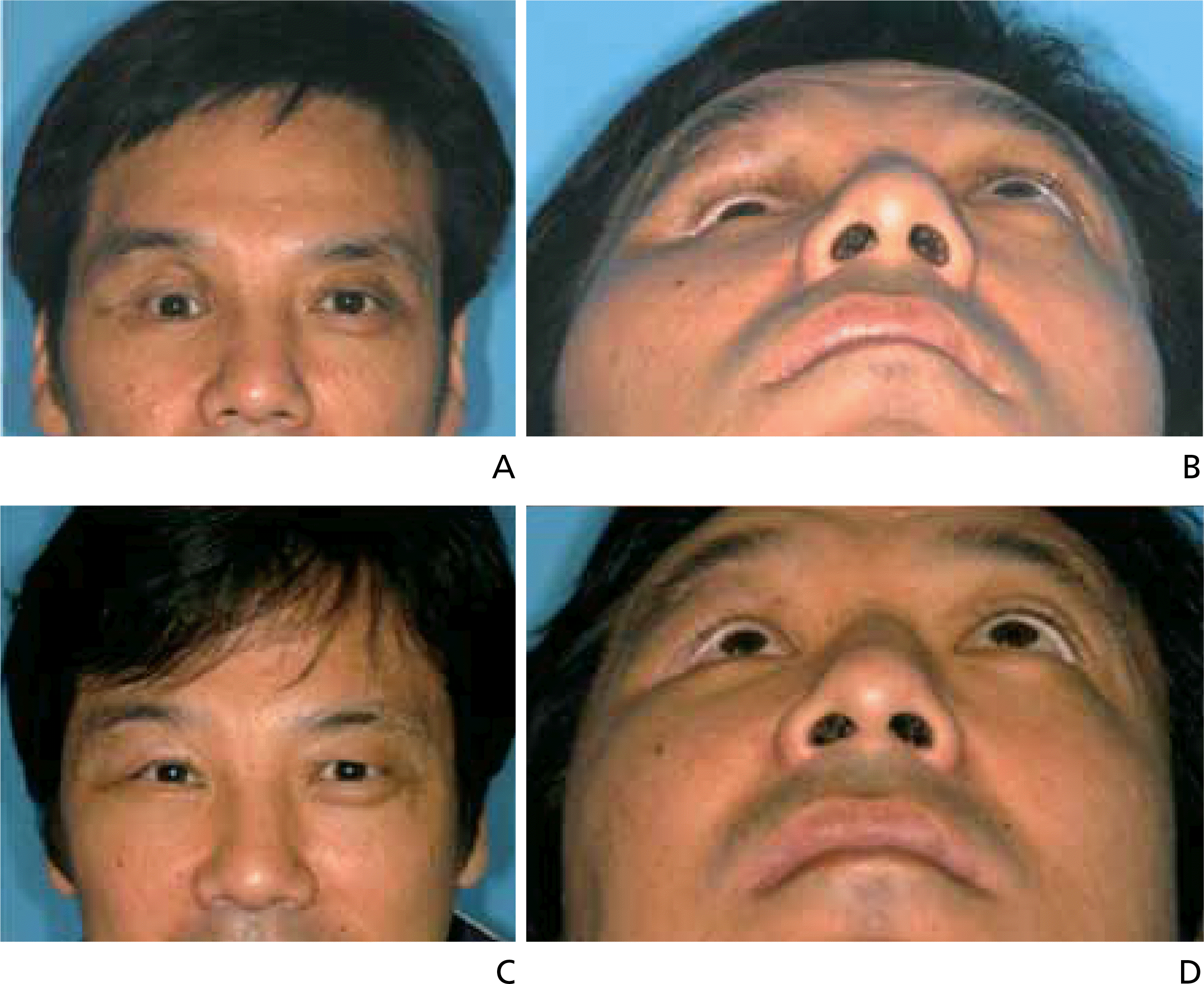
 | Figure 1.(A,B) Preoperative view of the patient. He showed 3 mm enophthalmos, 2 mm hypoophthal-mos, pseudoptosis, supratarsal fold deepening, mid face widening and retrusion, and cheek drooping. (C,D) Postoperative view after 6 months of surgery. Improvement facial deformities. Informed consent was obtained from the patient. |
 | Figure 2.(A) Preoperative view of the patient. This 12-year-old female patient was observed to have posttraumatic exotropia, hypotropia, and strabismus on the right eye. (B) Postoperative view after 12 months of surgery. (C) Postoperative view after 12 years of surgery (From Kim YH et al. J Craniofac Surg 2012;23:1005-1009, with permission from LWW Journals) [11]. |
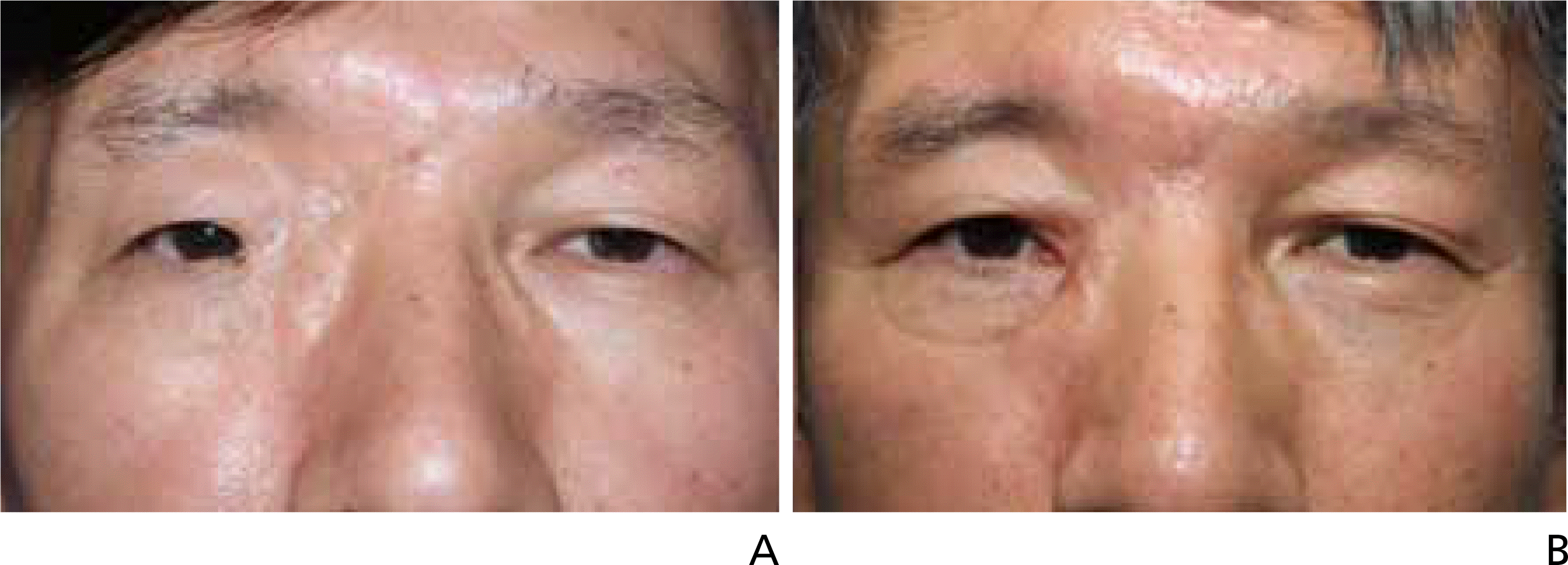
 | Figure 3.(A) Preoperative view of the patient. The distance of midline to medial canthus was 22 mm on the affected side and 35 mm on the other side. B. Postoperative view after 9 months of surgery. Telecanthus on the affected side was improved (From Kim TG et al. Ann Plast Surg 2014;72:164-168, with permission from LWW Journals) [46]. |


 PDF
PDF ePub
ePub Citation
 Print
Print


 XML Download