

 PDF
PDF ePub
ePub Citation
 Print
Print


Abstract
Methods
We reviewed the ocular records of 18 consecutive patients diagnosed with dermolipoma in our hospital between March 2016 and March 2018.
Results
The study population consisted of eight females and 10 males with a mean age of 61 months (range 3 months to 45 years old), with a pinkish conjunctival mass located at a mean of 4.7 mm (range 2–8 mm) from the temporal limbus and anterior to the lateral orbital rim. The mean visual acuity of eight patients in whom it was possible to measure visual acuity was 0.9 (range 0.6–1.0). Surgery was performed in two eyes. Pathological examination revealed conjunctival dermolipoma. Postoperatively, there was no evidence of visual changes or specific complications.
Go to : 
References
1. Mansour AM, Barber JC, Reinecke RD, Wang FM. Ocular choristomas. Surv Opthalmol. 1989; 33:339–58.

2. Cho BJ, Han YK, Hyon JY, et al. Two cases of cosmetic treatment of limbal dermoid. J Korean Ophthalmol Soc. 2001; 48:1419–24.
3. Spencer WH. Ophthalmic pathology: an atlas and textbook. 4th ed.Vol. 4. Philadelphia: W.B.Saunders;1996. p. 50.
4. Shields JA, Augsburger JJ, Donoso LA. Orbital dermoid cyst of conjunctival origin. Am J Ophthalmol. 1986; 101:726–9.
5. Lee SH, Kim KH, Kim SY. A case of conjunctival dermoid cyst of the orbit. J Korean Ophthalmol Soc. 2010; 51:601–5.
6. Shields JA, Kaden IH, Eagle RC Jr, Shields CL. Orbital dermoid cysts: clinicopathologic correlations, classification, and management. The 1997 Josephine E. Schueler Lecture. Ophthal Plast Reconstr Surg. 1997; 13:265–76.
7. Kim YD, Robert AG. Orbital fat prolapse and dermolipoma: two distinct entities. Korean J Ophthalmol. 1994; 8:42–3.
8. Kim E, Kim HJ, Kim YD, et al. Subconjunctival fat prolapse and dermolipoma of the orbit: Differentiation on CT and MR imaging. AJNR Am J Neuroradiol. 2011; 32:465–7.
9. Yoon CK, Kim JH, Kim SJ, Yu YS. The surgical outcomes of simple keratectomy for limbal dermoids. J Korean Ophthalmol Soc. 2008; 49:897–903.
10. Jeong J, Song YJ, Jung SI, Kwon JW. New surgical approach for limbal dermoids in children: simple excision, corneal tattooing and sutureless limboconjunctival autograft. Cornea. 2015; 34:720–3.
11. Zloto O, Rosner M. An unusual presentation of conjunctival dermolipoma with a skin tag. J Carniofac Surg. 2018; 29:e302–3.
12. Nevares RL, Mulliken JB, Robb RM. Ocular dermoids. Plast Reconstr Surg. 1988; 82:959–64.
13. Sa HS, Kim HK, Shin JA, et al. Dermolipoma surgery with rotational conjunctival flaps. Acta Ophthalmol. 2012; 90:86–90.
14. Choi YJ, Kim IH, Choi JH, et al. Early results of surgical management of conjunctival dermolipoma: partial excision and free conjunctival autograft. Br J Ophthalmol. 2015; 99:1031–6.
15. Glover AT, Grove AS Jr. Subconjunctival orbital fat prolapse. Ophthalmic Plast Reconstr Surg. 1987; 3:83–6.
Go to :
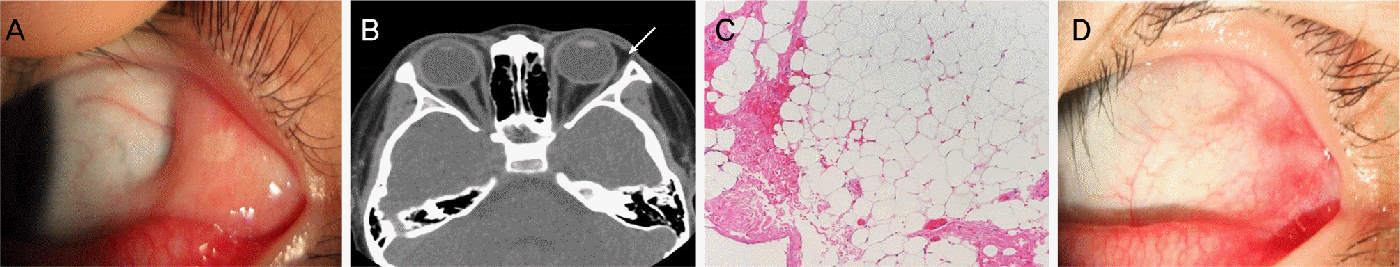 | Figure 1.Pre and Postoperative slit lamp photographs (A, D), preoperative orbital computed tomography (CT) (B) and histopathologic photographs (C) of the conjunctival mass in the left eye in 5-year-old boy. (A) At initial presentation, there is a dermolipoma in the temporal bulbar conjunctiva. (B) Preoperative orbital CT shows crescent-shaped fatty mass at the superotemporal aspect of the right epibulbar area, which abuts the lateral wall of the eyeball, anterior to the insertion of the lateral rectus muscle and medial to the lacrimal gland (arrow). (C) Histological examination shows abundant adipose tissue with collagenous bundles (hematoxylin and eosin [H&E] stain, ×200). (D) 1 month after surgery, there was clear excision site only with very slight conjunctival injection. A good cosmesis was achieved. |
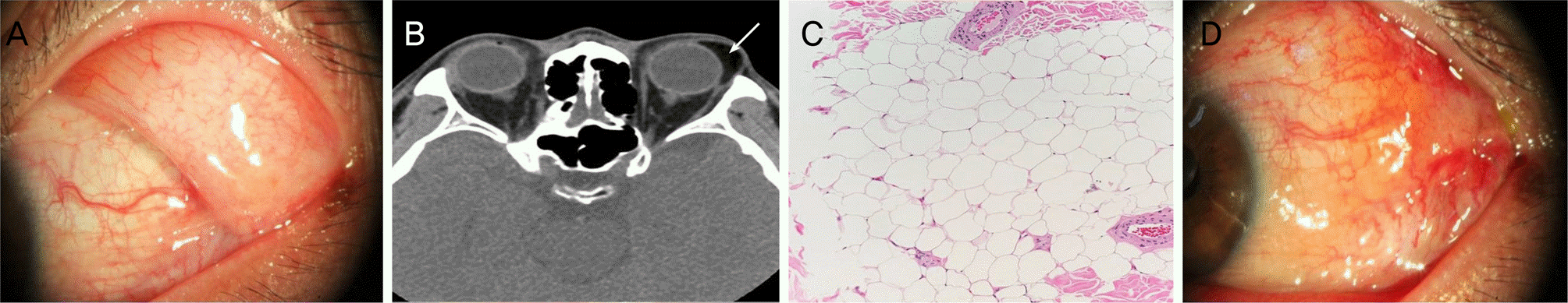 | Figure 2.Pre and postoperative slit lamp photographs (A, D), preoperative orbital computed tomography (CT) (B) and histopathologic photographs (C) of conjunctival mass in the left eye in 45-year-old man. (A) Temporal conjunctival mass in the superotemporal area is shown. (B) Preoperative orbital CT shows crescent-shaped fatty mass from lateral conjunctiva to the area of lateral rectus muscle (arrow). (C) Histological examination reveals abundant adipose tissue and collagen bundles (hematoxylin and eosin [H&E] stain, ×200). (D) 1 week after surgery, slight subconjunctival hemorrhage and injection were shown. |
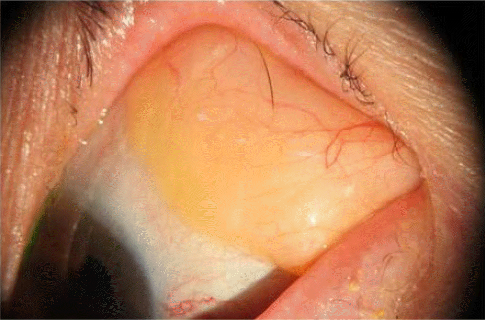 | Figure 3.A slit lamp photograph of 75-year-old women presented with a 1-year history of progressive swelling over the left eye. She had no other clinically significant medical history or trauma. There was a soft, yellowish mass in the outer temporal subconjunctival region. |
Table 1.
Dermographics and clinical characteristics of 18 patients who were clinically diagnosed with conjunctival dermolipoma
Table 2.
Comparison between orbital fat prolapse and conjunctival dermolipoma
 XML Download