

 PDF
PDF ePub
ePub Citation
 Print
Print


Abstract
Purpose
We report a case of unilateral, focal, pigmented paravenous retinochoroidal atrophy (PPRCA).
Case summary
A 46-year-old female visited our clinic in complaint of a vague problem with her right eye identified during a general medical examination. The visual acuity (without correction) of both eyes was 1.0. Slit-lamp examination of both eyes revealed no specific signs. Fundus examination of the right eye revealed focal, bony-spicule-shaped retinochoroidal atrophy with pigmentation along the course of the superior retinal vein. A fundus autofluorescence examination revealed principally hypofluorescence with some hyperfluorescence at the margin of the atrophic retinochoroidal lesion. Optical coherence tomography revealed mixed clumping and atrophy of the retinal pigment epithelium (RPE) layer and thinning of the choriocapillaris layer. Fluorescence angiography revealed a window defect and blockage at the site of the lesion (the fluorescent material did not enter the lesion). The site of the window defect was in correlation with the atrophic RPE region. The site of the blockage at lesion also matched with the site of the regional pigment clumping. No definite leakage was observed.
Figures and Tables
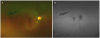 | Figure 1Wide fundus photography and fundus autofluorescence of the patient's right eye. (A) Focal bony- spicule shaped retinochoroidal atrophy with pigmentation along superior retinal vein was noted. (B) Besides some hyperfluorescence at margin of retinochoroidal atrophy, most part of the lesion showed hypofluorescence.
|
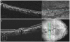 | Figure 2Optical coherence tomography of the patient's right eye. (A) Mixed signals of clumping and thinning of retinal pigment epithelium layer were noted. (B) Thinning of choriocapillaris layer was noted at the lesion site while choriocapillaris layer thickness was within normal range at locations other than the lesion site. S = superior; N = nasal; I = inferior; T = temporal.
|

References
1. Huang HB, Zhang YX. Pigmented paravenous retinochoroidal atrophy (review). Exp Ther Med. 2014; 7:1439–1445.

2. Aoki S, Inoue T, Kusakabe M, et al. Unilateral pigmented paravenous retinochoroidal atrophy with retinitis pigmentosa in the contralateral eye: a case report. Am J Ophthalmol Case Rep. 2017; 8:14–17.
3. Peduzzi M, Guerrieri F, Torlai F, Prampolini ML. Bilateral pigmented paravenous retino-choroidal degeneration following measles. Int Ophthalmol. 1984; 7:11–14.
4. Fernandez-Sanz G, Carreño E, Mall S, et al. Unilateral pigmented paravenous retinochoroidal atrophy associated with presumed ocular tuberculosis. Ophthalmic Surg Lasers Imaging Retina. 2017; 48:345–349.
5. Traboulsi EI, Maumenee IH. Hereditary pigmented paravenous chorioretinal atrophy. Arch Ophthalmol. 1986; 104:1636–1640.
6. Noble KG. Hereditary pigmented paravenous chorioretinal atrophy. Am J Ophthalmol. 1989; 108:365–369.
7. Cheung DS. Pigmented paravenous chorioretinal atrophy. Am J Ophthalmol. 1984; 97:113.
8. Charbel Issa P, Scholl HP, Helb HM, et al. Unilateral pigmented paravenous retinochoroidal atrophy. Klin Monbl Augenheilkd. 2007; 224:791–793.
9. Bozkurt N, Bavbek T, Kazokoğlu H. Hereditary pigmented paravenous chorioretinal atrophy. Ophthalmic Genet. 1998; 19:99–104.
10. Ahmad M, Leisy H, Carr RE, Smith RT. A rare case of unifocal, unilateral pigmented paravenous retinochoroidal atrophy (PPRCA). Am J Ophthalmol Case Rep. 2016; 4:41–44.
11. Barteselli G. Fundus autofluorescence and optical coherence tomography findings in pigmented paravenous retinochoroidal atrophy. Can J Ophthalmol. 2014; 49:e144–e146.
12. Skalka HW. Hereditary pigmented paravenous retinochoroidal atrophy. Am J Ophthalmol. 1979; 87:286–291.
13. Ratra D, Chandrasekharan DP, Aruldas P, Ratra V. Concurrent retinitis pigmentosa and pigmented paravenous retinochoroidal atrophy phenotypes in the same patient. Indian J Ophthalmol. 2016; 64:775–777.
14. Choi JY, Sandberg MA, Berson EL. Natural course of ocular function in pigmented paravenous retinochoroidal atrophy. Am J Ophthalmol. 2006; 141:763–765.
15. Pearlman JT, Heckenlively JR, Bastek JV. Progressive nature of pigmented paravenous retinochoroidal atrophy. Am J Ophthalmol. 1978; 85:215–217.
 XML Download