

 PDF
PDF ePub
ePub Citation
 Print
Print


Abstract
Purpose
To evaluate the availability of ultra-wide field fundus photography based on eye steering technique to diagnose retinal breaks in patients with symptomatic posterior vitreous detachment (PVD).
Methods
The medical records of patients with symptomatic PVD were reviewed. Retinal breaks were independently identified using four eye steering capture images of ultra-wide field fundus photographs. The sensitivity and specificity of eye steering capture imaging for diagnosing retinal breaks were calculated.
Results
A total of 94 eyes of 94 patients were included. Using fundus examination after pupil dilatation, retinal breaks were diagnosed in 42 (45%) eyes. The sensitivity of the eye steering capture imaging was 98% (95% confidence interval [CI]: 88–100%), and the specificity was 98% (95% CI: 90–100%). Of the 58 retinal tears, 28 (97%) involving the superior quadrant, 10 (100%) involving the inferior quadrant, 6 (100%) involving the nasal quadrant, and 13 (100%) involving the temporal quadrant were identified using eye steering capture images.
Figures and Tables
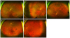 | Figure 1A standard capture image (A) and 4 eye steering capture images at superior (C), inferior (D), nasal (B), and temporal (E). A retinal break was detected at superior quadrant (C) with an eye steering capture image but not with the standard capture image (A).
|
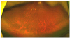 | Figure 2An eye steering capture image of a false positive case. At inferior quadrant, a lesion presumed to be retinal tear is identified. This lesion was described as the paving-stone degeneration in the medical record.
|

Notes
References
1. Hikichi T, Hirokawa H, Kado M, et al. Comparison of the prevalence of posterior vitreous detachment in whites and Japanese. Ophthalmic Surg. 1995; 26:39–43.

2. Oh JH, Oh J, Roh HC. Vitreous hyper-reflective dots in optical coherence tomography and retinal tear in patients with acute posterior vitreous detachment. Curr Eye Res. 2017; 42:1179–1184.
3. Shea M, Davis MD, Kamel I. Retinal breaks without detachment, treated and untreated. Mod Probl Ophthalmol. 1974; 12:97–102.
4. Campbell JP, Leder HA, Sepah YJ, et al. Wide-field retinal imaging in the management of noninfectious posterior uveitis. Am J Ophthalmol. 2012; 154:908–911.
5. Kornberg DL, Klufas MA, Yannuzzi NA, et al. Clinical utility of ultra-widefield imaging with the optos optomap compared with indirect ophthalmoscopy in the setting of non-traumatic rhegmatogenous retinal detachment. Semin Ophthalmol. 2016; 31:505–512.
6. Prasad PS, Oliver SC, Coffee RE, et al. Ultra wide-field angiographic characteristics of branch retinal and hemicentral retinal vein occlusion. Ophthalmology. 2010; 117:780–784.
7. Silva PS, Cavallerano JD, Sun JK, et al. Peripheral lesions identified by mydriatic ultrawide field imaging: distribution and potential impact on diabetic retinopathy severity. Ophthalmology. 2013; 120:2587–2595.
8. Soliman AZ, Silva PS, Aiello LP, Sun JK. Ultra-wide field retinal imaging in detection, classification, and management of diabetic retinopathy. Semin Ophthalmol. 2012; 27:221–227.
9. Spaide RF. Peripheral areas of nonperfusion in treated central retinal vein occlusion as imaged by wide-field fluorescein angiography. Retina. 2011; 31:829–837.
10. Theodoropoulou S, Ainsworth S, Blaikie A. Ultra-wide field imaging of retinopathy of prematurity (ROP) using Optomap-200TX. BMJ Case Rep. 2013; 2013:pii: bcr2013200734.
11. Wessel MM, Aaker GD, Parlitsis G, et al. Ultra-wide-field angiography improves the detection and classification of diabetic retinopathy. Retina. 2012; 32:785–791.
12. Lee DH, Kim SS, Kim M, Koh HJ. Identifiable peripheral retinal lesions using ultra-widefield scanning laser ophthalmoscope and its usefulness in myopic patients. J Korean Ophthalmol Soc. 2014; 55:1814–1820.
13. Mackenzie PJ, Russell M, Ma PE, et al. Sensitivity and specificity of the optos optomap for detecting peripheral retinal lesions. Retina. 2007; 27:1119–1124.
14. Khandhadia S, Madhusudhana KC, Kostakou A, et al. Use of optomap for retinal screening within an eye casualty setting. Br J Ophthalmol. 2009; 93:52–55.
15. Ahn HM, Rim TH, Chung EJ. Diagnostic availability of ultra-wide-field fundus imaging in Korean patient with retinal break. J Korean Ophthalmol Soc. 2016; 57:1254–1259.
16. Ghasemi Falavarjani K, Wang K, Khadamy J, Sadda SR. Ultrawide-field imaging in diabetic retinopathy; An overview. J Curr Ophthalmol. 2016; 28:57–60.
17. Mojana F, Kozak I, Oster SF, et al. Observations by spectral-domain optical coherence tomography combined with simultaneous scanning laser ophthalmoscopy: imaging of the vitreous. Am J Ophthalmol. 2010; 149:641–650.
 XML Download