Journal List > J Korean Ophthalmol Soc > v.59(12) > 1109301

Park, Son, Kim, Kim, Yoon, and Lee: Intravitreal Anti-vascular Endothelial Growth Factor Injections to Treat Neovascular Age-related Macular Degeneration: Long-term Treatment Outcomes

Abstract

Purpose

We assessed the visual and anatomical outcomes, and the safety profile of long-term intravitreal anti-vascular endothelial growth factor (VEGF) injections (aflibercept, ranibizumab, and bevacizumab) given to treat neovascular age-related macular degeneration (NAMD).

Methods

We analyzed medical records collected over 7 years of treatment-naive NAMD patients who received outpatient clinic-based intravitreal anti-VEGF injections. All were treated employing either “treat-and-extend” or “as needed” protocols at the discretion of the retinal specialist. The number of injections, adverse events associated with injection, and measures of visual acuity (VA), central foveal thickness (CFT), and intraocular pressure (IOP) were recorded.

Results

Overall, we assessed 196 eyes of 196 patients (average age 68.6 ± 9.6 years; 77 females). Patients received an average of 17.3 ± 13.5 injections over 78.0 ± 16.5 months of clinical follow-up. The initial mean VA (logMAR) was 0.75 ± 0.58 and the CFT was 349.7 ± 152.6 µm. Both parameters exhibited maximal improvements at the 6-month visit (p < 0.05). However, the clinical outcomes worsened over the 7-year clinical course; the best-corrected visual acuity (BCVA) was 0.91 ± 0.78 and the CFT was 284.5 ± 105.8 µm at 7 years. The BCVA at 7 years was significantly correlated with the initial BCVA. IOP-related events increased 11-fold and anterior chamber reactions increased 3-fold over the years, but no significant complications such as endophthalmitis were recorded.

Conclusions

The use of intravitreal anti-VEGF agents was associated with initial visual improvements over 6 months but did not prevent the worsening of NAMD over 5 years. The BCVA at the initial visit was a strong predictor of the final BCVA. A more intensive injection schedule might improve long-term outcomes.

Figures and Tables

Figure 1

Temporal changes in the best-corrected visual acuity (BCVA). (A) Temporal change of mean BCVA over 7 years (in ETDRS letters; Snellen visual acuity scores were converted to ETDRS letter scores). (B) Temporal change of mean BCVA over 7 years (in ETDRS letter; converted in same manner as above graph) after imputation of missing values by the last-observation-carried-forward method (n = 37). ETDRS = early treatment diabetic retinopathy study; n = number of patients at the time of BCVA measurements.

jkos-59-1142-g001
Figure 2

Temporal change in central foveal thickness. The central foveal thickness improved gradually during the first 6 months, and maintained over 5 years.

jkos-59-1142-g002
Figure 3

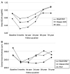
Visual outcome (A), anatomical outcome (B) for different CNV subtypes in the subgroup of 65 patients who completed the 7-year follow-up. Occult CNV (n = 45), classic CNV (n = 5), PCV (n = 15). There was no significant difference between groups during follow-up periods (repeated measured ANOVA). Mean visual acuity and central foveal thickness of each group decreased over time and stabilized. CNV = choroidal neovascularization; PCV = polypoidal choroidal vasculopathy; BCVA = best-corrected visual acuity; n = number of eyes with CNV subtype; ANOVA = analysis of variance.

jkos-59-1142-g003
Table 1

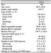
Baseline characteristics of patients with neovascular age-related macular degeneration who satisfied inclusion criteria

jkos-59-1142-i001

Values are presented as mean ± standard deviation or number (%) unless otherwise indicated.

CNV = choroidal neovascularization; RAP = retinal angiomatous proliferation; PCV = polypoidal choroidal vasculopathy; BCVA = best-corrected visual acuity; logMAR = logarithm of minimal angle of resolution; CFT = central foveal thickness; VEGF = vascular endothelial growth factor.

Table 2

Mean changes in BCVA (logMAR) from baseline after intravitreal anti-vascular endothelial growth factor injection

jkos-59-1142-i002

Values are presented as mean ± standard deviation unless otherwise indicated. (a) visual acuity of drop-outs is disregarded (b) visual acuity of drop-outs is carried forward and included in the calculations.

BCVA = best-corrected visual acuity; VA = visual acuity.

*Wilcoxon signed ranks test. p-value comparing each group to baseline BCVA (logMAR). There was no statistically significant difference in mean BCVA between the cohort without drop-outs and that with drop-outs included (all p > 0.05).

Table 3

Mean changes in central foveal thickness from baseline after intravitreal anti-vascular endothelial growth factor injection

jkos-59-1142-i003

Values are presented as mean ± standard deviation unless otherwise indicated.

CFT = central foveal thickness.

*Wilcoxon signed ranks test.

Table 4

Association between characteristics and BCVA at 7th year (n = 88)

jkos-59-1142-i004

BCVA = best-corrected visual acuity; logMAR = logarithm of minimal angle of resolution; r = Pearson correlation coefficient.

*Analysis was performed using Pearson correlation analysis.

Notes

Conflicts of Interest The authors have no conflicts to disclose.

References

1. Wong TY, Chakravarthy U, Klein R, et al. The natural history and prognosis of neovascular age-related macular degeneration: a systematic review of the literature and meta-analysis. Ophthalmology. 2008; 115:116–126.
2. Park SJ, Kwon KE, Choi NK, et al. Prevalence and incidence of exudative age-related macular degeneration in South Korea: a nationwide population-based study. Ophthalmology. 2015; 122:2063.e1–2070.e1.
3. Rosenfeld PJ, Brown DM, Heier JS, et al. Ranibizumab for neovascular age-related macular degeneration. N Engl J Med. 2006; 355:1419–1431.
crossref
4. Heier JS, Brown DM, Chong V, et al. Intravitreal aflibercept (VEGF trap-eye) in wet age-related macular degeneration. Ophthalmology. 2012; 119:2537–2548.
crossref
5. Grunwald JE, Daniel E, Huang J, et al. Risk of geographic atrophy in the comparison of age-related macular degeneration treatments trials. Ophthalmology. 2014; 121:150–161.
6. Krüger Falk M, Kemp H, Sørensen TL. Four-year treatment results of neovascular age-related macular degeneration with ranibizumab and causes for discontinuation of treatment. Am J Ophthalmol. 2013; 155:89.e3–95.e3.
crossref
7. Zhu M, Chew JK, Broadhead GK, et al. Intravitreal ranibizumab for neovascular age-related macular degeneration in clinical practice: five-year treatment outcomes. Graefes Arch Clin Exp Ophthalmol. 2015; 253:1217–1225.
crossref
8. Berg K, Roald AB, Navaratnam J, Bragadóttir R. An 8-year follow-up of anti-vascular endothelial growth factor treatment with a treat-and-extend modality for neovascular age-related macular degeneration. Acta Ophthalmol. 2017; 95:796–802.
crossref
9. Garweg JG, Zirpel JJ, Gerhardt C, Pfister IB. The fate of eyes with wet AMD beyond four years of anti-VEGF therapy. Graefes Arch Clin Exp Ophthalmol. 2018; 256:823–831.
crossref
10. Jang HJ, Song SJ, Bae JH. Long-term effect of intravitreal ranibizumab injection on choroidal neovascularization in age-related macular degeneration. J Korean Ophthalmol Soc. 2013; 54:1359–1364.
crossref
11. DiLoreto DA Jr, Bressler NM, Bressler SB, Schachat AP. Use of best and final visual acuity outcomes in ophthalmological research. Arch Ophthalmol. 2003; 121:1586–1590.
crossref
12. Chong V. Ranibizumab for the treatment of wet AMD: a summary of real-world studies. Eye (Lond). 2016; 30:270–286.
crossref
13. Frennesson CI, Nilsson SE. A three-year follow-up of ranibizumab treatment of exudative AMD: impact on the outcome of carrying forward the last acuity observation in drop-outs. Acta Ophthalmol. 2014; 92:216–220.
crossref
14. Rofagha S, Bhisitkul RB, Boyer DS, et al. Seven-year outcomes in ranibizumab-treated patients in ANCHOR, MARINA, and HORIZON: a multicenter cohort study (SEVEN-UP). Ophthalmology. 2013; 120:2292–2299.
15. Gillies MC, Campain A, Barthelmes D, et al. Long-term outcomes of treatment of neovascular age-related macular degeneration: data from an observational study. Ophthalmology. 2015; 122:1837–1845.
16. Comparison of Age-related Macular Degeneration Treatments Trials (CATT) Research Group. Maguire MG, Martin DF, et al. Five-year outcomes with anti-vascular endothelial growth factor treatment of neovascular age-related macular degeneration: the comparison of age-related macular degeneration treatments trials. Ophthalmology. 2016; 123:1751–1761.
17. Peden MC, Suñer IJ, Hammer ME, Grizzard WS. Long-term outcomes in eyes receiving fixed-interval dosing of anti-vascular endothelial growth factor agents for wet age-related macular degeneration. Ophthalmology. 2015; 122:803–808.
crossref
18. Holz FG, Tadayoni R, Beatty S, et al. Multi-country real-life experience of anti-vascular endothelial growth factor therapy for wet age-related macular degeneration. Br J Ophthalmol. 2015; 99:220–226.
crossref
19. Rasmussen A, Bloch SB, Fuchs J, et al. A 4-year longitudinal study of 555 patients treated with ranibizumab for neovascular age-related macular degeneration. Ophthalmology. 2013; 120:2630–2636.
crossref
20. Grunwald JE, Pistilli M, Ying GS, et al. Growth of geographic atrophy in the comparison of age-related macular degeneration treatments trials. Ophthalmology. 2015; 122:809–816.
crossref
21. Xu L, Mrejen S, Jung JJ, et al. Geographic atrophy in patients receiving anti-vascular endothelial growth factor for neovascular age-related macular degeneration. Retina. 2015; 35:176–186.
crossref
22. Sarks J, Tang K, Killingsworth M, et al. Development of atrophy of the retinal pigment epithelium around disciform scars. Br J Ophthalmol. 2006; 90:442–446.
crossref
23. Tanaka E, Chaikitmongkol V, Bressler SB, Bressler NM. Vision-threatening lesions developing with longer-term follow-up after treatment of neovascular age-related macular degeneration. Ophthalmology. 2015; 122:153–161.
crossref
24. Munk MR, Ceklic L, Ebneter A, et al. Macular atrophy in patients with long-term anti-VEGF treatment for neovascular age-related macular degeneration. Acta Ophthalmol. 2016; 94:e757–e764.
crossref
25. Bhisitkul RB, Mendes TS, Rofagha S, et al. Macular atrophy progression and 7-year vision outcomes in subjects from the ANCHOR, MARINA, and HORIZON studies: the SEVEN-UP study. Am J Ophthalmol. 2015; 159:915.e2–924.e2.
crossref
TOOLS
Similar articles