

 PDF
PDF ePub
ePub Citation
 Print
Print


Abstract
Purpose
We report the clinical outcomes of patients undergoing additional widening of the limbal incision to prevent disease recurrence after conjunctivo-limbal autograft combined with pterygial excision.
Methods
We retrospectively compared 95 eyes with primary and 12 eyes with recurrent pterygia randomized to treatment via two surgical methods: conjunctivo-limbal autograft alone and combined with widening of the limbal incision to allow for pterygial removal. We widened the limbal incisions by 1 mm on both the superior and inferior limbal margins and removed the pterygia. The outcomes were compared between 39 eyes undergoing conjunctivo-limbal autograft alone and 68 eyes undergoing conjunctivolimbal autograft with widening of the limbal incision.
Results
The mean overall postoperative follow-up period was 29.6 ± 10.5 months and conjunctivo-limbal autograft alone group was 27.4 ± 11.5 months, combined with widening of the limbal incision group was 30.7 ± 9.7 months. Six pterygia (15.4%; four primary and two recurrent) developed in those undergoing conjunctivo-limbal autograft alone and two (2.9%; one primary and one recurrent) in those undergoing additional widening of the limbal incision; the recurrence rate differed significantly between the two groups (p < 0.05). In the group treated with conjunctivo-limbal autograft alone, the mean time to development of a new primary pterygium was 6.3 ± 3.4 months and that to development of a recurrent pterygium 4.3 ± 2.5 months; the respective values for the group undergoing additional widening of the limbal incision were 12.1 ± 2.6 and 8.4 ± 4.6 months; the recurrence rates differed significantly (p < 0.05).
Go to : 
References
1. Taylor HR, West S, Muñoz B, et al. The long-term effects of visible light on the eye. Arch Ophthalmol. 1992; 110:99–104.

2. Di Girolamo N, Chui J, Coroneo MT, Wakefield D. Pathogenesis of pterygia: role of cytokines, growth factors, and matrix metalloproteinases. Prog Retin Eye Res. 2004; 23:195–228.
3. Chui J, Coroneo MT, Tat LT, et al. Ophthalmic pterygium: a stem cell disorder with premalignant features. Am J Pathol. 2011; 178:817–27.
4. Kim YJ, Yoo SH, Chung JK. Reconstruction of the limbal vasculature after limbal-conjunctival autograft transplantation in abdominal surgery: an angiography study. Invest Ophthalmol Vis Sci. 2014; 55:7925–33.
5. Rim H, Kang MJ, Choi M, et al. The incidence and prevalence of pterygium in South Korea: a 10-year population-based Korean abdominal study. PLoS One. 2017; 12:e0171954.
6. Kim DI, Kim MK, Wee WR, Oh JY. Surgical outcome of primary pterygium excision with conjunctival autograft. J Korean Ophthalmol Soc. 2015; 56:856–62.
7. Ang LP, Chua JL, Tan DT. Current concepts and techniques in pterygium treatment. Curr Opin Ophthalmol. 2007; 18:308–13.
8. Hong SM, Ko SJ, Kim SD. Effect of inferior conjunctival abdominal flap surgery for primary pterygium. J Korean Ophthalmol Soc. 2009; 50:1774–9.
9. Gris O, Güell JL, del Campo Z. Limbal-conjunctival autograft abdominal for the treatment of recurrent pterygium. Ophthalmology. 2000; 107:270–3.
10. Zheng K, Cai J, Jhanji V, Chen H. Comparison of pterygium abdominal rates after limbal conjunctival autograft transplantation and other techniques: meta-analysis. Cornea. 2012; 31:1422–7.
11. Tan DT, Chee SP, Dear KB, Lim AS. Effect of pterygium abdominal on pterygium recurrence in a controlled trial comparing abdominal autografting with bare sclera excision. Arch Ophthalmol. 1997; 115:1235–40.
12. Allan BD, Short P, Crawford GJ, et al. Pterygium excision with conjunctival autografting: an effective and safe technique. Br J Ophthalmol. 1993; 77:698–701.
13. Kwon SH, Kim HK. Analysis of recurrence patterns following pterygium surgery with conjunctival autografts. Medicine (Baltimore). 2015; 94:e518.
14. KEEDS. Cornea. 3rd ed.Seoul: Ilchokak Publishing Co., Ltd.;2013. p. 771–5.
15. Tseng SC. Concept and application of limbal stem cells. Eye (Lond). 1989; 3(Pt 2):141–57.
16. Anquria P, Carmichael T, Ntuli S, Kitinya J. Chronic inflammatory cells and damaged limbal cells in pterygium. Afr Health Sci. 2013; 13:725–30.
17. Sebban A, Hirst LW. Pterygium recurrence rate at the Princess Alexandra Hospital. Aust N Z J Ophthalmol. 1991; 19:203–6.
18. Han HC, Kim JH, Lee DH. The changes of corneal higher-order aberrations after surgery according to pterygium size. J Korean Ophthalmol Soc. 2014; 55:32–9.
19. Hirst LW, Axelsen RA, Schwab I. Pterygium and associated ocular surface squamous neoplasia. Arch ophthalmol. 2009; 127:31–2.
20. Hacı oğ lu D1, Erdöl H. Developments and current approaches in the treatment of pterygium. Int Ophthalmol. 2017; 37:1073–81.
21. Young AL, Ho M, Jhanji V, Cheng LL. Ten-year results of a randomized controlled trial comparing 0.02% mitomycin C and limbal conjunctival autograft in pterygium surgery. Ophthalmology. 2013; 120:2390–5.
22. Murube J. Pterygium: its treatment with beta therapy. Ocul Surf. 2009; 7:3–9.
23. Khan MS, Malik S, Basit I. Effect of intralesional 5 fluorouracil abdominal in primary pterygium. Pak J med Sci. 2016; 32:130–3.
24. Rúa O, Larráyoz IM, Barajas MT, et al. Oral doxycycline reduces pterygium lesions; results from a double blind, randomized, abdominal controlled clinical trial. PLoS One. 2012; 7:e52696.
25. Chen KH, Hsu WM. Intraoperative ethanol treatment as an adjuvant therapy of pterygium excision. Int J Biomed Sci. 2006; 2:414–21.
26. Kim YI, Lee GY, Kim EJ, et al. The Effect of subconjunctival abdominal Injection before conjunctival autograft for pterygium. J Korean Ophthalmol Soc. 2015; 56:847–55.
27. Besharati Manaviat, Souzani A. Subconjunctival bevacizumab abdominal in treatment of pterygium. Acta Med Iran. 2011; 49:179–83.
28. Kim CH, Lee JK, Park DJ. Recurrence rates of amniotic membrane transplantation, conjunctival autograft and conjunctivolimbal abdominal in primary pterygium. J Korean Ophthalmol Soc. 2009; 50:1780–8.
29. Gazzard G, Saw SM, Farook M, et al. Pterygium in Indonesia: prevalence, severity and risk factors. Br J Ophthalmol. 2002; 86:1341–6.
30. Kim KW, Kim JC. The clinical effect of micro-multiporous abdominal polyeterafluoroethylene insertion for recurrent pterygium. J Korean Ophthalmol Soc. 2013; 54:416–26.
31. Kenyon KR, Tseng SC. Limbal autograft transplantation for ocular surface disorders. Ophthalmology. 1989; 96:709–22.
32. Hirst LW. Prospective study of primary pterygium surgery using pterygium extended removal followed by extended conjunctival transplantation. Ophthalmology. 2008; 115:1663–72.
33. Hirst LW. Recurrence and complications after 1,000 surgeries abdominal pterygium extended removal followed by extended abdominal transplant. Ophthalmology. 2012; 119:2205–10.
34. Lee JS, Ha SW, Yu S, et al. Efficacy and safety of a large abdominal autograft for recurrent pterygium. J Korean Ophthalmol Soc. 2017; 31:469–78.
35. Barraquer JI. Etiology, pathogenesis, and treatment of the pterygium. Trans New Orleans Acad Ophthalmol. 1980; 28:167–78.
36. Kria L, Ohira A, Amemiya T. Growth factors in cultured pterygium fibroblasts: immunohistochemical and ELISA analysis. Graefes Arch Clin Exp Ophthalmol. 1998; 236:702–8.
37. Kria L, Ohira A, Amemiya T. TNP-470 (a fungus-derived inhibitor of angiogenesis) reduces proliferation of cultured fibroblasts abdominal from primary pterygia: a possible drug therapy for pterygia. Curr Eye Res. 1998; 17:986–93.
38. John T. Pterygium excision and conjunctival mini-autograft: preliminary report. Eye (Lond). 2001; 15(Pt 3):292–6.
39. Hwang HS, Cho KJ, Rand G, et al. Optimal size of pterygium abdominal for limbal conjunctival autograft using fibrin glue in primary pterygia. BMC Ophthalmol. 2018; 18:135.
40. Bazzazi N, Ramezani A, Rabiee MA. A comparative study of abdominal autograft and minimally invasive pterygium surgery in primary pterygia. Pak J Biol Sci. 2010; 13:409–12.
41. Masters JS, Harris DJ. Low recurrence rate of pterygium after abdominal with conjunctival Limbal autograft: a retrospective study with long-term follow-up. Cornea. 2015; 34:1569–72.
Go to :
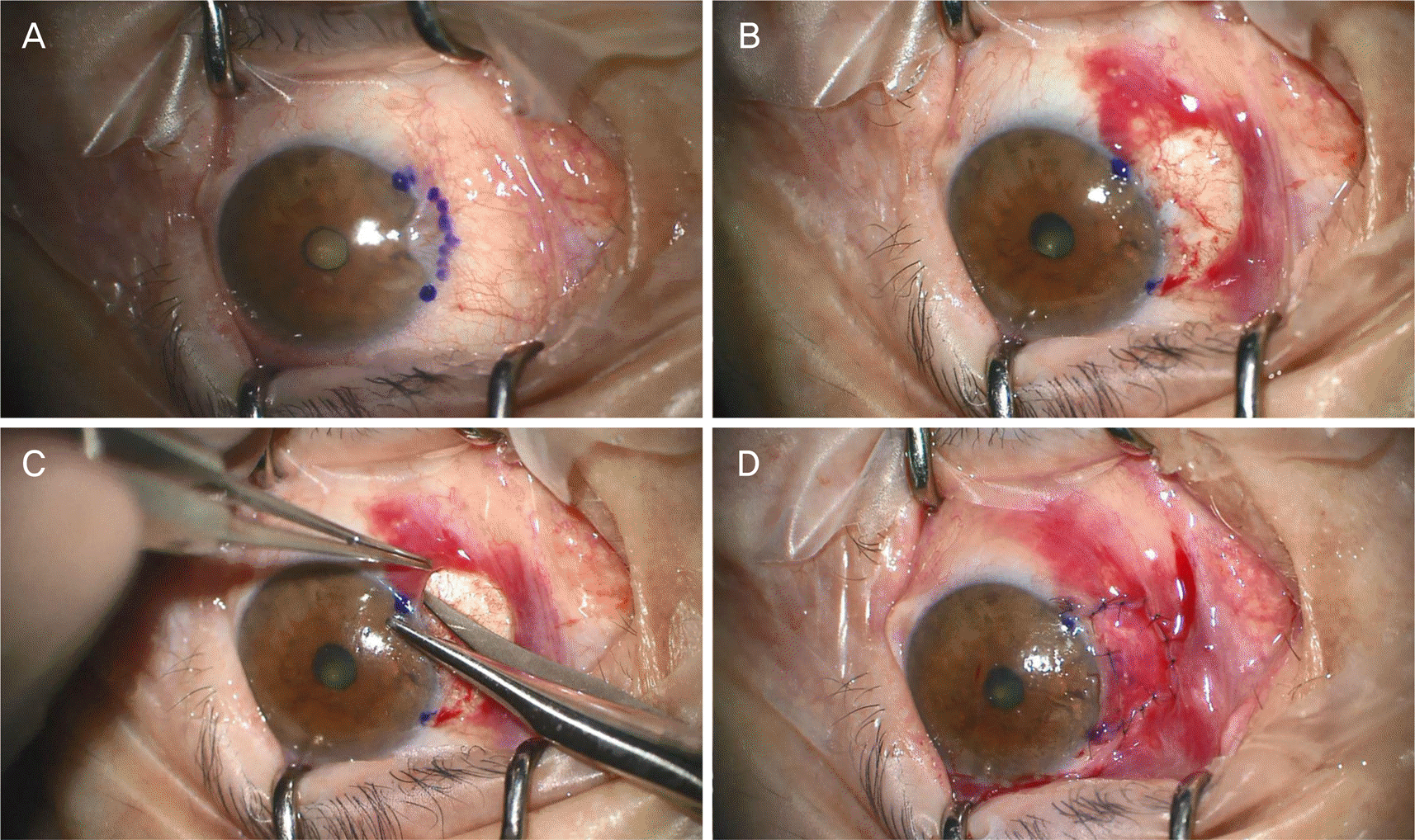 | Figure 1.Surgical technique of conjunctivo-limbal autograft combined with widening of the limbal incision in pterygium. (A) Pterygium incision area was marked by Gentian violet and the area where pterygium's body met limbus was also marked by Gentian violet to locate additional widening of limbal incision area. On each superior and inferior margin of limbus where the pterygium was removed. (B) Fibrovascular tissues that built up on the exposed sclera surface were removed sufficiently using Westcott scissor and #15 blades to eliminate any residuals in limbus. (C) Extra 1 mm incision was carried out in the additional widening limbal incision area where it was marked in (A). (D) After suture of both margins of limbus, 10–0 Nylon was grafted by interrupted suture and then fixation suture was carried out in episclera parallel to limbus to prevent grafted area from moving. |
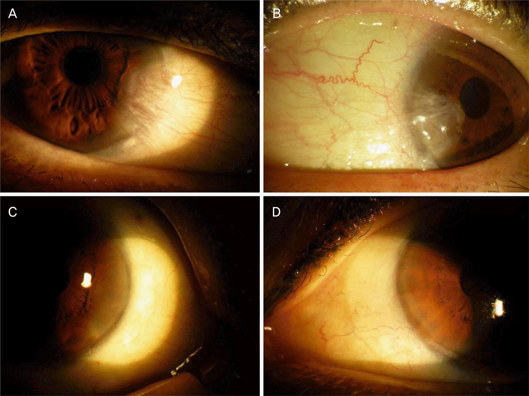 | Figure 2.The preoperative and postoperative photographs of patients who have pterygium with conjunctivo-limbal autograft alone and combined with widening of the limbal incision. (A, B) Preoperative anterior segment photos of pterygium. (C) Postoperative 6 months after conjunctivo-limbal autograft alone. (D) postoperative 6 months after conjunctivo-limbal autograft combined with widening of the limbal incision. |
Table 1.
Characteristics of patients
| Conjunctivolimbal autograft alone (n = 39) | Combined with widening of the limbal incision (n = 68) | p-value* | |
|---|---|---|---|
| Age (years) | 64.15 ± 9.47 | 63.46 ± 12.47 | 0.763 |
| Follow up period | 27.4 ± 11.5 | 30.7 ± 9.7 | 0.683 |
| Gender (male:female) | 18:16 | 26:35 | 0.334 |
| Primary:recurrence | 37:2 | 58:10 | 0.242 |
Table 2.
Comparison of recurrence rates and time to recurrece in patients with pterygium using conjunctivo-limbal autograft alone and combined with widening of the limbal incision
| Conjunctivolimbal autograft alone (n = 39) | Combined with widening of the limbal incision (n = 68) | p-value | |
|---|---|---|---|
| Primary | 4/37 | 1/58 | 0.049* |
| Mean time to recurrence (months) | 6.3 ± 3.4 | 12.1 ± 2.6 | 0.000† |
| Recurrent | 2/2 | 1/10 | 0.045* |
| Mean time to recurrence (months) | 4.3 ± 2.5 | 8.4 ± 4.6 | 0.000† |
| Total | 6/39 | 2/68 | 0.049* |
| Mean time to recurrence (months) | 5.5 ± 3.1 | 10.6 ± 3.4 | 0.000† |
 XML Download