

 PDF
PDF ePub
ePub Citation
 Print
Print


Abstract
Purpose
To evaluate the clinical effect of intense regulated pulse light (IRPL) and changes in tear film thickness in dry eye patients.
Methods
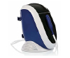
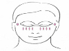
A retrospective study was conducted on 25 patients with Meibomian gland dysfunction who had subjective dry eye discomfort and whose tear film break-up time (TBUT) was < 10 seconds. All patients were treated with IRPL three times on days 1, 15, and 45. TBUT, Schirmer 1 test results, cornea staining score with fluorescein, ocular surface disease index (OSDI), and lipid layer thickness were measured and compared before and after the procedure.
Results
TBUT was significantly increased from 3.7 ± 1.2 s to 4.4 ± 2.1 s after IRPL (p = 0.03). Schirmer 1 test and cornea staining scores changed significantly (p < 0.01 and p = 0.01, respectively). OSDI also showed a statistically significant improvement from 39.5 ± 17.5 points to 30.4 ± 18.7 points (p = 0.01). However, no significant difference in lipid layer thickness was observed (p = 0.49).
Figures and Tables
References
1. Uchino M, Schaumberg DA, Dogru M, et al. Prevalence of dry eye disease among Japanese visual display terminal users. Ophthalmology. 2008; 115:1982–1988.

2. Guo B, Lu P, Chen X, et al. Prevalence of dry eye disease in Mongolians at high altitude in China: the Henan eye study. Ophthalmic Epidemiol. 2010; 17:234–241.
3. Han SB, Hyon JY, Woo SJ, et al. Prevalence of dry eye disease in an elderly Korean population. Arch Ophthalmol. 2011; 129:633–638.
4. The definition and classification of dry eye disease: report of the definition and classification subcommittee of the international dry eye workShop (2007). Ocul Surf. 2007; 5:75–92.
5. Nelson JD, Shimazaki J, Benitez-del-Castillo JM, et al. The international workshop on meibomian gland dysfunction: report of the definition and classification subcommittee. Invest Ophthalmol Vis Sci. 2011; 52:1930–1937.
6. Nichols KK. The international workshop on meibomian gland dysfunction: introduction. Invest Ophthalmol Vis Sci. 2011; 52:1917–1921.
7. Hyon JY, Kim HM, Lee D, et al. Korean guidelines for the diagnosis and management of dry eye: development and validation of clinical efficacy. Korean J Ophthalmol. 2014; 28:197–206.
8. Craig JP, Chen YH, Turnbull PR. Prospective trial of intense pulsed light for the treatment of meibomian gland dysfunction. Invest Ophthalmol Vis Sci. 2015; 56:1965–1970.
9. Vora G, Gupta P. Intense pulsed light therapy for the treatment of evaporative dry eye disease. Curr Opin Ophthalmol. 2015; 26:314–318.
10. Piccolo D, Di Marcantonio D, Crisman G, et al. Unconventional use of intense pulsed light. Biomed Res Int. 2014; 2014:618206.
11. Papageorgiou P, Clayton W, Norwood S, et al. Treatment of rosacea with intense pulsed light: significant improvement and longlasting results. Br J Dermatol. 2008; 159:628–632.
12. Knop E, Knop N, Millar T, et al. The international workshop on meibomian gland dysfunction: report of the subcommittee on anatomy, physiology, and pathophysiology of the meibomian gland. Invest Ophthalmol Vis Sci. 2011; 52:1938–1978.
13. Nichols KK, Foulks GN, Bron AJ, et al. The international workshop on meibomian gland dysfunction: executive summary. Invest Ophthalmol Vis Sci. 2011; 52:1922–1929.
14. Freedman HL, Preston KL. Heat retention in varieties of warm compresses: a comparison between warm soaks, hard-boiled eggs and the re-heater. Ophthalmic Surg. 1989; 20:846–848.
15. Toyos R, McGill W, Briscoe D. Intense pulsed light treatment for dry eye disease due to meibomian gland dysfunction: a 3-year retrospective study. Photomed Laser Surg. 2015; 33:41–46.
16. Moon IH, Kim TI, Seo KY, et al. The relationship between subjective ocular discomfort and blepharitis severity in dry eye patients. J Korean Ophthalmol Soc. 2016; 57:1507–1513.
17. Isreb MA, Greiner JV, Korb DR, et al. Correlation of lipid layer thickness measurements with fluorescein tear film break-up time and Schirmer's test. Eye (Lond). 2003; 17:79–83.
18. Nichols JJ, Nichols KK, Puent B, et al. Evaluation of tear film interference patterns and measures of tear break-up time. Optom Vis Sci. 2002; 79:363–369.
19. Korb DR, Blackie CA. Meibomian gland diagnostic expressibility: correlation with dry eye symptoms and gland location. Cornea. 2008; 27:1142–1147.
20. Mishima S, Maurice DM. The oily layer of the tear film and evaporation from the corneal surface. Exp Eye Res. 1961; 1:39–45.
 XML Download