Abstract
Pigmented villonodular synovitis (PVNS) is a proliferative disease that affects the synovial joint, tendon and bursa. PVNS can form a nodular structure in any joint, but it most commonly affects the knee joint and is rare in the foot and ankle joint. PVNS is divided into two types. Localized-type PVNS exhibits focal involvement with a nodular mass, while diffuse-type PVNS involves the entire synovium. Synovitis of the affected joint can also destroy cartilage and bone. Diffuse type accounts for 75% of PVNS and has a reported recurrence rate of 12.2% to 46%; aggressive synovectomy is recommended as the most effective treatment. In localized-type PVNS, only arthroscop-ic partial synovectomy is effective with a lower recurrence rate. We report a patient with severe ankle joint arthritis induced by diffuse-type PVNS. The patient was treated by lateral malleolar ostectomy and ankle arthrodesis with a plate and screws via a lateral approach.
REFERENCES
1.Saxena A., Perez H. Pigmented villonodular synovitis about the ankle: a review of the literature and presentation in 10 athletic patients. Foot Ankle Int. 2004. 25:819–26.

2.Mendenhall WM., Mendenhall CM., Reith JD., Scarborough MT., Gibbs CP., Mendenhall NP. Pigmented villonodular synovitis. Am J Clin Oncol. 2006. 29:548–50.

3.Myers BW., Masi AT. Pigmented villonodular synovitis and tenosynovitis: a clinical epidemiologic study of 166 cases and literature review. Medicine (Baltimore). 1980. 59:223–38.
4.Muramatsu K., Iwanaga R., Tominaga Y., Hashimoto T., Taguchi T. Diffuse pigmented villonodular synovitis around the ankle. J Am Podiatr Med Assoc. 2018. 108:140–4.

5.Korim MT., Clarke DR., Allen PE., Richards CJ., Ashford RU. Clinical and oncological outcomes after surgical excision of pigmented villonodular synovitis at the foot and ankle. Foot Ankle Surg. 2014. 20:130–4.

6.Ma X., Shi G., Xia C., Liu H., He J., Jin W. Pigmented villonodular synovitis: a retrospective study of seventy five cases (eighty one joints). Int Orthop. 2013. 37:1165–70.

7.Li X., Xu Y., Zhu Y., Xu X. Surgical treatment for diffused-type giant cell tumor (pigmented villonodular synovitis) about the ankle joint. BMC Musculoskelet Disord. 2017. 18:450.

8.Mollon B., Lee A., Busse JW., Griffin AM., Ferguson PC., Wunder JS, et al. The effect of surgical synovectomy and radiotherapy on the rate of recurrence of pigmented villonodular synovitis of the knee: an individual patient meta-analysis. Bone Joint J. 2015. 97:550–7.
Figure 1.
An ankle anteroposterior radiograph with weightbearing (A) shows severe arthritis with varus tilting while lateral radiograph (B) shows anterior translation of the talus. (C, D) Computed tomographic images show joint space obliteration with bony cyst in talus and fibula. (E, F) T2-weighted coronal magnetic resonance images show bony infiltrated synovial mass with low signal intensity.

Figure 2.
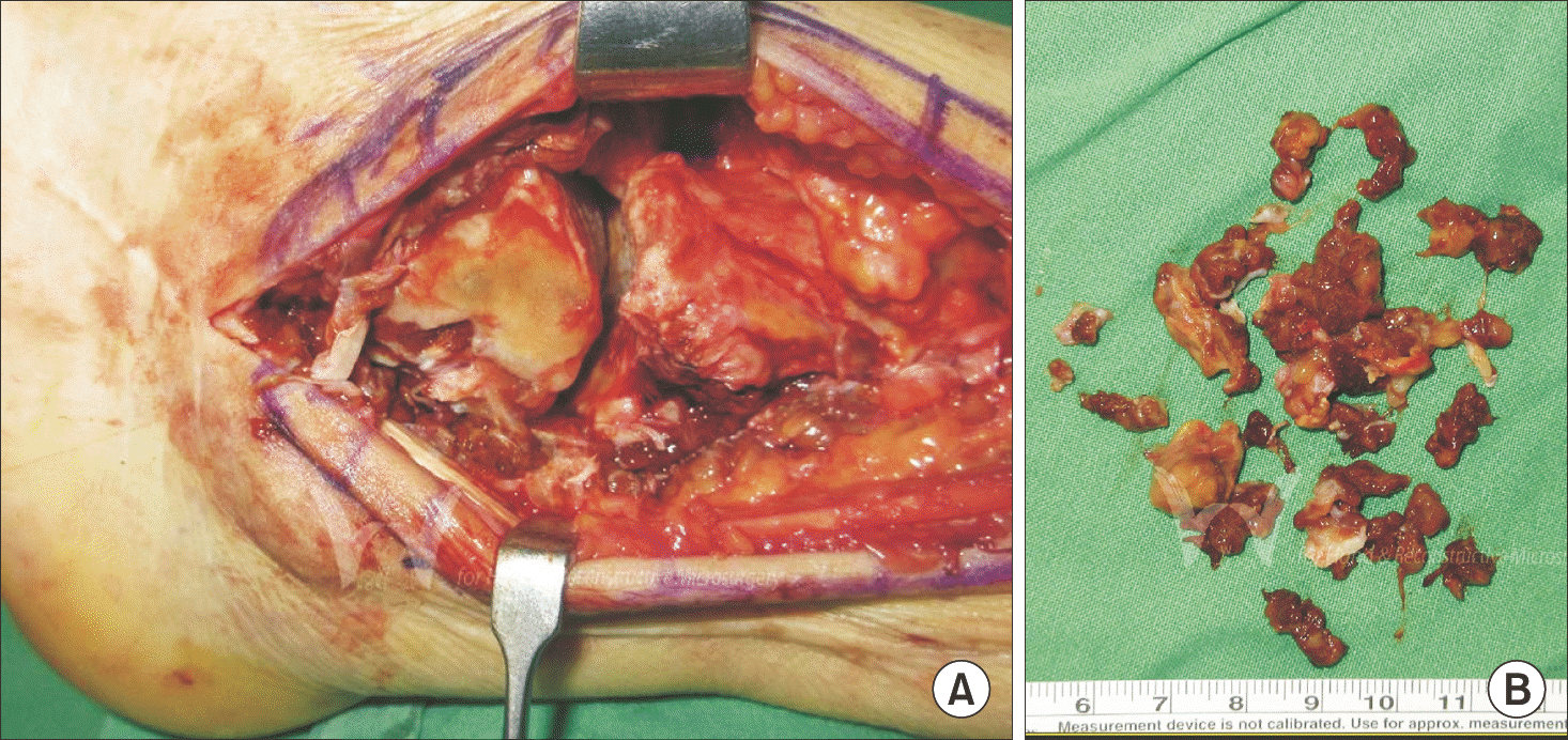
(A) Reddish synovial mass around the ankle joint. (B) Near complete synovectomy was performed through lateral approach. (C) Synovial mass was also infiltrated into tibiofibular articular surface of lateral malleolus.

Figure 3.
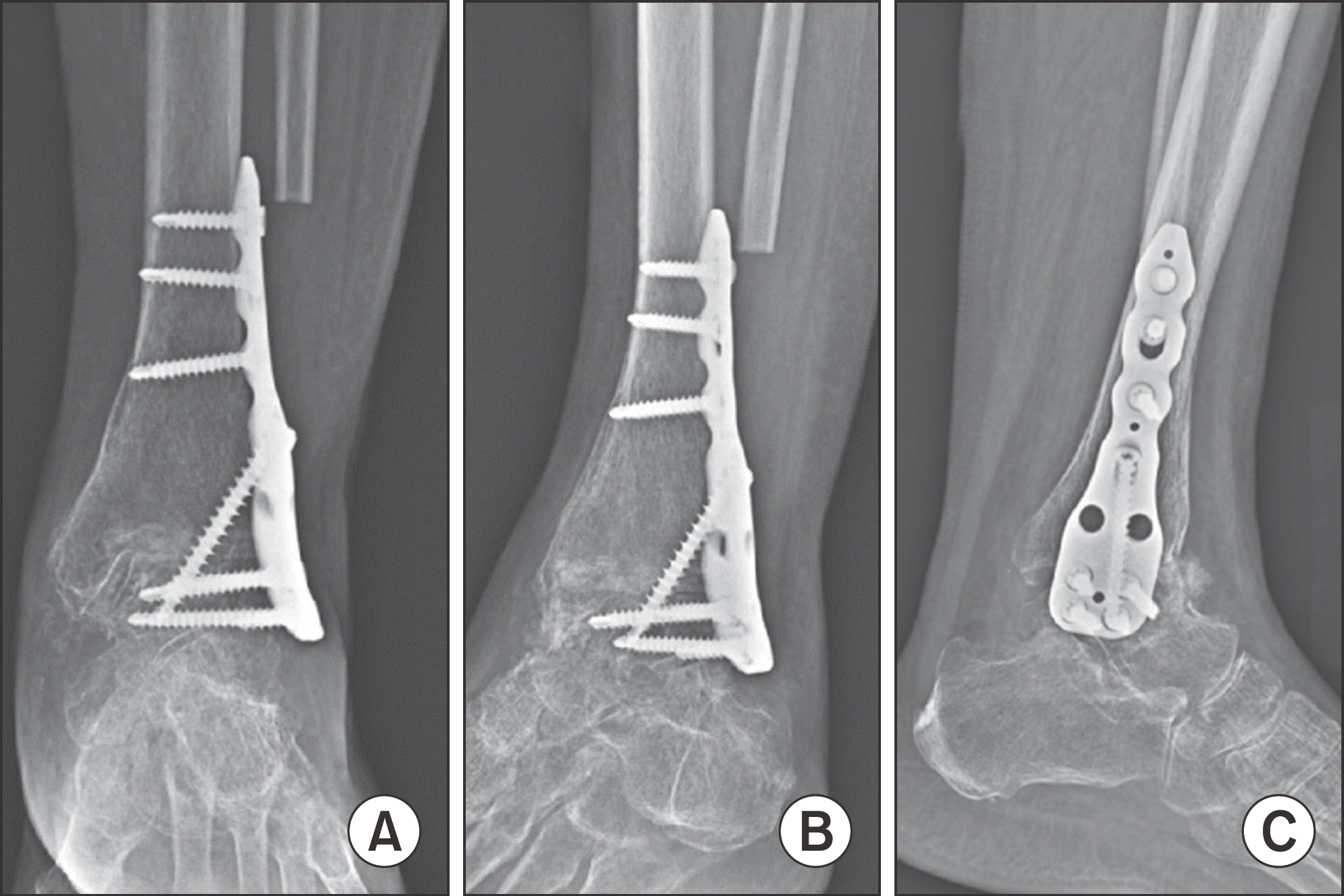
Immediate postoperative radiographs. Anteroposterior (A), mortise (B), and lateral (C) radiographs show a stable plate and screws fixation state.


 PDF
PDF ePub
ePub Citation
 Print
Print


 XML Download