

 PDF
PDF ePub
ePub Citation
 Print
Print


One of the most frequent injuries during sports activities is an ankle sprain. Previously, it was reported to account for up to 40% of all sports injuries.1) Patients presenting with ankle sprains account for 10% of emergency room visits in the United States with an incidence of 30,000 ankle sprains a day.2) However, severe open ankle sprain (SOAS) is known to be a rare injury, found in 0.22% of ankle injuries.3) We encountered a patient with SOAS and report the case with a review of the literature.
CASE REPORT
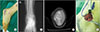
A 27-year-old woman was admitted to the hospital due to an inversion ankle injury while running the day before her visit. There was focal tenderness and an open wound of approximately 1-cm diameter over the lateral malleolus.
We could not identify a prominent fracture on the initial images of a simple radiograph. Considering the open wound site and the type of injury, we decided to perform computed tomography (CT) scans for additional information. Coronal CT images demonstrated bone fragment avulsion from the anteroinferior aspect of the distal tip of the fibula. Sagittal CT images demonstrated an irregular bone fragment in the anteroinferior aspect of the lateral malleolus. There was also an elevation of the periosteum at the tip of fibula. The injury location and imaging findings were suggestive of anterior talofibular ligament (ATFL) injury with an avulsed bone fragment from the tip of fibula. We decided to perform surgical treatment (Fig. 1).
After general anesthesia, the patient was placed in a lateral position. Routine aseptic drape was done. Curved longitudinal lateral incision, including the open wound, was performed. To avoid additional damage to the soft tissue, deep dissection was performed. There was an avulsed fragment from the distal tip of the fibula with ATFL tear. There was a connection between the skin wound and the bone fragment. The fractured fragment was too small to be fixed by a screw, so the fragment was fixed using an anchor with ATFL. After irrigation and hemostasis, we performed layered sutures.
The patient was asked not to bear weight for two weeks postoperatively. After that, gradual walking and ankle range of motion was allowed, followed by return to active daily living.
The report has been reviewed and approved by Institutional Review Board of Chungnam National University Hospital.
DISCUSSION
The ligaments of the lateral ankle are the ATFL, the calcaneofibular ligament, and the posterior talofibular ligament. The ATFL attaches to the distal end of the fibula and the lateral surface of the talus bone, having its center approximately 10 mm above the apex of the lateral malleolus.4) Histological observations of the structure and molecular composition of the ATFL helps to understand the pattern of injury to which the attachment sites of the ligament are subjected to during sporting activities. At the talar end, the ligament wraps with increased metachromasia to protect the talar enthesis from avulsion. Therefore, fewer avulsion fractures are observed at the talar end of ATFL.5) In this case, ATFL avulsion fracture occurred at the fibular end, and it caused an open wound due to the stretching force on the skin and the strong traction of the fragment.
Soubeyrand et al.3) described the hypermobility of the ankle joint as the reason due to which an open wound appears in case of ankle injury. The ligament around the ankle gets torn, resulting in hypermobility of the ankle joint, which causes the skin to tear because of excessive tension on the skin. SOAS is defined as a tear in a medial or lateral collateral ankle ligament combined with a tear in the overlying skin. SOAS has been reported to be very rare, occurring in 0.22% (9/4,142) of ankle injuries. SOAS generally occurs in high energy impact injuries, such as motor vehicle accidents or fall from heights, and is usually accompanied by a transverse skin wound, generally more than 3 cm in length. In recent years, however, there have been several reports of SOAS cases in relatively less severe injuries.67) SOAS occurred in a less severe injury in the patient discussed here, and was accompanied by a 1-cm long open wound. However, we could correctly diagnose the case due to the ATFL tear.
If an open wound without fracture or dislocation is present, SOAS should be considered as a differential diagnosis. This is because there may be invisible nerve or vascular injuries accompanying ligament injuries in SOAS.3) Operative treatment is needed to confirm the accompanying injuries and to treat the injured ligaments, along with appropriate antibiotics. Therefore, in case of open wound on the lateral or the medial malleolus, physicians must consider performing magnetic resonance imaging (MRI) or ultrasonographic examination after confirming the damage mechanism. MRI and ultrasonography can be helpful for a more accurate diagnosis of combined injuries (fracture, cartilage, tendon, or another ligament).8)
In conclusion, if an open wound is present without fracture or dislocation following an ankle sprain injury, SOAS should be considered as a differential diagnosis. This is because there is a large difference in treatment and prognosis between such an injury and a simple penetrating wound.
 XML Download