Background and Objectives
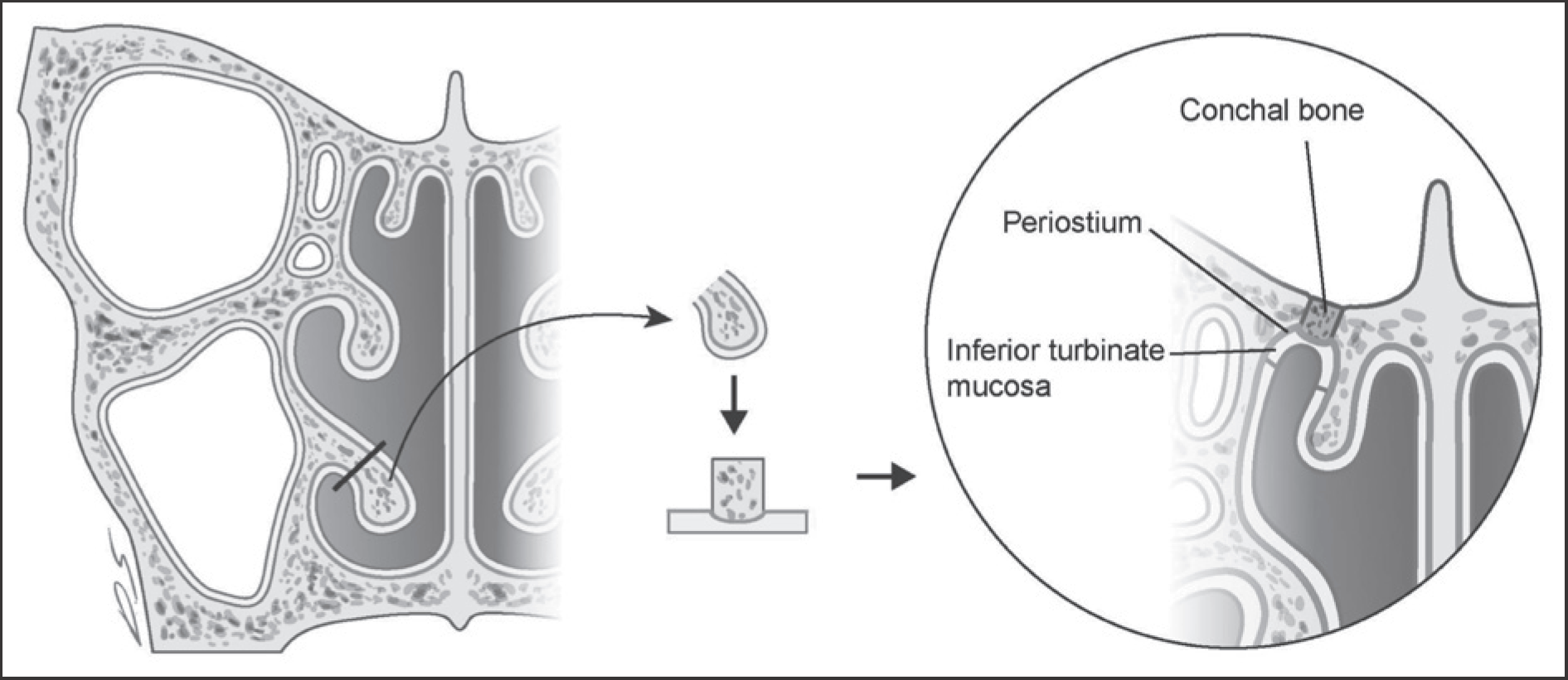
Endoscopic repair of cerebrospinal fluid (CSF) leak can avoid morbidity of open approaches and has shown a favorable success rate. Free mucosal graft is a good method, and multilayered repair is more favorable. The inferior turbinate has been commonly utilized for the free mucosal graft, but we newly designed it as a bone-periosteal-mucosal composite graft for multilayered reconstruction.
Subjects and Method
Four subjects with a skull base defect were treated with this method. The inferior turbinate was partially resected including the conchal bone and was trimmed according to defect size. Both bony parts and periosteum were preserved on the basolateral side of the mucosa as a composite graft. The graft was applied to the defect site using an overlay technique.
REFERENCES
1). Loew F., Pertuiset B., Chaumier EE., Jaksche H. Traumatic, spontaneous and postoperative CSF rhinorrhea. Adv Tech Stand Neurosurg. 1984. 11:169–207.

2). Stankiewicz JA. Cerebrospinal fluid fistula and endoscopic sinus surgery. Laryngoscope. 1991. 101:250–6.

3). Mirza S., Thaper A., McClelland L., Jones NS. Sinonasal cerebrospinal fluid leaks: management of 97 patients over 10 years. Laryngoscope. 2005. 115:1774–7.

4). Wigand ME. Transnasal ethmoidectomy under endoscopical control. Rhinology. 1981. 19:7–15.
5). Alexander A., Mathew J., Varghese AM., Ganesan S. Endoscopic Repair of CSF Fistulae: A Ten Year Experience. J Clin Diagn Res. 2016. 10:MC01–04.

6). Ibrahim AA., Okasha M., Elwany S. Endoscopic endonasal multilayer repair of traumatic CSF rhinorrhea. Eur Arch Otorhinolaryn-gol. 2016. 273:921–6.

7). Senior BA., Jafri K., Benninger M. Safety and efficacy of endoscopic repair of CSF leaks and encephaloceles: a survey of the members of the American Rhinologic Society. Am J Rhinol. 2001. 15:21–5.

8). Turgut S., Ercan I., Pinarci H., Beskonakli E. First experience with transnasal and transseptal endoscopic and microscopic repair of anterior skull base CSF fistulae. Rhinology. 2000. 38:195–9.
9). Harvey RJ., Smith JE., Wise SK., Patel SJ., Frankel BM., Schlosser RJ. Intracranial complications before and after endoscopic skull base reconstruction. Am J Rhinol. 2008. 22:516–21.

10). Bernal-Sprekelsen M., Bleda-Vazquez C., Carrau RL. Ascending meningitis secondary to traumatic cerebrospinal fluid leaks. Am J Rhinol. 2000. 14:257–9.

11). Bernal-Sprekelsen M., Alobid I., Mullol J., Trobat F., Tomas-Barberan M. Closure of cerebrospinal fluid leaks prevents ascending bacterial meningitis. Rhinology. 2005. 43:277–81.
12). Harvey RJ., Parmar P., Sacks R., Zanation AM. Endoscopic skull base reconstruction of large dural defects: a systematic review of published evidence. Laryngoscope. 2012. 122:452–9.

13). Patel MR., Stadler ME., Snyderman CH., Carrau RL., Kassam AB., Germanwala AV, et al. How to choose? Endoscopic skull base reconstructive options and limitations. Skull base: Official Journal of North American Skull Base Society. 2010. 20:397–404.

14). Kassam AB., Prevedello DM., Carrau RL., Snyderman CH., Thomas A., Gardner P, et al. Endoscopic endonasal skull base surgery: analysis of complications in the authors' initial 800 patients. Journal of Neurosurgery. 2011. 114:1544–68.

15). Harvey RJ., Nogueira JF., Schlosser RJ., Patel SJ., Vellutini E., Stamm AC. Closure of large skull base defects after endoscopic transnasal craniotomy. Clinical article. Journal of Neurosurgery. 2009. 111:371–9.
16). Pinheiro-Neto CD., Prevedello DM., Carrau RL., Snyderman CH., Mintz A., Gardner P, et al. Improving the design of the pedicled nasoseptal flap for skull base reconstruction: a radioanatomic study. Laryngoscope. 2007. 117:1560–9.

17). Chin D., Harvey RJ. Endoscopic reconstruction of frontal, cribiform and ethmoid skull base defects. Advances in oto-rhino-laryngology. 2013. 74:104–18.

18). Fortes FS., Carrau RL., Snyderman CH., Prevedello D., Vescan A., Mintz A, et al. The posterior pedicle inferior turbinate flap: a new vascularized flap for skull base reconstruction. Laryngoscope. 2007. 117:1329–32.

19). Amit M., Cohen J., Koren I., Gil Z. Cadaveric study for skull base reconstruction using anteriorly based inferior turbinate flap. Laryngoscope. 2013. 123:2940–4.

20). Cassano M., Felippu A. Endoscopic treatment of cerebrospinal fluid leaks with the use of lower turbinate grafts: a retrospective review of 125 cases. Rhinology. 2009. 47:362–8.

21). Zweig JL., Carrau RL., Celin SE., Schaitkin BM., Pollice PA., Snyderman CH, et al. Endoscopic repair of cerebrospinal fluid leaks to the sinonasal tract: predictors of success. Otolaryngology—head and neck surgery: official journal of American Academy of Otolaryngology-Head and Neck Surgery. 2000. 123:195–201.

22). Heo SJ., Kim JS. Endoscopic Management of Cerebrospinal Fluid Rhinorrhea. J Rhinol. 2014. 21(1):15–21.
Fig. 1
Schematic illustration showing procedures of inferior turbinate bone-periosteum-mucosal composite free graft for skull base defect.

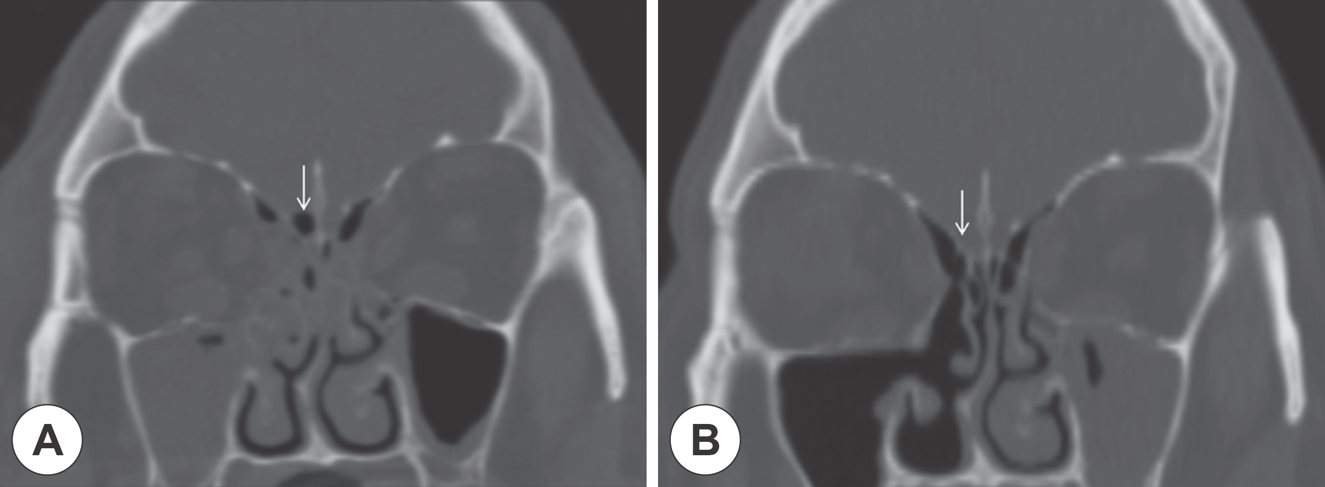
Fig. 2
Intraoperative photos of inferior turbinate bone-periosteum-mucosa free graft for skull base defect.


 PDF
PDF ePub
ePub Citation
 Print
Print


 XML Download