

 PDF
PDF ePub
ePub Citation
 Print
Print


Abstract
Intraoperative identification and localization of parathyroid glands are crucial step in preventing postoperative hypocalcemia during thyroid and parathyroid surgery. If there is a method to predict the parathyroid's location rather than detecting and verifying with naked eye, it would make the operator easier to find and identify the parathyroid. Recently, near-infrared light imaging technologies have been introduced in the fields of thyroid and parathyroid surgery to predict the localization of the parathyroid. These are being conducted in two ways: autofluorescence imaging with a unique intrinsic fluorophore in the parathyroid tissues and fluorescence imaging with external fluorescence materials specially absorbed into parathyroid tissues. We are suggest that parathyroid glands can be detected by surgeon with NIR autofluorescence imaging even if they are covered by fibrofatty tissues before they are detected by surgeon's naked eye. These novel techniques are very useful to identify and preserve parathyroid glands during thyroidectomy. In this article, we reviewed the latest papers that describe autofluorescence imaging and exogenous ICG fluorescence imaging of parathyroid glands during thyroid and parathyroid surgery.
Figures and Tables
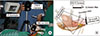 | Fig. 1Photos of set up and a graphically illustration. (A) A photo of equipment setting in the operating room. (B) Schematic diagram for detecting autofluorescence of parathyroid tissue with simultaneous illumination of the background tissues.
|
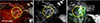
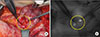 | Fig. 2Images after thyroid lobe lateralization. (A) Inferior and superior parathyroid glands is localized to be in the circle with visible light. (B) Autofluorescence with NIR illuminator image. Autofluorescence emissions coincided with parathyroid glands. Whole surgical field could be visualized by NIR illumination.
|
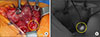 | Fig. 3Images that is taken immediately after paratracheal exposure without middle thyroid vein ligation. (A) Superior parathyroid underneath the fatty connective tissues was expected (circle), and still covered by background tissues with visible light. (B) Parathyroid glands was not yet exposed, but that was navigated and confirmed with NIR autofluorescence (circle).
|
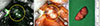
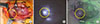 | Fig. 4Images after thyroid medialization. (A) Visible image by a normal digital camera the superior parathyroid gland was presumed (circle). (B) Near-infrared image was taken with room light-off. Parathyroid autoflorescence signal was stronger than thyroid signal. (C) Dual wavelength image by auto-para viewer (camera used in this experiment) with room light-on. The surgical field was visualized in its original form because part of the visible light entered the camera sensor.
|
 | Fig. 5Intraoperative images showing identification of normal parathyroid gland during retroauricular robotic (da Vinci Si system) thyroidectomy. (A) A right inferior parathyroid gland observed under visible light imaging (circle). (B) Ahead of administration of indocyanine green. The parathyroid gland was not clearly distinguishable with NIR mode imaging. (C) After administration of ICG. The fluorescence was observed from the both parathyroid gland (circle) and thyroid gland. The intensity of the parathyroid gland is not as prominent as thyroid gland. Parathyroid fluorescence become gradually marked the parathyroid gland was separated from the thyroid gland.
|
References
1. Hundahl SA, Fleming ID, Fremgen AM, Menck HR. A National Cancer Data Base report on 53,856 cases of thyroid carcinoma treated in the U.S., 1985–1995 [see comments]. Cancer. 1998; 83(12):2638–2648.

2. Suh YJ, Choi JY, Chai YJ, Kwon H, Woo JW, Kim SJ, et al. Indocyanine green as a near-infrared fluorescent agent for identifying parathyroid glands during thyroid surgery in dogs. Surg Endosc. 2015; 29(9):2811–2817.
3. Ahn D, Sohn JH, Kim JH, Park JY, Park J. Inadvertent parathyroidectomy during thyroid surgery for papillary thyroid carcinoma and postoperative hypocalcemia. J Korean Thyroid Assoc. 2012; 5(1):65–72.
4. Lee NJ, Blakey JD, Bhuta S, Calcaterra TC. Unintentional parathyroidectomy during thyroidectomy. Laryngoscope. 1999; 109(8):1238–1240.
5. Lin DT, Patel SG, Shaha AR, Singh B, Shah JP. Incidence of inadvertent parathyroid removal during thyroidectomy. Laryngoscope. 2002; 112(4):608–611.
6. Sasson AR, Pingpank JF Jr, Wetherington RW, Hanlon AL, Ridge JA. Incidental parathyroidectomy during thyroid surgery does not cause transient symptomatic hypocalcemia. Arch Otolaryngol Head Neck Surg. 2001; 127(3):304–308.
7. McWade MA, Paras C, White LM, Phay JE, Mahadevan-Jansen A, Broome JT. A novel optical approach to intraoperative detection of parathyroid glands. Surgery. 2013; 154(6):1371–1377. discussion 7.
8. Patel HP, Chadwick DR, Harrison BJ, Balasubramanian SP. Systematic review of intravenous methylene blue in parathyroid surgery. Br J Surg. 2012; 99(10):1345–1351.
9. Tummers QR, Schepers A, Hamming JF, Kievit J, Frangioni JV, van de Velde CJ, et al. Intraoperative guidance in parathyroid surgery using near-infrared fluorescence imaging and low-dose Methylene Blue. Surgery. 2015; 158(5):1323–1330.
10. Prosst RL, Gahlen J, Schnuelle P, Post S, Willeke F. Fluorescence-guided minimally invasive parathyroidectomy: a novel surgical therapy for secondary hyperparathyroidism. Am J Kidney Dis. 2006; 48(2):327–331.
11. Grubbs EG, Mittendorf EA, Perrier ND, Lee JE. Gamma probe identification of normal parathyroid glands during central neck surgery can facilitate parathyroid preservation. Am J Surg. 2008; 196(6):931–935. discussion 5-6.
12. Hyun H, Park MH, Owens EA, Wada H, Henary M, Handgraaf HJ, et al. Structure-inherent targeting of near-infrared fluorophores for parathyroid and thyroid gland imaging. Nat Med. 2015; 21(2):192–197.
13. Paras C, Keller M, White L, Phay J, Mahadevan-Jansen A. Near-infrared autofluorescence for the detection of parathyroid glands. J Biomed Opt. 2011; 16(6):067012.
14. Ladurner R, Sommerey S, Arabi NA, Hallfeldt KKJ, Stepp H, Gallwas JKS. Intraoperative near-infrared autofluorescence imaging of parathyroid glands. Surg Endosc. 2017; 31(8):3140–3145.
15. Falco J, Dip F, Quadri P, de la Fuente M, Rosenthal R. Cutting edge in thyroid surgery: autofluorescence of parathyroid glands. J Am Coll Surg. 2016; 223(2):374–380.
16. De Leeuw F, Breuskin I, Abbaci M, Casiraghi O, Mirghani H, Ben Lakhdar A, et al. Intraoperative near-infrared imaging for parathyroid gland identification by auto-fluorescence: A feasibility study. World J Surg. 2016; 40(9):2131–2138.
17. Brown EM, MacLeod RJ. Extracellular calcium sensing and extracellular calcium signaling. Physiol Rev. 2001; 81(1):239–297.
18. McWade MA, Sanders ME, Broome JT, Solorzano CC, Mahadevan-Jansen A. Establishing the clinical utility of autofluorescence spectroscopy for parathyroid detection. Surgery. 2016; 159(1):193–202.
19. McWade MA, Paras C, White LM, Phay JE, Solorzano CC, Broome JT, et al. Label-free intraoperative parathyroid localization with near-infrared autofluorescence imaging. J Clin Endocrinol Metab. 2014; 99(12):4574–4580.
20. Kim SW, Song SH, Lee HS, Noh WJ, Oak C, Ahn YC, et al. Intraoperative real-time localization of normal parathyroid glands with autofluorescence imaging. J Clin Endocrinol Metab. 2016; 101(12):4646–4652.
21. Kim SW, Lee HS, Ahn YC, Park CW, Jeon SW, Kim CH, et al. Near-infrared autofluorescence image-guided parathyroid gland mapping in thyroidectomy. J Am Coll Surg. 2018; 226(2):165–172.
22. Kim Y, Kim SW, Lee KD, Ahn YC. Real-time localization of the parathyroid gland in surgical field using Raspberry Pi during thyroidectomy: a preliminary report. Biomed Opt Express. 2018; 9(7):3391–3398.
23. Vahrmeijer AL, Hutteman M, van der Vorst JR, van de Velde CJ, Frangioni JV. Image-guided cancer surgery using nearinfrared fluorescence. Nat Rev Clin Oncol. 2013; 10(9):507–518.
24. Marshall MV, Rasmussen JC, Tan IC, Aldrich MB, Adams KE, Wang X, et al. Near-infrared fluorescence imaging in humans with indocyanine green: A review and update. Open Surg Oncol J. 2010; 2(2):12–25.
25. Alander JT, Kaartinen I, Laakso A, Patila T, Spillmann T, Tuchin VV, et al. A review of indocyanine green fluorescent imaging in surgery. Int J Biomed Imaging. 2012; 2012:940585.
26. Frangioni JV. In vivo near-infrared fluorescence imaging. Curr Opin Chem Biol. 2003; 7(5):626–634.
27. Miwa M. The principle of ICG fluorescence method. Open Surg Oncol J. 2010; 2(2):26–28.
28. Fox IJ, Wood EH. Indocyanine green: physical and physiologic properties. Proc Staff Meet Mayo Clin. 1960; 35:732–744.
29. Hope-Ross M, Yannuzzi LA, Gragoudas ES, Guyer DR, Slakter JS, Sorenson JA, et al. Adverse reactions due to indocyanine green. Ophthalmology. 1994; 101(3):529–533.
30. Vidal Fortuny J, Belfontali V, Sadowski SM, Karenovics W, Guigard S, Triponez F. Parathyroid gland angiography with indocyanine green fluorescence to predict parathyroid function after thyroid surgery. Br J Surg. 2016; 103(5):537–543.
31. Zaidi N, Bucak E, Okoh A, Yazici P, Yigitbas H, Berber E. The utility of indocyanine green near infrared fluorescent imaging in the identification of parathyroid glands during surgery for primary hyperparathyroidism. J Surg Oncol. 2016; 113(7):771–774.
32. Yu HW, Chung JW, Yi JW, Song RY, Lee JH, Kwon H, et al. Intraoperative localization of the parathyroid glands with indocyanine green and Firefly(R) technology during BABA robotic thyroidectomy. Surg Endosc. 2017; 31(7):3020–3027.
33. Lang BH, Wong CK, Hung HT, Wong KP, Mak KL, Au KB. Indocyanine green fluorescence angiography for quantitative evaluation of in situ parathyroid gland perfusion and function after total thyroidectomy. Surgery. 2017; 161(1):87–95.
34. Sound S, Okoh A, Yigitbas H, Yazici P, Berber E. Utility of indocyanine green fluorescence imaging for intraoperative localization in reoperative parathyroid surgery. Surg Innov. 2015; [Epub ahead of print].
 XML Download