

 PDF
PDF ePub
ePub Citation
 Print
Print


초록
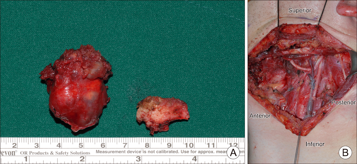
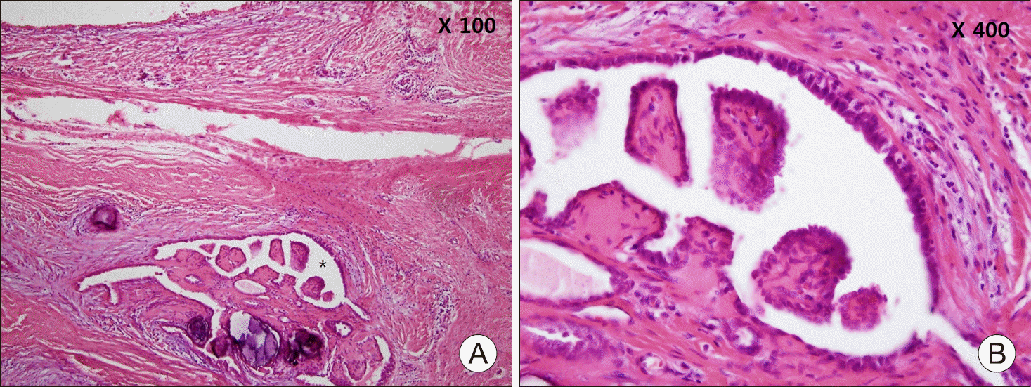
A thyroglossal duct cyst (TGDC) is the most common congenital anomaly of the neck. However, carcinoma arising from TGDC is extremely rare. We report 2 cases of TGDC carcinoma. In the first case, a 21-year-old male patient complained of an anterior cervical mass; computed tomography (CT) and sonography revealed cystic mass that was suspected to be a TGDC. Sistrunk operation was performed. Papillary carcinoma was confirmed in pathologic examination. Additionally, he underwent total thyroidectomy and central neck dissection. After radioactive iodine ablation (RAI) was performed. In the second case, a 28-year-old male patient visited our outpatient department complaining of submental mass. He had already been diagnosed TGDC carcinoma 13 years ago and had undergone Sistrunk operation and total thyroidectomy. Malignancy was confirmed using fine-needle aspiration; thus, lateral neck dissection was performed and following this, he underwent RAI. Till date, no evidence of recurrence has been observed in these patients.
REFERENCES
1). Mazzaferri EL. Thyroid cancer in thyroglossal duct remnants: a diagnostic and therapeutic dilemma. Thyroid. 2004; 14(5):335–6.

2). Wood CB, Bigcas JL, Alava I, Bischoff L, Langerman A, Kim Y. Papillary-type carcinoma of the thyroglossal duct cyst: the case for conservative management. Ann Otol Rhinol Laryngol. 2018; 127(10):710–6.
3). Kim CH, Han DH, Kim JH, Rho YS. A case of papillary carcinoma in thyroglossal duct cyst with mandibular invasion. Korean J Otolaryngol-Head Neck Surg. 2006; 49(12):1217–9.
4). LiVolsi VA, Perzin KH, Savetsky L. Carcinoma arising in median ectopic thyroid (including thyroglossal duct tissue). Cancer. 1974; 34(4):1303–15.
5). Weiss SD, Orlich CC. Primary papillary carcinoma of a thyroglossal duct cyst: report of a case and literature review. Br J Surg. 1991; 78(1):87–9.
6). Klubo-Gwiezdzinska J, Manes RP, Chia SH, Burman KD, Stathatos NA, Deeb ZE, et al. Clinical review: ectopic cervical thyroid carcinoma–review of the literature with illustrative case series. J Clin Endocrinol Metab. 2011; 96(9):2684–91.
7). Patel SG, Escrig M, Shaha AR, Singh B, Shah JP. Management of well-differentiated thyroid carcinoma presenting within a thyroglossal duct cyst. J Surg Oncol. 2002; 79(3):134–9. discussion 40-1.
8). Rayess HM, Monk I, Svider PF, Gupta A, Raza SN, Lin HS. Thyroglossal duct cyst carcinoma: a systematic review of clinical features and outcomes. Otolaryngol Head Neck Surg. 2017; 156(5):794–802.
9). Shemen L, Sherman CH, Yurovitsky A. Imaging characteristics and findings in thyroglossal duct cyst cancer and concurrent thyroid cancer. BMJ Case Rep. 2016; 2016.
10). Iftikhar H, Ikram M, Rizwan Nathani K, Yar Muhammad A. Papillary thyroid carcinoma within thyroglossal duct cyst: case series and literature review. Int Arch Otorhinolaryngol. 2018; 22(3):253–5.
Fig. 1.
Profile photo. There was 5×3 cm-sized, movable, round mass in left anterior neck, hyoid level (black arrows).
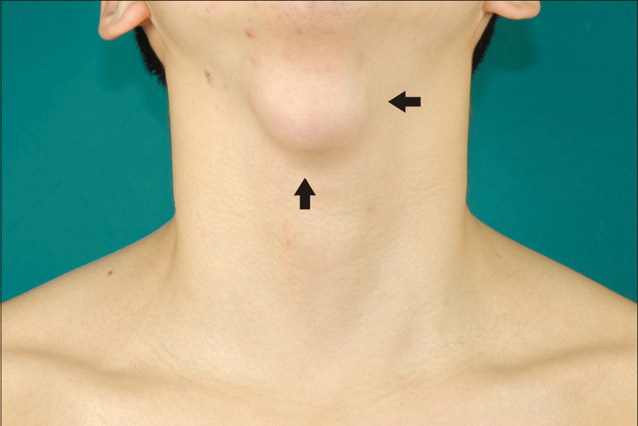
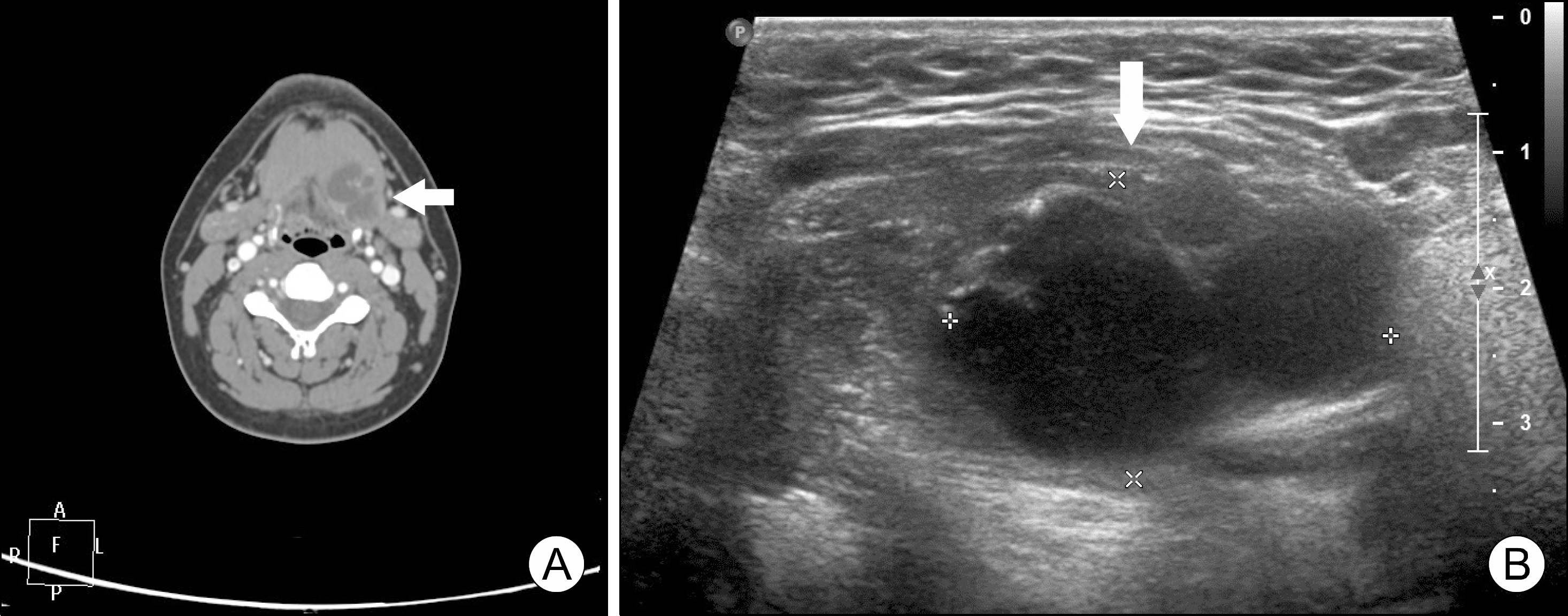
Fig. 2.
CT (A) and sono-graphic (B) finding. 4.1×2.6×2.8 cm nearly complete cystic nodule and 1.1 cm focal enhancing soft tissue within the cyst with stippled calcifi-cation were identified (white arrows).
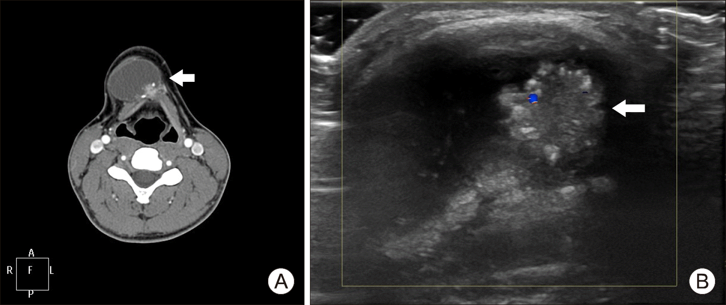
Fig. 4.
Profile photo. There was no other lesion except scars of previous Sistrunk operation and total thyroidectomy (black arrows).
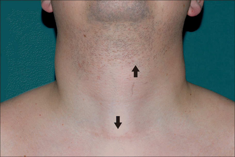
Fig. 5.
CT (A) and sono-graphic (B) finding. 2 cm- sized left submental cystic lesion (white arrows).
 XML Download