

 PDF
PDF ePub
ePub Citation
 Print
Print


Introduction
Antithyroid drugs (ATDs) including propylthiouracil (PTU), methimazole (MMI) and carbimazole (CMZ), a precursor of MMI, belong to the thionamide family, and are commonly used as first-line therapy for the management of Graves' disease (GD). Although ATDs are relatively safe, patients who are prescribed ATDs should be cautioned in regard to the potential side effects. These include rashes and joint pain, as well as major adverse effects such as agranulocytosis and hepatotoxicity, which typically occur early in the course of treatment.12) The side-effects of MMI are dose-dependent, whereas those of PTU do not show the same dose dependence. Vasculitis is the third most common major toxic reaction seen with ATD treatment, and is more often associated with PTU than with MMI. Anti-neutrophil cytoplasmic antibody (ANCA)-associated vasculitis has also been reported in connection with ATDs, especially in Asian patients treated with PTU. The risk of developing ANCA-associated vasculitis is increased with time, in contrast to other adverse effects seen with ATDs.23) The clinical features of drug-induced ANCA-associated vasculitis are highly variable and include a variety of symptoms, such as fever, myalgia, arthralgia and flu-like syndrome, as well as demonstrating multisystem involvement most commonly involving the kidneys, respiratory tract and skin. Vessels in the joints, eyes, skeletal muscle, gastrointestinal tract and central and peripheral nerves may also be involved.4) Although this syndrome generally resolves after drug cessation, high-dose glucocorticoid therapy or cyclophosphamide may be needed in severe cases.3)
Here we describe a case of ANCA-associated vasculitis manifesting as peripheral neuropathy in the lower extremities in a GD patient taking MMI.
Case Report
A 66-year-old woman presented to our endocrinologic outpatient clinic with palpitations, hand tremor, general weakness, weight loss (3 kg), chills and pain in both knees, which had developed over the previous 3 weeks. She had been diagnosed with GD 5 years previously, and was treated with MMI for the first 2 years in a local clinic. Her GD relapsed 1 year after discontinuing MMI treatment, and was then retreated with MMI for an additional 2 years. Several days prior to presenting to the clinic, her MMI dose was increased from 5 to 10 mg/day due to aggravation of thyrotoxicosis. She had also received acupuncture in both knees due to pain. Her past history included percutaneous intervention for acute myocardial infarction (AMI) 15 years prior and endoscopic submucosal dissection (ESD) for early-stage stomach cancer 4 years prior. She reported no specific diseases in her family history, including autoimmune thyroid disease, and was both a non-smoker and non-drinker. Upon physical examination, the patient was afebrile with a blood pressure of 98/58 mmHg and pulse rate of 93 beats/minute with sinus rhythm. Her thyroid gland was diffusely enlarged to three-times its normal size, and she exhibited a fine hand tremor and no ophthalmopathy. Thyroid function test results were as follows: free thyroxine (fT4) 2.57 ng/dL (0.93–1.71 ng/dL), thyrotropin (TSH) 0.005 mIU/L (0.27–4.20 mIU/L), anti-TSH receptor antibody (anti-TSH-R) 14.43 IU/L (0–1.75 IU/L), anti-thyroid peroxidase antibody (anti-TPO) 600.0 IU/mL (0–34 IU/mL), and anti-thyroglobulin antibody (anti-Tg) 522.3 IU/mL (0–115 IU/mL). Laboratory results showed normal white blood cell (WBC) count (8230/µL, 68.4% neutrophils) and normal hepatic and renal function. Her random plasma glucose level was 175 mg/dL and hemoglobin A1c was 6.1%. The MMI dose was increased from 10 to 20 mg daily along with 10 mg betaxolol daily as an additional beta-adrenergic blocker. However, 6 days later she was admitted for further evaluation and treatment due to aggravated general weakness, poor appetite and chills, especially at night, despite taking the medications.
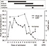
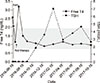
Upon admission, the vital signs of the patient (height 155 cm, body weight 55.5 kg) were as follows: blood pressure 106/50 mmHg, pulse rate 50 beats/min, respiratory rate 20/min, and body temperature 36.5℃. She had also been taking isosorbide-5-mononitrate 40 mg/day, trimetazidine dihydrochloride 40 mg/day, atorvastatin 10 mg/day and valsartan/hydrochlorothiazide 80/12.5 mg/day for previous AMI. After admission, she continued to be administered MMI 20 mg/day as well as all other medications except betaxolol and valsartan/hydrochlorothiazide due to her low pulse rate and blood pressure. Routine laboratory blood and urine analyses revealed normal findings, except for normocytic and normochromic anemia (hemoglobin 9.0 g/dL, 10.6 g/dL on ESD 4 years ago), increased total protein (TP): albumin ratio (TP 6.8 g/dL, albumin 2.8 g/dL, 3.4–4.8 g/dL) and an increased C-reactive protein (CRP; 4.78 mg/dL, 0–0.5 mg/dL) level. Serum iron (28 µg/dL, 37–158 µg/dL) and total iron-binding capacity (205 µg/dL, 228–428 µg/dL) were low with normal reticulocyte, ferritin, vitamin B12 and folate levels. Occult blood in stool was negative. Polyclonal gammopathy due to an acute-phase reaction, such as infection or inflammation, was suggested to explain the increased TP:albumin ratio, with immuglobulin G (IgG) 2560 mg/dL (700–1600 mg/dL), IgA 610 mg/dL (70–400 mg/dL), IgM 165 mg/dL (40–230 mg/dL), free kappa light chain 86.06 mg/L (3.3–19.4 mg/L), free lambda light chain 62.01 mg/L (5.71–26.3 mg/L), K/L ratio 1.33 (0.26–1.65) and suspicious polyclonal gammopathy, combined with increased acute-phase reactants in serum and urine protein electrophoresis. Rapid adrenocorticotropic hormone (ACTH) stimulation test was normal. Neck ultrasonography showed diffuse thyroid disease, and the fT4 level was 2.07 ng/dL with TSH 0.005 mIU/L. From the day after admission, she had chills and a fever reaching 39℃, usually at night, and also complained of a loss of sensation over the dorsum of the right foot and paresthesia in both dorsal feet, especially on the right side. She also described pain in both knees and morning stiffness lasting 1 month. She displayed no motor weakness, but showed hypoactive deep tendon reflex in the left ankle joint and distal hypoesthesia to pain upon neurological examination. No joint tenderness, swelling, local heating or redness was observed. There was only diffuse degeneration in both knee X-rays with negative bone scans. Blood, urine and sputum cultures were obtained, and ceftriaxone was initiated to control the fever. Computed tomographic images of the chest and abdomen and cultures for work-up of fever were normal. Serologic tests were positive for anti-nuclear antibody (Ab; 1:80, homogeneous type), rheumatoid factor (273 IU/mL; 0–15 IU/mL) and ANCA, and negative for anti-citrullinated protein (anti-CCP) Abs. Work-up for HIV, hepatitis B and hepatitis C were negative. The nerve conduction velocity (NCV) study suggested a right superficial peroneal nerve lesion with normal electromyography. Her fever and numbness on both dorsal feet still continued for 1 week and left foot drop suddenly developed. Motor weakness in the left ankle and left first toe were observed. She had a WBC count of 10,390/µL (76.9% neutrophils), CRP 5.37 mg/dL and ESR 63 mm/h (0–30 mm/h). She was referred to the rheumatology department with the diagnosis of peripheral neuropathy due to systemic vasculitis. The ANCA screen was positive for myeloperoxidase (MPO) Ab (3.20, negative ≤0.90) and negative for proteinase-3 (PR3) Ab. Anti-double stranded DNA Ab, lupus anticoagulant screening, cardiolipin IgM and IgG Abs, SS-B/La Ab, SS-A/Ro Ab, Scl-70 Ab, Smith Ab, Sm/ribonucleoprotein Ab, centromere Ab and beta-2 glycoprotein 1 Ab IgM were all negative. Tests for antibodies against leptospira, tsutsugamushi and Hantaan virus were all negative. The complement C3 and C4 levels were normal, as well as the anti-streptolysin O titer. Considering all of the clinical manifestations, the patient was strongly suspected to have peroneal nerve involvement associated with MPO-ANCA-associated vasculitis induced by MMI. To prevent disability due to permanent nerve damage, methylprednisolone 500 mg was administered intravenously as a high dose of glucocorticoid pulse therapy for 3 days, after which the dose was tapered, followed by intravenous administration of 500 mg of cyclophosphamide. In addition, after MMI treatment was stopped, free T4 and TSH levels were 1.15 ng/dL and 0.005 mIU/L respectively, and radioactive iodine (15 mCi of 131I) was administered for the definitive therapy of GD 1 week after MMI withdrawal. Fever and CRP levels quickly improved after glucocorticoid administration (Fig. 1), and the lower extremity paresthesia and pain and foot drop gradually recovered. She was subsequently stabilized with azathioprine 50 mg/day as a steroid-sparing drug with subsequent relief of symptoms, and the glucocorticoid was gradually tapered. Since then, she was prescribed methylprednisolone 2.5 mg/day, and is in a healthy, euthyroid state without medication (Fig. 2) for an 18-month period after the diagnosis of MPO-ANCA-positive vasculitis.
Discussion
ANCA are autoantibodies directed against primary azurophil granules in polynuclear neutrophils and lysosomes in monocytes.5) There are two major patterns of ANCA immunostaining, as determined by indirect immunofluorescence, cytoplasmic (c-ANCA) and perinuclear (p-ANCA). As uncovered using enzymelinked immunosorbent assay (ELISA), PR3 is the major target of c-ANCA (PR3-ANCA), which is associated with Wegener's granulomatosis, whereas p-ANCA reacts with MPO as well as other myeloid proteins (MPO-ANCA) and is mainly associated with microscopic polyangiitis and Churg-Strauss syndrome.456) A number of drugs including ATDs, have been implicated in the induction of ANCA production, and are generally reported to be associated with p-ANCA and MPO-ANCA.5)
ANCA-associated vasculitis is histologically characterized by inflammatory and necrotic processes that selectively affect the walls of small vessels such as small arteries and arterioles, capillaries and venules.5) Cases of ANCA-associated vasculitis are rare, but can be triggered by chemicals, infections and certain drugs.6) The pathogenic role of ANCA in the onset of small-vessel vasculitis has been demonstrated by the involvement of proinflammatory cytokines, stimulation of the expression of adhesion molecules on endothelial cells, and the extracellular translocation of granular contents to the outside of the cell membrane. In the vessel wall, neutrophil activation leads to the production and release of toxic contents that induce cellular alterations. Finally, complement activation induced by ANCA and MPO or PR3 interactions further reinforce the inflammatory events, leading to damage of the blood vessels.56)
All ATDs can induce asymptomatic production of ANCA, but PTU is most often associated with ANCA-associated vasculitis.7) A recent review including approximately 260 case reports of ANCA-associated vasculitis related to ATDs showed that 75% of these cases were associated with the use of PTU compared with only 25% with MMI.5) The estimated incidence ANCA-associated vasculitis for PTU was calculated to be 39.2-times that for MMI.8) The prevalence of ANCA-positive cases caused by ATDs varied between 4% and 64% for PTU (median 30%) and 0% and 16% with MMI (median 6%).5) During ATD administration in patients with GD, 21% of patients were found to be for ANCA positivity irrespective of the type of ATD administered. Only 15% of ANCA-positive patients who received ATD treatment actually presented with clinical manifestations of ANCA-associated vasculitis, corresponding to 3% of all GD patients who received ATD treatment.5) The estimated annual incidence of ANCA-associated vasculitis by ATDs was between 0.53 and 0.79 patients per 10,000 people.8) In addition, there was an overall trend between the onset of ANCA positivity and the duration of ATD administration.5) The median time until onset of ANCA-associated vasculitis was 42 months after starting ATD treatment as a whole, specifically 39 months for PTU and 60 months for MMI.8) The median dose at onset was 200 mg/day for PTU and 15 mg/day for MMI.8) Clinical vasculitis was more frequent when the MPO-ANCA titers were high. There was a trend toward a decrease in ANCA titers after ATD discontinuation, although these titers remained positive in 40–60% of cases.5) As GD is an autoimmune disease, it has a tendency to develop along with other autoimmune conditions. However, the prevalence of ANCA positivity in GD patients before initiating ATD therapy was shown to be between 0% to 13%, which was strikingly lower than the prevalence observed during (or after) ATD therapy.5) Also, the presence of ANCAs in patients with GD before ATD treatment was predominantly with anti-bactericidal/permeability-increasing protein (BPI)-ANCA and anticathepsin-ANCA, and were different from the ANCAs, usually MPO-ANCA, that developed after initiating ATD therapy.5) These observations clearly highlight the specific role of ATD in the development of ANCA-associated vasculitis.
The clinical features related to ANCA-associated vasculitis are polymorphic and usually severe, potentially affecting several organs such as the skin, kidneys, upper and lower respiratory tract, brain, nerves, musculoskeletal tissue, gastrointestinal tract, ear, nose and throat tissues and eyes.5) The clinical and serological profiles of idiopathic and drug-induced ANCA-associated vasculitis are similar. However, patients with ATD-induced vasculitis usually have a milder course with less frequent renal, pulmonary and neurologic manifestations than with idiopathic vasculitis.79) In addition, patients with ATD-induced vasculitis had more frequent skin manifestations, resulting in a shorter duration of symptoms before diagnosis.7) In general, prognosis of ATD-induced vasculitis is good. All 56 patients with idiopathic vasculitis were treated with immunosuppressive therapy, of which six patients died during the first episode and 47 patients out of the remaining 50 had at least one relapse, while 16 ATD-treated patients stopped ATD treatment and only six patients were treated with immunosuppressive therapy with no disease relapse.7)
The treatment of ANCA-associated vasculitis induced by ATDs is to stop ATD therapy immediately, which often leads to rapid clinical improvement and an overall good prognosis. When no improvement is observed after ATD withdrawal, the treatment is similar to those treatments for primary vasculitis in general, including high-dose glucocorticoids and immunosuppressive drugs such as cyclophosphamide and cyclosporine.5)
Recent data highlight the mechanisms leading to ANCA-associated vasculitis secondary to the administration of ATD.5) Researchers have suggested that there may be PTU-induced alterations in the configuration of granules present in polymorphonuclear neutrophils containing MPO, leading to the formation of anti-MPO antibodies. Other authors have proposed that PTU could bind MPO within neutrophils, thereby inducing the formation of intracellular cytotoxic products and leading to cellular alterations within vascular walls and promotion of autoimmune production of ANCA in susceptible individuals. Another hypothesis for the predominance of PTU-associated ANCA production is based on the finding that PTU (and not MMI) accumulates in neutrophils and is oxidized by MPO to a highly reactive intermediate form, which, in turn, induces T-cell sensitization.5) An alternative hypothesis stems from the 40% molecular homology between MPO and thyroperoxidase, and claims that epitopic crossover between the two molecules could promote ANCA production. Finally, a recent study reported the abnormal conformation of neutrophil extracellular traps (NETs) generated in the presence of PTU and neutrophil cell death.57)
The present patient presented general weakness, poor appetite, chills, pain in both knees as well as thyrotoxic symptoms including palpitations, hand tremor and weight loss during MMI treatment for GD. These symptoms may be manifestations of GD itself, as side effects of ATD or those of other diseases, especially in elderly patients. Her clinical manifestations, excluding the thyrotoxic symptoms, were aggravated despite the improved thyroid function, and she showed no agranulocytosis or hepatotoxicity. Her fever was unresponsive to antibiotics, and work-up for fever showed normal results. Elevated CRP levels and positivity for MPO-ANCA as well as her history of ATD use suggested peripheral neuropathy caused by ATD-induced MPO-ANCA positive vasculitis, although we did not perform a nerve biopsy due to fear of nerve damage. A few cases of ATD-induced vasculitis with neurological involvement have been reported. The first patient who developed polyneuropathy during CMZ treatment for thyrotoxicosis was reported in 1984.10) In a series of 16 ATD-induced vasculitis patients (12 receiving PTU and four receiving MMI), none manifested central nervous system (CNS) effects, while only one patient had peripheral sensory polyneuropathy that regressed upon drug withdrawal.7) There have been two separate reports of cases of CNS vasculitis caused by MMI and PTU.911) In a series of 92 Japanese patients with ATD-induced vasculitis, three patients had CNS manifestations, all of whom were taking PTU, but none involved the peripheral nervous systems (PNS).8) In addition, there were two reports of neurological system involvement by PTU-induced vasculitis.1213) To the best of our knowledge, our case is the third case of PNS involvement and the first case of PNS involvement associated with MMI.
In conclusion, ATD-induced vasculitis is an extremely rare adverse effect with various clinical manifestations. Clinicians should be aware of this uncommon side effect of ATDs, which should be included in differential diagnosis. Early recognition of this adverse effect is important because immediate withdrawal of ATD is needed to prevent serious sequelae.
 XML Download