

 PDF
PDF ePub
ePub Citation
 Print
Print


Abstract
Synovial chondromatosis is an uncommon disorder characterized by cartilaginous proliferation within the synovial membrane of the articular joint. Smaller joints are rarely affected and it may be progressed to osteochondromatosis after ossification or calcification of metaplastic cartilage. It is commonly presented in the third to fourth decade of life, but rarely presented in adolescence. We report a unique case of synovial osteochondromatosis of the subtalar joint in 14-year-old baseball player. Arthroscopic removal of loose body and complete excision of the osteochondral mass with concomitant synovectomy resulted in satisfactory outcome without recurrence at final follow-up.
Figures and Tables
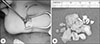
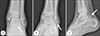 | Fig. 1Preoperative plain radiographs of left ankle. (A) Anteroposterior view showed radio-opaque area near the subtalar joint (arrow). Mortis (B) and lateral (C) views showed innumerable subcentimeter loose bodies within posterior subtalar joint (arrows).
|
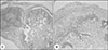
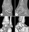 | Fig. 2Computed tomographic images of the left ankle. Multiple high-density loose bodies were demonstrated in subtalar joint space on coronal (A) and sagittal (B) images. (C, D) Three-imensional reconstruction images showed the main clusters of multiple loose bodies were in posterior subtalar joint and the remainders were scattered into sinus tarsi.
|
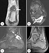 | Fig. 3Magnetic resonance imaging of the left ankle. (A) The T1-weighted coronal image demonstrated a lobulated low signal intensity mass (arrow). (B) T2-weighted axial image showed multiple osteochondral and chondral loose bodies which demonstrated signal void or low signal intensity (arrow) surrounded by an area of high signal intensity effusion. Low signal intensity cyst-like synovial lining (arrowheads) was extended from posterior subtalar joint on (C) T2-weighted and (D) proton density image, and signal void loose bodies were identified in sinus tarsi.
|
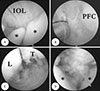 | Fig. 4Subtalar arthroscopic photographs through the lateral portal (A, B) and the posterolateral portal (C, D). (A) Multiple cartilaginous loose bodies (asterisks) and minimal synovial proliferation were shown around talocalcaneal interosseous ligament (IOL). After removal of loose bodies, (B) synovectomy was performed in sinus tarsi. Yellowish nodular cartilaginous metaplasia and accompanying loose bodies were shown in (C) lateral and (D) medial gutters of posterior subtalar joint. Asterisks indicate multiple loose bodies. PFC: posterior articular facet of calcaneus, L: lateral malleolus, T: talus.
|
References
1. Jacob RA, Campbell WP, Niemann KM. Synovial chondrometaplasia: a case report. Clin Orthop Relat Res. 1975; (109):152–154.
2. Maurice H, Crone M, Watt I. Synovial chondromatosis. J Bone Joint Surg Br. 1988; 70:807–811.

3. Kim JB, Song IS, Shin SY, Yoon JY. Primary synovial chondromatosis of the talonavicular joint: a case report. Foot Ankle Surg. 2016; 22:e25–e28.
4. Suh JS, Kim JH, Kim JI, Kim HS. Synovial chondromatosis of the Ankle Joint. J Korean Foot Ankle Soc. 2009; 13:14–18.
5. Hocking R, Negrine J. Primary synovial chondromatosis of the subtalar joint affecting two brothers. Foot Ankle Int. 2003; 24:865–867.
6. Milgram JW. Synovial osteochondromatosis: a histopathological study of thirty cases. J Bone Joint Surg Am. 1977; 59:792–801.
7. Hsueh CJ, Huang GS, Juan CJ, et al. Synovial chondroma of the ankle in a young child after recent trauma: CT and MR features. Clin Imaging. 2001; 25:296–299.
8. Hentges MJ, Lee MS. Chronic ankle and subtalar joint instability in the athlete. Clin Podiatr Med Surg. 2011; 28:87–104.
9. Kadakia AP, Sarkar J. Osteochondritis dissecans of the talus involving the subtalar joint: a case report. J Foot Ankle Surg. 2007; 46:488–492.
10. Samson L, Mazurkiewicz S, Treder M, Wisniewski P. Outcome in the arthroscopic treatment of synovial chondromatosis of the knee. Ortop Traumatol Rehabil. 2005; 7:391–396.
 XML Download