

 PDF
PDF ePub
ePub Citation
 Print
Print


Abstract
Purpose
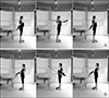
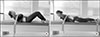
This study analyzed the muscle activity changes induced by motions of reaching forward and chest expansion that were examined from the bilateral muscles with rectus abdominis, external oblique, multifidus, and longissimus thoracic using Pilates cadillac instrument.
Methods
Nine young adult women, who have no musculoskeletal disorder and any of chronic diseases, were participated. Surface electromyography system was used for recording of all signals produced by muscles, and then normalized as percentage of maximum voluntary isometric contraction (%MVIC). The paired t-test and repeated measures of analysis of variance was performed.
Results
Reaching-forward motion showed a higher muscle activity from non-dominant external oblique muscle than that of the chest-expansion motion. During both reaching-forward motion and chest-expansion motion, MVIC values collected from dominant side of external oblique muscle were shown a significantly lower than the values obtained from non-dominant side (p<0.05). Conversely, %MVIC values in external oblique muscle collected from dominant side showed a significantly higher than the values obtained from non-dominant side of the same oblique muscle (p<0.05). Reaching-forward motion was caused a higher %MVIC on non-dominant external oblique muscle than that of the chest-expansion motion (p<0.05). Regardless of dominant or non-dominant sides, external oblique muscle was shown the highest activation rate of all the other muscles during reaching forward action, and longissimus thoracic muscle was shown the highest activation rate of all the other muscles during chest expansion action.
Figures and Tables
 | Fig. 2A measurement of voluntary maximal isometric contraction. (A) Rectus abdominis and external obliques. (B) Multifidus and longissimus thoracis.
|
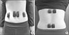 | Fig. 3Location of surface electromyography sensor attachment. (A) Rectus abdominis and external obliques. (B) Multifidus and longissimus thoracis.
|
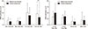 | Fig. 4Comparison of electromyography (EMG) activation from the muscles between dominant and non-dominant, and between ventral region and dorsal region. Rec Abd: rectus abdominis, D: dominant, N: non-dominant, Ext Obl: external obliques, Longi Thor: longissimus thoracis, Multi: multifidus. **Significant difference to the mode of operation (p<0.01).
|
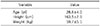
Table 2
Comparison of muscle activations during reaching forward movement between dominant and non-dominant body sides while maximal voluntary isometric contraction was performing
![]()
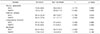
Table 3
Comparison of muscle activations during the chest expansion movement between dominant and non-dominant body sides while maximal voluntary isometric contraction was performing
![]()
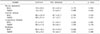
Table 5
%MVIC comparison between muscles according to movement type (n=7)

Values are presented as mean±standard deviation. The unit of values is %MVIC.
%MVIC, percentage of maximum voluntary isometric contraction, D: dominant, N: non-dominant.
aSignificantly different to rectus abdominis (p<0.05); bSignificantly different to external oblique (p<0.05); abSignificantly different to rectus abdominis and external oblique (p<0.05).
![]()
References
1. Ferreira PH, Ferreira ML, Hodges PW. Changes in recruitment of the abdominal muscles in people with low back pain: ultrasound measurement of muscle activity. Spine (Phila Pa 1976). 2004; 29:2560–2566.
2. Cholewicki J, VanVliet JJ 4th. Relative contribution of trunk muscles to the stability of the lumbar spine during isometric exertions. Clin Biomech (Bristol, Avon). 2002; 17:99–105.

3. Aruin AS, Latash ML. Directional specificity of postural muscles in feed-forward postural reactions during fast voluntary arm movements. Exp Brain Res. 1995; 103:323–332.
4. Muscolino JE, Cipriani S. Pilates and the “powerhouse”-I. J Bodyw Mov Ther. 2004; 8:15–24.
5. Latey P. The Pilates method: history and philosophy. J Bodyw Mov Ther. 2001; 5:275–282.
7. Isacowitz R, Clippinger KS. Pilates anatomy. Champaign: Human Kinetics;2011.
8. Siqueira Rodrigues BG, Ali Cader S, Bento Torres NV, Oliveira EM, Martin Dantas EH. Pilates method in personal autonomy, static balance and quality of life of elderly females. J Bodyw Mov Ther. 2010; 14:195–202.
9. Herrington L, Davies R. The influence of pilates training on the ability to contract the transversus abdominis muscle in asymptomatic individuals. J Bodyw Mov Ther. 2005; 9:52–57.
10. Sekendiz B, Altun O, Korkusuz F, Akin S. Effects of pilates exercise on trunk strength, endurance and flexibility in sedentary adult females. J Bodyw Mov Ther. 2007; 11:318–326.
11. Park HS. The effects of pilates mat exercise in 12 weeks on middle-aged womens' blood lipid level, body composition and lumbar muscle [dissertation]. Cheongju: Seowon University;2009.
12. Kloubec JA. Pilates for improvement of muscle endurance, flexibility, balance, and posture. J Strength Cond Res. 2010; 24:661–667.
13. Lee J, Seo SJ. A study of middle aged women, on their muscle activity and subjective analysis of the body according to pilates positions. Official J Korean Soc Dance Sci. 2016; 33:109–123.
14. Kim WK. The effect of pilates reformer exercise on body composition, electromyogram of abdominal muscles and blood free oxygen radical in middle age women [dissertation]. Gwangju: Chosun University;2017.
15. Cram JR, Kasman GS, Holtz J. Introduction to surface electromyography. Gaithersburg: Aspen;1998.
16. Won M, Kim M, Kim S, Lee J. The effect of visual information provision on the changes of electromyogram activity in trunk and lower leg muscles during dynamic balance control. Korean J Sports Med. 2014; 32:44–54.
17. Im SY, Kim SJ, Hur SH, An KJ, Lee JS. The effect of regular aquatic exercise on balance capacity, physical fitness and performance level, and muscular activity in elderly women arthritis patients. J Korean Phys Educ Assoc Girls Women. 2014; 28:37–54.
18. Kim SJ, Lee JS. The effect of multi-axis sling suspension exercise on trunk-muscle activation. Exerc Sci. 2008; 17:317–330.
19. Finni T, Cheng S. Variability in lateral positioning of surface EMG electrodes. J Appl Biomech. 2009; 25:396–400.
20. Carpes FP, Reinehr FB, Mota CB. Effects of a program for trunk strength and stability on pain, low back and pelvis kinematics, and body balance: a pilot study. J Bodyw Mov Ther. 2008; 12:22–30.
21. Hibbs AE, Thompson KG, French D, Wrigley A, Spears I. Optimizing performance by improving core stability and core strength. Sports Med. 2008; 38:995–1008.
22. Kerr A. Introductory biomechanics e-book. Milton: Elsevier Health Sciences;2010.
23. Sahrmann SA. Diagnosis and treatment of movement impairment syndromes. St. Louis: Mosby;2002.
24. Knapik JJ, Reynolds KL, Harman E. Soldier load carriage: historical, physiological, biomechanical, and medical aspects. Mil Med. 2004; 169:45–56.
25. Kim JJ, Lee MH, Kim YJ, Chae WS, Han YS, Kwon SO. Comparison of the maximum EMG levels recorded in maximum effort isometric contractions at five different knee flexion angles. Korean J Sports Biomech. 2005; 15:197–206.
26. Schega L, Bertram D, Folsch C, Hamacher D, Hamacher D. The influence of visual feedback on the mental representation of gait in patients with THR: a new approach for an experimental rehabilitation strategy. Appl Psychophysiol Biofeedback. 2014; 39:37–43.
27. Nam HS, Kim JH. Effects of arm swing with a light weight on anticipatory postural adjustment and postural stability in standing. J Korea Soc Neurotherapy. 2017; 21:31–37.
28. Hall CM, Brody LT. Therapeutic exercise: moving toward function. 1st ed. Philadelphia: Lippincott Williams and Wilkins;1999.
29. Stevens VK, Coorevits PL, Bouche KG, Mahieu NN, Vanderstraeten GG, Danneels LA. The influence of specific training on trunk muscle recruitment patterns in healthy subjects during stabilization exercises. Man Ther. 2007; 12:271–279.
 XML Download